
Abstract
Human lung cancer cell lines A549 and A549/DDP, and ovarian cancer cell lines SKOV3 and SKOV3/DDP were subjected to thioridazine (Thio), cisplatin, or the combination; A549/DDP and SKOV3/DDP were the cisplatin-resistant sublines. Cell viability, apoptosis, and cell cycle were detected; the mitochondrial membrane potential and proteins related to mitochondrial apoptosis were determined. Thio induced cell death, and the combination of Thio and cisplatin led to the highest percentage of dead cells in four cells lines. Thio and the combined modality led to cell apoptosis by inducing G0/G1 arrest. The collapse of mitochondrial membrane potential, activation of caspase 9, upregulation of Bax protein, and downregulation of Bcl-2 protein demonstrated that apoptosis was mitochondria dependent. These data indicated that Thio could be used to modulate cisplatin-based chemotherapeutic regimen in lung and ovary cancers.
Introduction
Cisplatin (DDP)
Thioridazine (Thio) is an antischizophrenic drug (Ly et al., 2002; Fenton et al., 2007). Recent data have manifested that Thio can suppress cancer cells of ovary, uterine cervix, esophagus, melanoma, and glioblastoma, including tumor stem cells (Cheng et al., 2015; Mao et al., 2015; Shen et al., 2017; Jiang et al., 2018; Chu et al., 2019). Thio binds to the dopamine receptor, and then causes cytotoxicity through the apoptosis and/or autophagy pathways. Dopamine receptors express in cell membranes of lung and ovary cancers, and therefore these cells can respond to Thio (Spengler et al., 2016). However, the responsive difference between sensitive and resistant cells remains unclear.
In this study, Thio was employed to enhance DDP in lung and ovary cancer cells, and the response between sensitive and resistant cells was compared. Preliminary data indicated that Thio can deactivate resistant cells and that Thio can sensitize DDP in both cell types.
Materials and Methods
Cells
Human cancer cell lines A549, A549/DDP, SKOV3, and SKOV3/DDP were cultured in RPMI-1640 medium (GIBCO, Beijing, China) supplemented with 10% fetal bovine serum (GIBCO), at 37°C and 5% CO2. Tissue type of A549 was lung cancer and that of SKOV3 was ovarian cancer; A549/DDP and SKOV3/DDP were the resistant sublines, which can grow in 2 and 0.75 μg/mL of DDP, respectively. Cells were transferred into DDP-free medium for 7 days before experiments, thereby avoiding interferences due to residual DDP (Yu et al., 2016).
Cell viability
Cells were seeded in a 96-well plate (5 × 10³ cells/well) and exposed to Thio (Sigma-Aldrich, Darmstadt, Germany) and/or DDP (Qilu Pharm. Co. Ltd., Jinan, China). Cells were subjected to DDP in group DDP, to Thio in group Thio, and to Thio combined with DDP in group Thio + DDP.
Cells were subjected to Thio (0, 2.5, 5, 10, 20, and 40 μM) or DDP (0, 5, 10, 20, 40, 80, 160, and 320 μM) for 4 h, and then drugs were washed away. In the combined regimen, the level of Thio was 20 μM and that of DDP was 40 μM, which were determined according to the cell-survival curve. Therefore, both the drug level and value of “drug concentration × exposure time” were within the range of human pharmacokinetics, having clinical relevancy (Yu et al., 2016). Cells viability was determined with a CCK-8 assay after 24 h (Dojindo Lab., Kumamoto, Japan).
Detection of apoptotic cells and cell cycle
Apoptotic cells were detected using the Annexin V assay (Keygen Biotech., Nanjing, China), and cell cycle was determined with flow cytometry after propidium iodide staining.
Quantifying the combined effect
Percentages of dead cells (1 percentage of survival cells) were used to calculate the combination index (CI), thereby evaluating the drug interaction (He et al., 2012).
EA+B was the effect of combination, and EA /EB was the effect of single agent. A CI of >1.15, 0.85–1.15, or <0.85 indicated synergy, addition, or antagonism, respectively.
Determination of mitochondrial membrane potential
The mitochondrial membrane potential (MMP) was fluorophotometrically determined with the JC-1 assay (Invitrogen, Eugene, OR). Cells were seeded in a 6-well plate (2 × 10⁵ cells/well) for 24 h and then treated with Thio (20 μM) and/or DDP (40 μM) for 4 h. Cells were stained with JC-1 (5 μg/mL) for 30 min and washed with phosphate-buffered saline for three times. λex was 485 nm and λem was 529 or 590 nm. The ratio of red to green fluorescence reflected the potential (Perelman et al., 2012).
Determination of the activity of caspase 9
The activity of caspase 9 was determined using a luminescent assay after 24 h (Promega, Madison, WI).
Western blot
Proteins were extracted using the RIPA kit (Beyotime Biotechnol., Shanghai, China). Proteins (30 μg/well) were separated by 15% sodium dodecyl sulfate-polyacrylamide gel electrophoresis and transferred onto a polyvinylidene fluoride membrane. Rabbit monoclonal antibodies against Bax, Bcl-2, and caspase-3 (Cell Signaling Technol., Danvers, MA) were used; β-actin served as the reference, with a rabbit polyclonal antibody (Cell Signaling Technol.). The secondary antibody was a goat-anti-rabbit IgG antibody (Sigma-Aldrich). Proteins were visualized using an enhanced chemiluminescence kit (Pierce Biotechnol., Rockford, IL).
Statistics
The statistical analysis was performed with the software SPSS 17.0 (Chicago, IL). Data were expressed as means ± standard deviation, and analysis of variance (ANOVA) was used. The critical value was set p < 0.05.
Results
Thio enhanced DDP in both native and resistant cells
DDP led to a higher percentage of dead cells in A549 cells compared with A549/DDP cells (p < 0.001), with the half-maximal inhibitory concentration (IC50) of 42.8 versus 181.8 μM (Fig. 1A). The response difference was noted in ovarian cancer cells (p < 0.001); IC50 were 42.0 and 107.0 μM in SKOV3 and SKOV3/DDP cells, respectively (Fig. 1D). Therefore, 40 μM of DDP was chosen for the following trials.
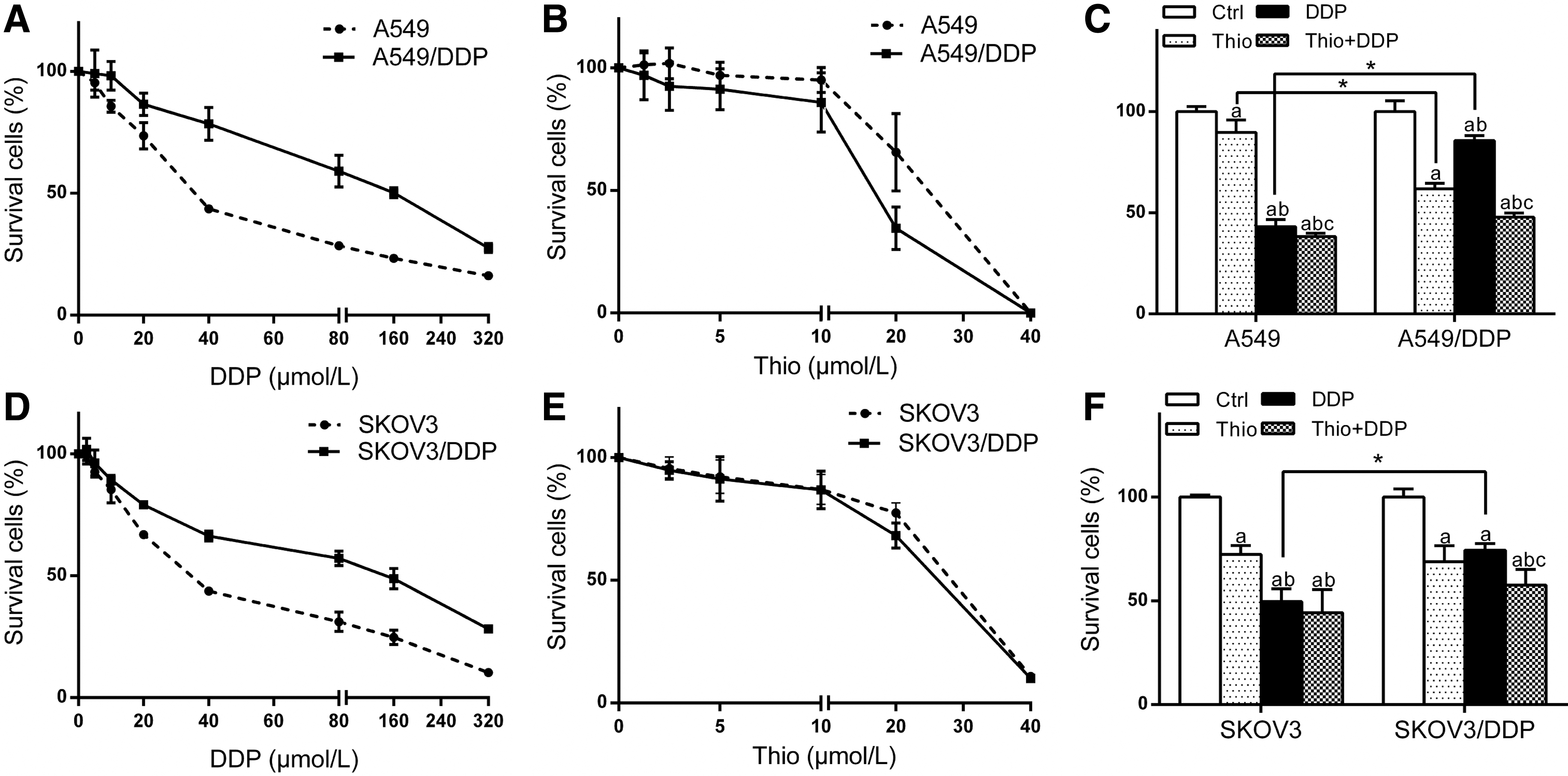
Cytotoxicity of Thio, DDP, or the combination. DDP led to a less percentage of dead cells in A549/DDP and SKOV3/DDP cells, compared with A549 and SKOV3 cells, respectively
Thio produced cytotoxicity in all cell lines, with IC50 of 19.1 versus 17.1 μM in A549 and A549/DDP, and 23.5 versus 21.3 μM in SKOV3 and SKOV3/DDP, respectively (Fig. 1B, E). Thus, 20 μM of Thio was employed in the combination regimen.
The combination of Thio and DDP resulted in the highest percentage of dead cells in both A549 and A549/DDP cells (Fig. 1C). Noticeably, Thio alone led to a higher percentage of dead cells in A549/DDP cells compared with A549 cells (p = 0.002). However, this trend was not detected in SKOV3 and SKOV3/DDP cells (p = 0.516) (Fig. 1F). The interaction between DDP and Thio was qualified using CI. CI were 0.97 and 1.08 in A549 and A549/DDP cells, and 0.87 and 0.87 in SKOV3 and SKOV3/DDP cells, respectively, indicating addition. These data demonstrated that Thio can enhance DDP in both sensitive and resistant cells.
The combined regimen induced G0/G1 arrest
The percentage of cells at G0/G1 phase was increased in group Thio, DDP or Thio + DDP, in A549 (p < 0.001, p < 0.001, p < 0.001), A549/DDP (p < 0.001, p = 0.004, p = 0.001) and SKOV3 (p = 0.002, p = 0.003, p = 0.001) cells. The percentage of G0/G1 cells was increased in group Thio or Thio + DDP in SKOV3/DDP cells (p = 0.021, p = 0.006) (Fig. 2). These findings manifested that the combined regimen induced G0/G1 arrest.
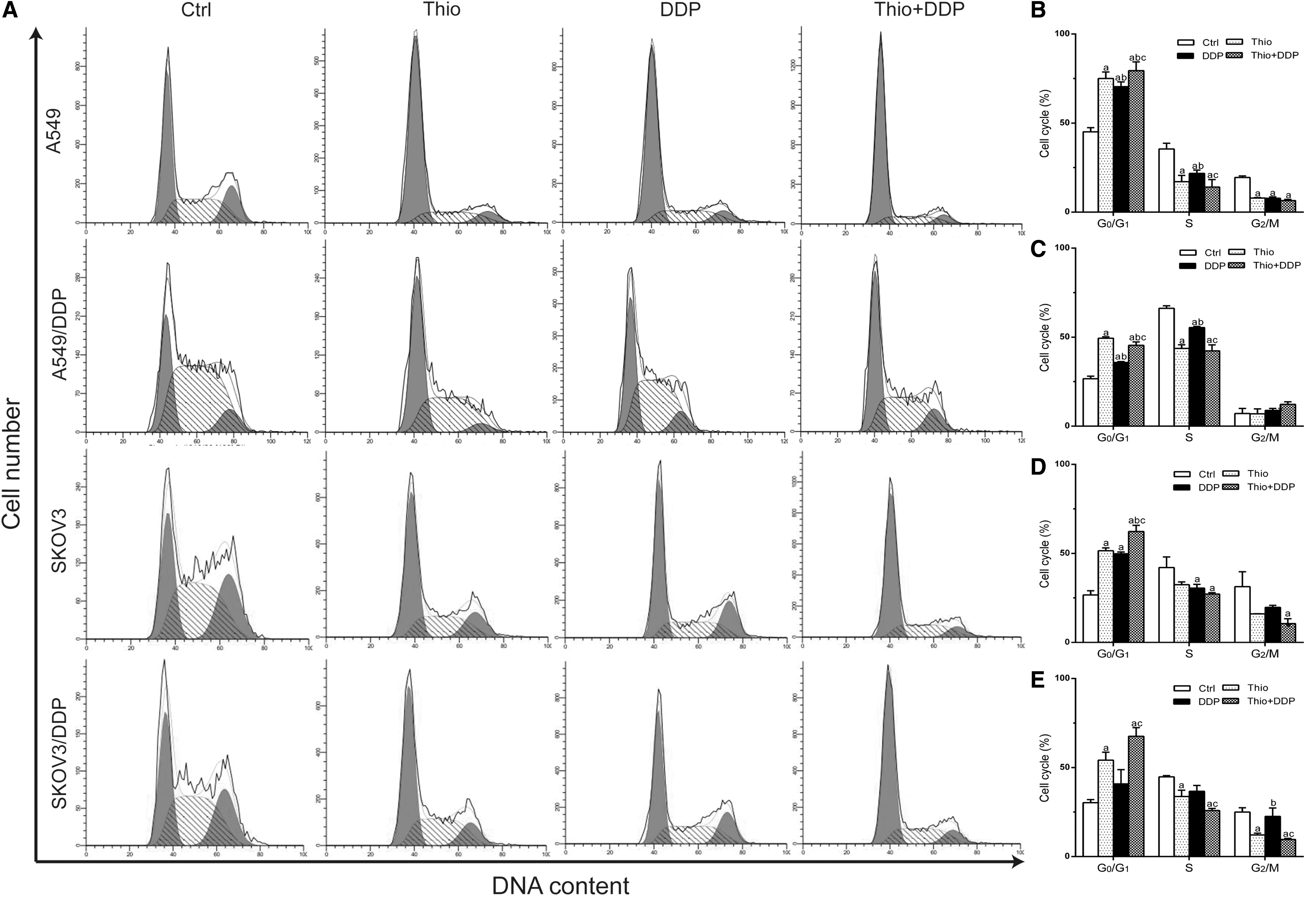
Cell cycle distribution in A549
Thio enhanced mitochondrial apoptosis attributable to DDP
Thio or DDP induced apoptosis in A549 (p = 0.034, p = 0.002) and SKOV3 (p = 0.030, p = 0.006) cells. The combination of Thio and DDP led to the highest apoptotic percentage in A549 (p = 0.001) and SKOV3 (p = 0.001) cells. The percentage of apoptotic cells in group DDP was not increased in A549/DDP (p = 0.050) and SKOV3/DDP (p = 0.053) cells, demonstrating insufficiency of apoptosis; the highest apoptotic percentage was detected in group Thio + DDP (p < 0.001, p < 0.001) (Fig. 3). Interestingly, for group Thio, the apoptotic fraction in A549/DDP cells was higher than that in A549 cells (p = 0.028) (Fig. 3).

Apoptosis in A549, A549/DDP
MMP was determined using the JC-1 assay. MMP was decreased after Thio and/or DDP exposure in A549, A549/DDP, SKOV3, and SKOV3/DDP cells (p = 0.004, p < 0.001, p < 0.001, p < 0.001) (Fig. 4A, B).
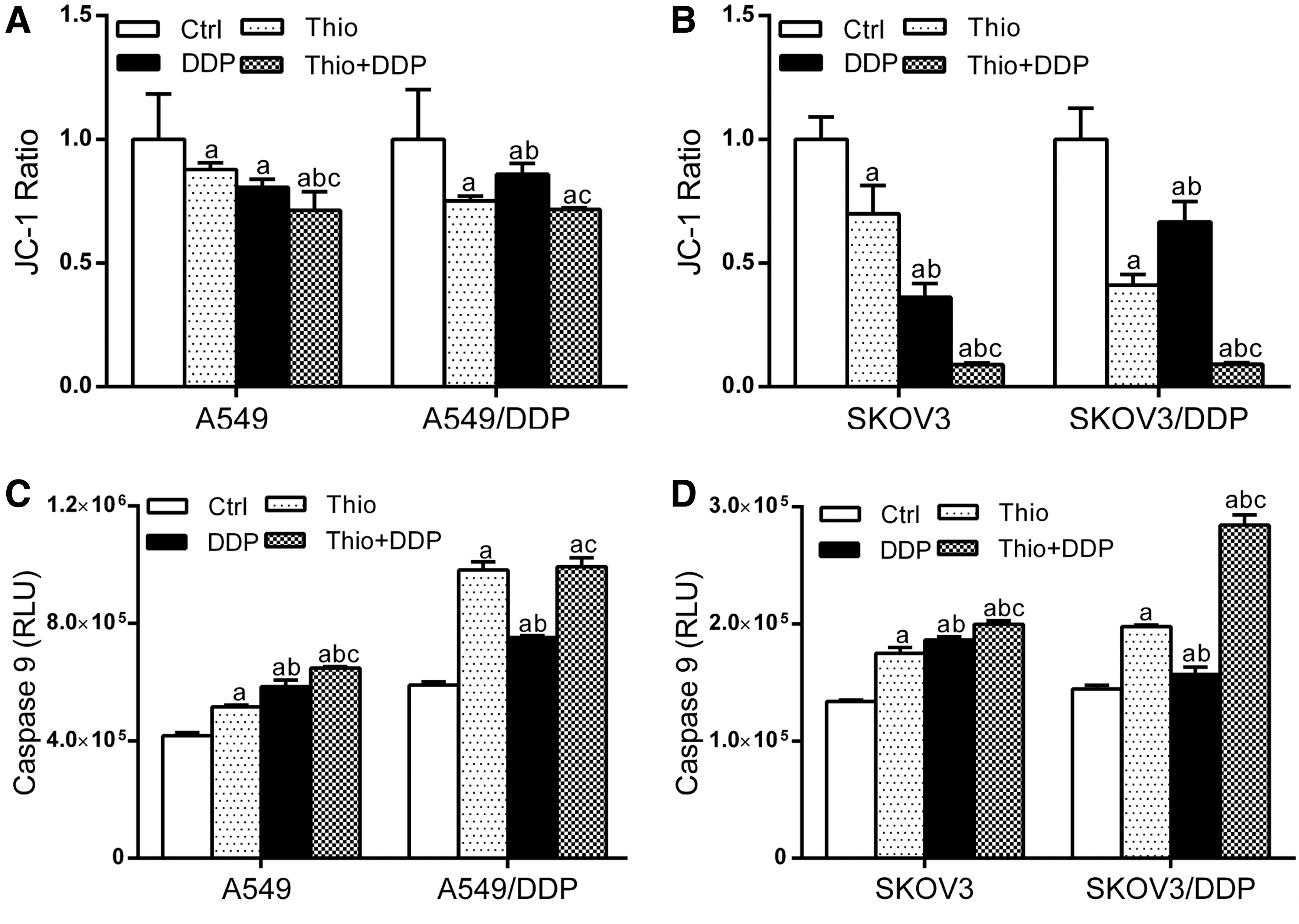
Mitochondrial membrane potential and the activity of caspase 9 in A549, A549/DDP, SKOV3, and SKOV3/DDP cells. A less JC-1 ratio was detected in cells exposed to Thio and/or DDP, demonstrating collapse of potential
The activity of caspase 9 in groups Thio, DDP, and Thio + DDP was increased in four cell lines (p = 0.001, p < 0.001, p < 0.001, p < 0.001) (Fig. 4C, D).
Apoptosis-related proteins Bax, Bcl-2 and caspase 3 were determined with western blot. The Bax level was increased and the Bcl-2 level was decreased, after treatment with Thio and/or DDP in A549, A549/DDP, SKOV3, and SKOV3/DDP cells (p < 0.001, p = 0.047, p < 0.001, p < 0.001; p < 0.001, p < 0.001, p < 0.001, p < 0.001). The level of cleaved caspase 3 was increased, accompanied by a decrease of intact caspase 3 (p < 0.001, p = 0.019, p < 0.001, p < 0.001; p < 0.001, p = 0.017, p < 0.001, p < 0.001) (Fig. 5). These data manifested that Thio can enhance apoptosis due to DDP in both native and resistant cancer cells, mainly through the mitochondria-dependent pathway.
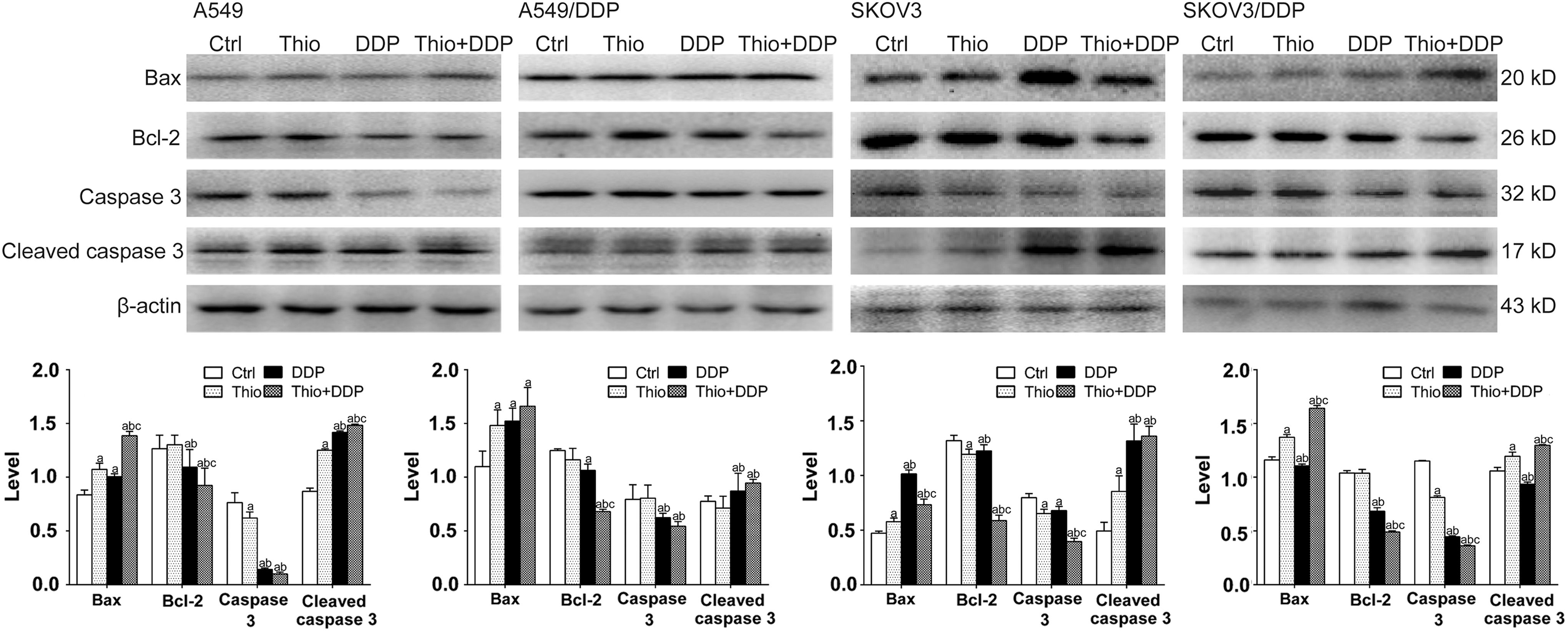
Levels of caspase 3, Bax, and Bcl-2 proteins validated by western blot. Levels of Bax and cleaved caspase 3 were increased in group Thio + DDP in all cell lines, with a decrease of intact caspase 3 and Bcl-2. Bcl-2 level was decreased after DDP exposure, but the downregulation was only noted in SKOV3 cells after Thio exposure. Data were mean ± standard deviation for three independent experiments. a: versus group Ctrl, p < 0.05; b: versus group Thio, p < 0.05; c: versus group DDP, p < 0.05.
Discussion
Thio was a widely used antipsychotic agent. Recent data manifested that Thio can promote the differentiation of tumor stem cell and deactivate them without affecting normal cells, indicating that Thio may be an anticancer drug (Sachlos et al., 2012). Thio can induce autophagy and sensitize glioblastoma cells to temozolomide (Johannessen et al., 2019). Autophagy was involved in the DDP resistance (Wan et al., 2018). Therefore, whether Thio can be a sensitizer for DDP was explored in this study. Both lung and ovary cancer cell lines were adopted to improve the clinical relevancy, considering that mechanisms of chemoresistance drastically varied between cancer types.
The present data manifested that Thio caused cytotoxicity in lung and ovary cancer cells. This was consistent with our previous data that Thio induced apoptosis in human ovarian cancer cells SKOV3 and A2780, and caused cell death in uterine cervical cancer cells SiHa (Mao et al., 2015; Yong et al., 2017). Interestingly, Thio alone induced a higher percentage of dead cells in A549/DDP cells in comparison with A549 cells, which was not observed in SKOV3 and SKOV3/DDP cells. These data demonstrated that the response varied between tissue types. This may be related to the expression level of dopamine receptors, which varied among tissue types (Chen et al., 2014). Therefore, the therapeutic regimen should be specifically tailored for a specific cancer type, according to the expression status of dopamine receptors. A higher percentage of dead cells in A549/DDP cells suggested that Thio alone can be a therapeutic strategy for certain resistant cancers.
Thio combined DDP induced the highest percentage of dead cells, and that the interaction was additive. Thio can inhibit P-glycoprotein to decrease the drug efflux (Choi et al., 2014). A higher intracellular drug level resulted in more severe cytotoxicity, thereby enhancing the action of DDP. Thus, Thio can be employed to modulate DDP-based chemotherapy regimen against cancers.
DDP resulted in a less apoptotic percentage in A549/DDP and SKOV3/DDP cells, demonstrating that apoptotic insufficiency was a mechanism of resistance. Cytotoxicity of DDP was frequently mediated by apoptosis (Brozovic et al., 2010). The combination of Thio and DDP led to the highest apoptotic fraction in A549, A549/DDP, SKOV3, and SKOV3/DDP cells. Collapse of MMP, and the pattern of Bcl-2, Bax, caspase 9, and caspase 3 indicated that apoptosis was mitochondria dependent. DDP caused DNA damages, leading to G0/G1 arrest for DNA repair. Certain damages were repaired and cells entered the S phase. Certain damages were unrepairable; the signal was transmitted to mitochondria, and eventually apoptosis was initiated (De Zio et al., 2013). Therefore, Thio was a strategy to modulate apoptotic insufficiency in DDP-resistant cancer cells.
Conclusion
Thio can deactivate certain chemoresistant cells, and can enhance DDP in both sensitive and resistant cancer cells. Chemosensitization was mediated by inducing the G0/G1 arrest, thereby resulting in apoptosis mainly through the mitochondria-dependent pathway. Therefore, Thio can be used to modulate DDP-based chemotherapy regimen in lung and ovary cancers.
Footnotes
Acknowledgment
This study was supported with a grant from the Science and Technology Commission of Qianjiang District, Chongqing (no. 2018016).
Disclosure statement
No competing financial interests exist.