
Abstract
Background:
Non-small cell lung cancer is the most common type of lung cancer and is a frequent cause of death. In our research, A549 and SK-MES-1 were used to assess the effect of three-dimensional (3D) culture compared to that of two-dimensional (2D) monolayers in cell proliferation, migration, and invasion, response to chemotherapy, as well as the expression of epithelial to mesenchymal transition (EMT) and cancer stem cell (CSC)-related markers. As tadalafil is a phosphodiesterase type 5 (PDE5) inhibitor with the potential to target CSC maintenance in multiple cancer cell lines, we assessed its function in 3D culture and detected the downstream pathway genes.
Methods:
We compared the viability and proliferation capacity of A549 and SK-MES-1 cells in 2D and 3D culture by cell counting kit (CCK)-8, foci formation, and Live/Dead double stain (Operetta CLS High content screening). Migration, invasion, and other functions were also assessed. To elucidate the underlying mechanisms, the expression of EMT and CSC markers was analyzed by quantitative real-time PCR (qPCR) and Western blot.
Results:
A549 and SK-MES-1 cells formed spheroids heterogeneous in shape and size. In our 3D spheroid systems, cells went through EMT, and were also capacitated with higher stemness and chemoresistance. Combination use of tadalafil and cisplatin showed higher chemotherapy efficiency in the 3D model, compared to that of the 2D cell culture.
Conclusion:
Our research aims at the notable differences between these two cellular systems in terms of cell functions, EMT, and stemness-associated gene expression and chemo-response. We showed that a commonly used drug, tadalafil, showed more pronounced inhibitory effects when cells were cultured in the 3D model. Since the 3D culture system could imitate the in vivo behavior of cancer cells within the tumor, we advocate that this system is superior to traditional 2D culture and should be used in the investigation of new therapeutic compounds.
Introduction
Non-small cell lung cancer (NSCLC), which accounts for 80–90% of all lung cancers (Cao and Chen, 2019), is one of the leading causes of cancer-related deaths worldwide. Its incidence rates increased from 2000 to 2011, making it the most frequent cancer type, while the 5-year survival rate of NSCLC remains as low as 15%, despite the availability of multiple new therapies and numerous studies (Siegel et al., 2019). Patients may show satisfactory initial responses, but within 1 year, many of them develop relapse and drug resistance. Increasing data demonstrate that cancer stem cells (CSCs) play an important role in cancer progression, metastasis, and drug resistance; thus, it may be beneficial to target key molecular drivers of CSCs in combination with standard treatments (Zhao et al., 2018).
The model used in basic and translational studies is of paramount importance to their conclusions. Researchers have devised a vast array of model systems to study the complex components of tumors and their treatment, each with unique advantages and disadvantages. The most frequent method in cancer research is traditional two-dimensional (2D) culture; however, it cannot fully simulate the in vivo status since important signals are lost when cells are cultured ex vivo on a 2D plastic substrate (He et al., 2017). In addition, it is hard for it to manipulate a CSC model in 2D models.
An increasing number of researchers have adopted the use of three-dimensional (3D) cell culture models, due to their easy establishment and stability. Therefore, in the past decade, 3D cell culture technology has become a focus of research in tumor cell biology. In comparison to conventional 2D culture, 3D models provide an ideal spatial environment for the cells, being promising and suitable systems for various purposes, ranging from disease modeling to drug target identification and identification of potential therapeutic substances.
In this regard, tadalafil is a phosphodiesterase type 5 (PDE5) inhibitor that has been widely used for the treatment of erectile dysfunction (ED). It has shown a beneficial effect in a variety of clinical conditions, such as pulmonary arterial hypertension, prostate hyperplasia, and pulmonary arterial hypertension, while recent studies reveal that tadalafil can benefit cancer patients (Huang et al., 2020).
Therefore, in this study, we optimized the 3D culture conditions and compared the differences between 3D and 2D culture conditions. We found that cells cultured in a 3D system were correlated with higher stemness and went through epithelial to mesenchymal transition (EMT). Also, 3D culture could promote tumor cell chemoresistance. We further confirmed the efficiency of tadalafil in our model.
Materials and Methods
Cell culture
Cell lines: four NSCLC cell lines, A549, SK-MES-1, H520, and H23, were chosen for the study. The A549 cell line is a lung adenocarcinoma cell line. It was first developed in 1972 through the removal and culturing of cancerous lung tissue in the explanted tumor of a 58-year-old Caucasian male. The SK-MES-1 cell line is a human lung cancer cell line that displays epithelial morphology and grows as monolayers in tissue culture. It was derived from the pleural effusion of a 65-year-old Caucasian male with squamous cell carcinoma of the lung. These cells exhibit a cytokeratin expression pattern typical of simple epithelia. H23 is a human lung adenocarcinoma cell line and H520 is a lung squamous cell line.
2D culture: NSCLC cell lines were maintained in Dulbecco's modified eagle media (DMEM) (GIBCO, Gaithersburg, MD) with 10% fetal bovine serum (FBS) (GIBCO). All cells were cultured at 37°C with 5% CO2 and were authenticated by STR profiling. SK-MES-1 is a human lung cancer cell line that displays epithelial morphology and grows as monolayers in tissue culture. It was extracted from a metastatic site (pleural effusion) of a male patient with lung squamous cell carcinoma.
3D culture: we prepared the medium as indicated. Cells were suspended in 75% BME matrix (1 mg/mL; prepared according to the manufacturer's instructions), and liquid gels were cast in either 24-well (for histologic and molecular analyses), 96-well plates (for cell viability assays; described below), or Perkin Elmer Cell Carrier 96-well plates (for Live/Dead analyses). The detail and chemicals used in the culturing medium are listed in Supplementary Table S1.
To extract cells from 3D culture, we disrupted the gels mechanically in cold phosphate-buffered saline (PBS), and then the spheroids were isolated by centrifugation and incubated in TrypLE™ Express (cat. 12604-021; GIBCO) for 20 to 30 min to generate a single-cell solution. To obtain the spheroids without destroying their structure, we used Gentle Cell Dissociation Reagent (cat. 07174; STEMCELL) instead of TrypLE Express, and left the suspension on ice for 10 min before centrifugation. To obtain the 3D hematoxylin and eosin (H&E) slides, we placed the collected spheroids into 3% agarose before embedding them for sectioning. The staining procedure for H&E followed a standard protocol.
3D suspension culture system: cells were cultured in six-well Ultra-Low Attachment plate (Corning, cat. CLS3471) to avoid them from attaching to the plate. Serum-free 1640 medium was supplemented with 20 ng/mL epidermal growth factor (EGF), 10 ng/mL fibroblast growth factor (FGF), and 4 μg/mL insulin. The cells were digested and passaged using recovery solution and dispased upon expiration.
Western blotting
Western blotting was performed according to the standard protocols of our laboratory. Total cell protein was lysed in radioimmunoprecipitation assay (RIPA) buffer (Thermo Fisher, Waltham, MA) with 1% proteinase inhibitor (Beyotime Biotechnology, China). Equal amounts of proteins were added onto a 10% or 12.5% polyacrylamide gel. Proteins were electrophoretically transferred to a polyvinylidene difluoride membrane (Millipore). Antibodies used were: GAPDH, β-actin, E-cadherin, vimentin, PDE5A, PARP, BAX, cleaved caspase3 (Cell Signaling Technology, Danvers, MA), and ALDH1A1 (Proteintech, China).
RNA extraction, cDNA synthesis, and quantitative real-time PCR
The mRNA from A549 and SK-MES-1 cell lines was isolated in TRIZOL (Invitrogen, Waltham, MA). cDNA synthesis was made with Takara PrimeScript II 1st strand cDNA synthesis kit (cat. #6210A; Takara Biotechnology, Mountain View, CA) according to manufacturer's instructions. The cDNA fragments were amplified with primers (listed in Supplementary Table S2) and TB Green® Fast qPCR Mix (cat. #RR430A; Takara Biotechnology). Specific primers for EMT-related genes, stemness-elated genes, and PDE5 signaling-related genes were designed with Oligo 7 and purchased from TIANYI HUIYUAN (Guangzhou, China). Normalization was performed using β-actin as an internal reference. Data were analyzed by the change-in-threshold (2−ΔΔCT) method of the specific gene of interest over 18S or β-actin.
Immunofluorescence assay
We seeded cells on the coverslips for immunofluorescence (IF) analyses. First, cells were fixed with 3.2% paraformaldehyde for about 2 h and permeabilized with 0.1% Triton X-100 in PBS, and then incubated with primary antibody overnight at 4°C. After washing and incubation with secondary antibody for 1 h, cells were subsequently counterstained with DAPI (Life Technologies) for 3 min at room temperature. Images were taken by Leica BX53 fluorescent microscope (Olympus Corporation, Japan).
Wound healing
3D cultured cells were digested to 2D and cultured on six-well dish plates until 95% confluence and then wounded with a 10 μL pipette tip. Migration photos were captured at 0 and 48 h after scratching.
Cell invasion and migration assays
Migration and invasion assays were performed in 24-well 8 μm BioCoat Matrigel transwells and Invasion Chambers (Corning, Franklin Lakes, NJ). Briefly, 8 × 10⁴ cells/well were seeded to the top chamber with 200 μL DMEM without FBS, while 10% FBS medium was added to the bottom chamber as a chemoattractant. After 18 or 24 h of incubation, the bottom chambers were fixed with 75% ethanol. Cells were stained with crystal violet and counted under an optic microscope. Three independent experiments were performed for both migration and invasion assays.
Foci formation assay
For foci formation, 1.0 × 10³cells/well were digested and seeded in six-well plates. After 15 days, cell-formed colonies were fixed for 30 min and stained with crystal violet (Sigma-Aldrich) and the number of foci was counted. Three independent experiments were performed.
Cell cycle assay
Cells in the logarithmic growth phase were seeded in 6-well plates (3D cells seeded in 96-well plates were first digested to single cells with trypLE express), washed twice with cold PBS, and fixed in 70% precooled ethanol at 4°C for 2 h. After fixation, cells were washed with PBS and stained with PI/RNase A for 30 min at room temperature. Tubes were shaken every 10 min during the incubation. Cells were washed, resuspended in cold PBS to a final concentration of 1 × 10⁶ cells/mL, and subjected to flow cytometry (CytoFLEX, BeckmanCoulter) for cell cycle distribution analysis.
Apoptosis assay
To evaluate the extent of early and late apoptosis, we used the Annexin V-FITC/PI kit from Genestar. We carried out the detection according to the manufacturer's instructions. In brief, cells were washed, collected, and resuspended, and then 5 μL annexin V-FITC were added and mixed gently. After 20 min, 4 μL PI were added to the tubes and mixed gently. After incubation for another 20 min in the dark, cells were subjected to flow cytometry (CytoFLEX, Beckman Coulter). The numbers of living, dead, early, and late apoptotic cells were determined and compared.
LIVE/DEAD staining
This experiment was performed on 3D cells using LIVE/DEAD® Viability/Cytotoxicity kit (L3224; Thermo Fisher) containing calcein-AM and Propidium Iodide. 3D Spheroids were washed twice with PBS and stained for 20 min with a mixture of 4 μM calcein and 10 μM PI, and stained with 5 μg/mL Hoechst33342 for 10 min, and fluorescent imaging was captured by Operetta CLS from Perkin Elmer.
Gene expression and correlation
We used the online database Gene Expression Profiling Interactive Analysis (GEPIA) to further confirm the correlation between PDE5A and candidate influenced genes: PRKACA, PRKG1, CREB1, and ALDH1A1. GEPIA (Tang et al., 2017) is an interactive web that includes 9736 tumors and 8587 normal samples from TCGA and the GTEx projects, which analyzes the RNA sequencing expression. The correlation analysis was performed for given sets of TCGA expression data and Spearman method was used to determine the correlation coefficient.
Statistical analysis
One-way analysis of variance was used to compare statistical differences between more than two groups. Other statistical differences were evaluated by Student's unpaired t-tests using GraphPad Prism (San Diego). p-Values <0.05 were considered significant.
Results
Spheroid morphology of NSCLC cells cultured in a 3D model
During the development of 3D culture, many methods have been adopted. Up to now, there are currently two main types of 3D culture systems, namely 3D scaffold-free culture and 3D scaffold culture. The spheroid method is a type of 3D scaffold-free culture system widely used to assess the stemness of cancer, but since there is no scaffold to support cells, some cell lines die during the process. Liu et al. (2017) have optimized a scaffold 3D system and showed that IGF1 is essential for A549 3D culturing. Based on Liu's protocol, we adopted the 3D culture system. Figure 1A presents spheroids from 4 NSCLC cell lines cultured for 3, 5, and 8 days in 3D culture conditions in comparison to their parental 2D cells. Morphology differences were observed already after 3 days of culture and were obvious thereafter. As measured in diameter, the spheroids of A549 grow faster compared with SK-MES-1 (Fig. 1A). Cell counting kit (CCK)-8 and foci formation were performed to compare the survival ability of A549 and SK-MES-1.
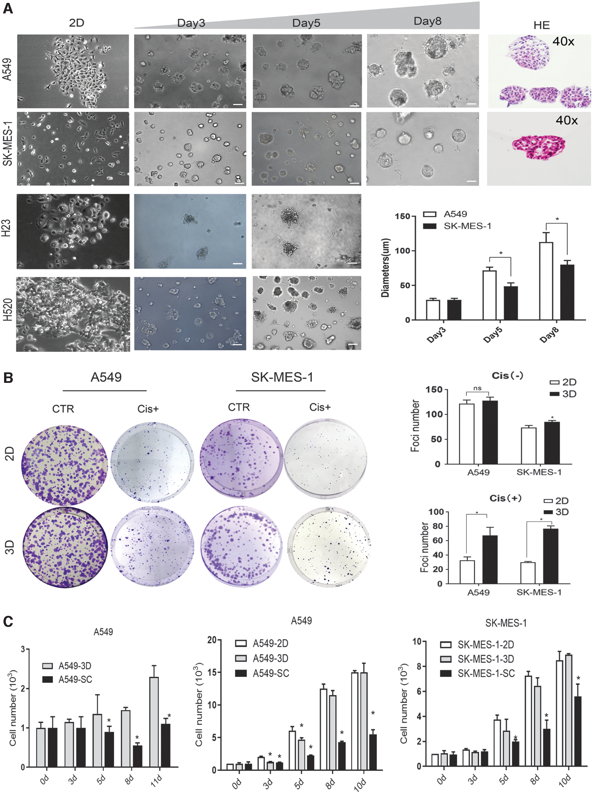
Morphology of NSCLC 3D cultured tumor spheroids.
As measured by CCK-8, the two groups had little differences in growing speed, but when pretreated with 2 μg/mL cisplatin, cells in 3D culture formed more foci when compared to those formed in 2D cultured cells (Fig. 1B), indicating that they may be endowed with stronger survival ability. Further experiments were carried out to compare the efficiency between our BME-based 3D culture system, 2D culture, and typical spheroid method that was used to grow CSC from lung cancer cell line (stem cell [SC]). We found that the proliferation rate of A549-3D was low in our 3D culture system, but is still much faster compared with the spheroid method (Fig. 1C, left). To compare the proliferation potentials among 2D, SC, and 3D cells, we seeded the same number of different cells in the same 96-well plates and cultured for 3, 5, 8, and 10 days. We found that the proliferation potential was much higher in 3D culture compared with the spheroid method (Fig. 1C, right).
A549 and SK-MES-1 cultured in 3D exhibited higher migration and invasion ability
To investigate the effect of our 3D culture system on cell function, wound healing and transwell assays were performed to assess migration and invasion, respectively. As shown in Fig. 2A, the 3D cultured group was more proficient in repairing wounds at 24 and 48 h after the wound (the two cell lines, A549 and SK-MES-1, differed in wound healing speed). Then we carried out migration and invasion assay to further investigate the effect of culture condition on the invasion ability. Using the transwell chambers, we further showed that 3D cultured A549 and SK-MES-1exhibited elevated migration and invasion rates relative to those of their parental 2D cultured cells (Fig. 2B).
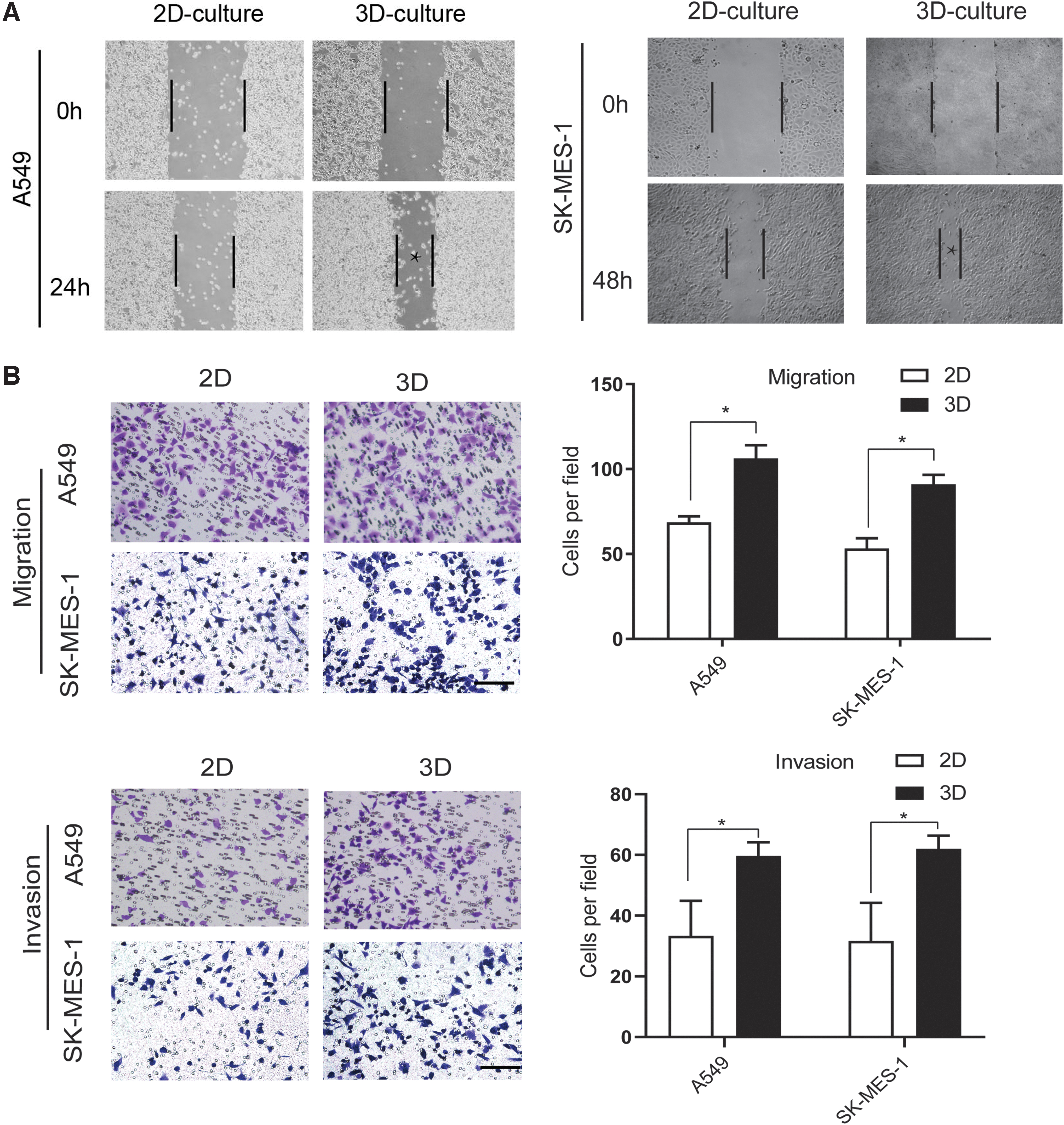
A549 and SK-MES-1 3D cultured cells exhibit higher migration and invasion ability.
Long-term 3D culture caused changes in cell cycle phase and led to antiapoptosis ability
A key characteristic of CSCs is their slow growth rate and chemoresistance. At present, most chemical drugs act when tumor cells are in S or G2 phase. Through cell cycle experiments, cells cultured in 2D or 3D models, we found that the G1 phase increased, while the S phase decreased in 3D culture cells, compared to 2D ones (Fig. 3A). We also demonstrated that this process is reversible. After re-attaching to the 2D culture, it was found that the G1 ratio of the 3D cultured cells decreased and that these cells re-entered the S phase, which is consistent with the characteristics of CSCs and the results of previous studies (Fig. 3B).
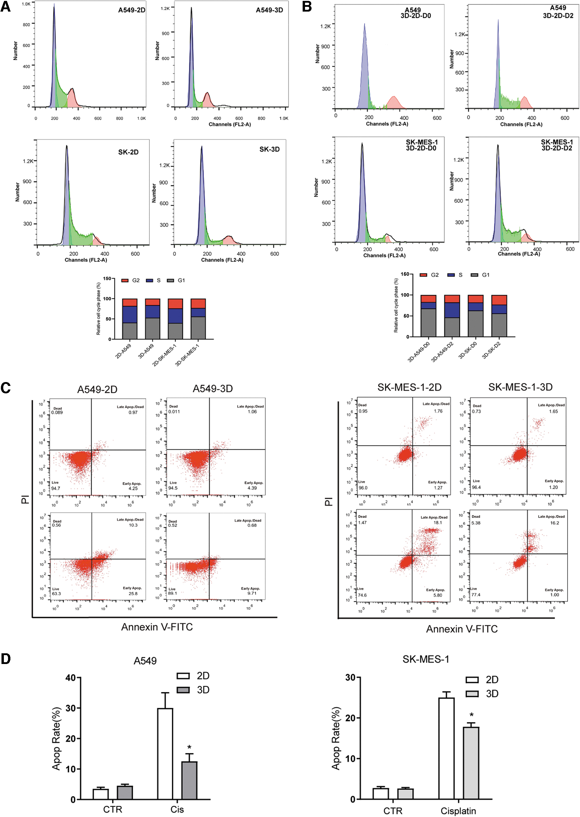
Long-term 3D culture causes cell cycle phase change and endows cells with antiapoptosis ability.
Using flow cytometry (Fig. 3C), we detected the ratio of early and late cell apoptosis and found that after 2 days of treatment with 5 μg/mL of cisplatin, the proportion of cell apoptosis in the 3D group was lower compared with the 2D group (Fig. 3D).
A549 and SK-MES-1 cells go through EMT under 3D conditions
To investigate the underlying mechanisms of our 3D culture model, EMT and signal transduction pathways were analyzed. Expression of EMT-related genes showed a progressive decrease in epithelial marker (CDH1) along with a simultaneous increase in mesenchymal markers (VIM, SNAI1, SNAI2, and ZEB2) in 3D cultured cells. Then Western blot (Fig. 4B) was carried out, too. IF staining EMT markers (E-cadherin and vimentin) were performed to evaluate the EMT phenotype. As shown in Figure 4C, 3D culture condition was positively associated with an EMT phenotype (Fig. 4C). Studies have demonstrated cancer cells that acquire stemness features of CSCs in response to signals may undergo EMT (Zhang et al., 2019); thus, we examined stemness-related markers, including genes that code cell surface proteins: CD44, PROM1, ICAM1, ALCAM, and IGF1R. We also test other important stemness-related markers that have been reported to play an important part in regulation of lung cancer CSCs: FAK2, Hes1, and ALDH1A1, in which we found that ALDH1A1 was upregulated in both A549 and SK-MES-1 during 3D culturing (Fig. 4D). These results indicate that the anti-apoptotic changes may be caused by EMT and enhancement of stemness during the 3D culture.
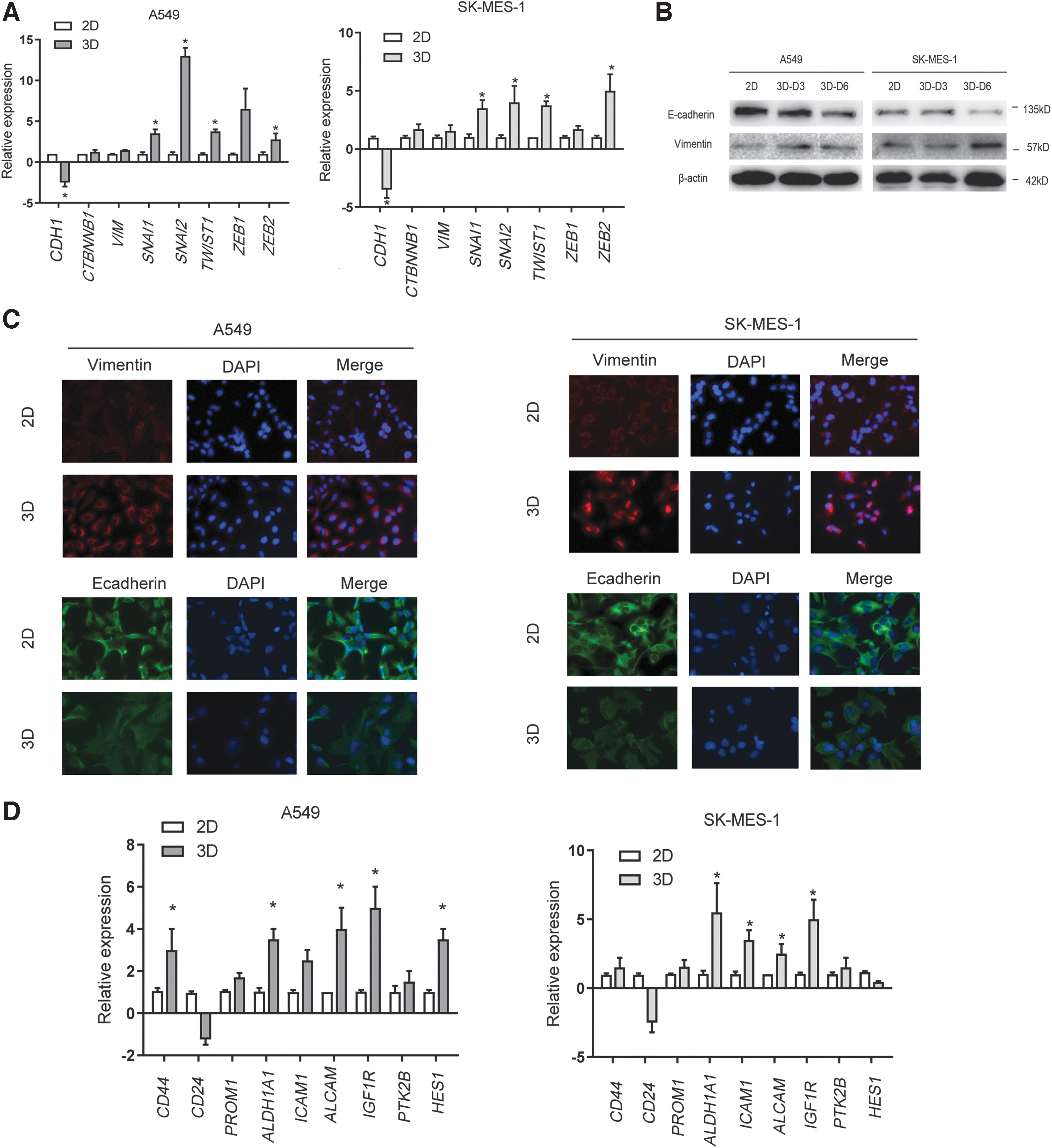
A549 and SK-MES-1 go through EMT under 3D conditions.
PDE5 inhibiton promoted chemotherapy through suppressing the tumorigenesis of stem-like tumor cells
CSCs are characteristically resistant to conventional therapies. Therefore, to determine whether 3D cultured cells possessed a CSC chemoresistant phenotype, cells of the two groups were treated in the same serum-free medium for 24 h before measuring the cytotoxic activity of cisplatin at different doses. As expected, A549 and SK-MES-1 in 3D culture for 7 days were chemoresistant (Fig. 5A).
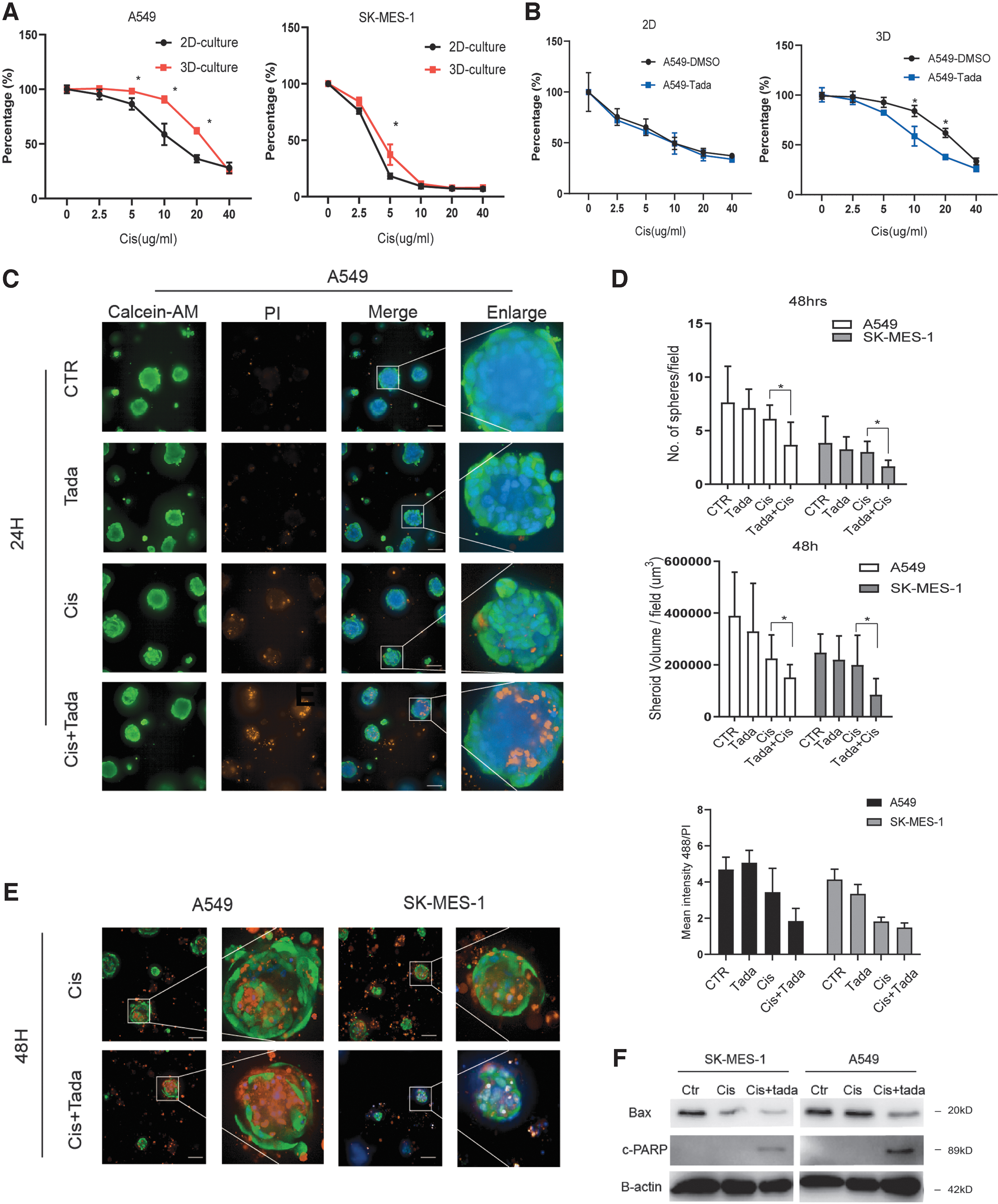
PDE5 inhibitor promoted chemotherapy in 3D culture, which induced acquired resistance to cisplatin.
As PDE5 inhibitors were shown to benefit patients with multiple types of cancer, we assessed if these inhibitors would be effective in our culture conditions. Tadalafil was adopted to test whether it alone or in combination use has effect on cell viability. First, tadalafil alone showed good tolerance at a dose of 40 μM. Tadalafil did not show much influence on cisplatin's efficacy on a 2D manner, but notably, in 3D cultred cells, the combination treatment of tadalafil and cisplatin resulted in significant reduction in cell viability (p < 0.05) than cisplatin monotherapy (Fig. 5B). Western blot of apoptotic markers confirmed our results (Fig. 5F).
The effect of tadalafil was also determined by staining with live-dead two-color fluorescent dyes (Calcein-AM/PI) in A549 and SK-MES-1 cells. We analyzed cell viability to evaluate the response of 3D tumor spheroids to anticancer drugs. Results showed that after 24 h, nontreated cells were mostly viable, while 40 μM tadalafil in combination use with 5 μg/mL cisplatin increased cell apoptosis (Fig. 5C). Sphere numbers and cell volume were also analyzed (Fig. 5D).
PDE5 inhibitor suppressed the tumorigenesis of stem-like tumor cells through PKA signaling
PDE5 inhibitors were shown to impair cancer stemness by increasing cGMP and cAMP levels, and in turn activate PKA and induce the differentiation of CSCs (Klutzny et al., 2018). Our results demonstrated that 3D cultured cells were able to induce high rates of tumor incidence due to their stem-like features. To further investigate the effect of PDE5 inhibition on the maintenance of CSCs in 3D cultured A549 and SK-MES-1 cells, we performed quantitative real-time PCR (qPCR) for different stemness-associated marker genes. Indeed, as expected, treatment with tadalafil decreased the expression of ALDH1A1 (Fig. 6A), and reduced the expression of ALCAM, KLF4, and PTK2B (Fig. 6B). These genes code CSC-associated markers in lung cancer. Western blot confirmed the reversal of EMT induced by tadalafil treatment. We next confirmed our results by searching the TCGA data on GEPIA, using the correlation module of this website. We assessed the relationship between PDE5A and the related genes: PRKACA, CREB1, PRKG1, and ALDH1A1. We found that PDE5A was correlated with PRKG1 in LUAD (Fig. 6C), while it was weakly correlated with PRKACA and ALDH1A1 (p < 0.05). The schematic flow of our data is summarized in Figure 6D.
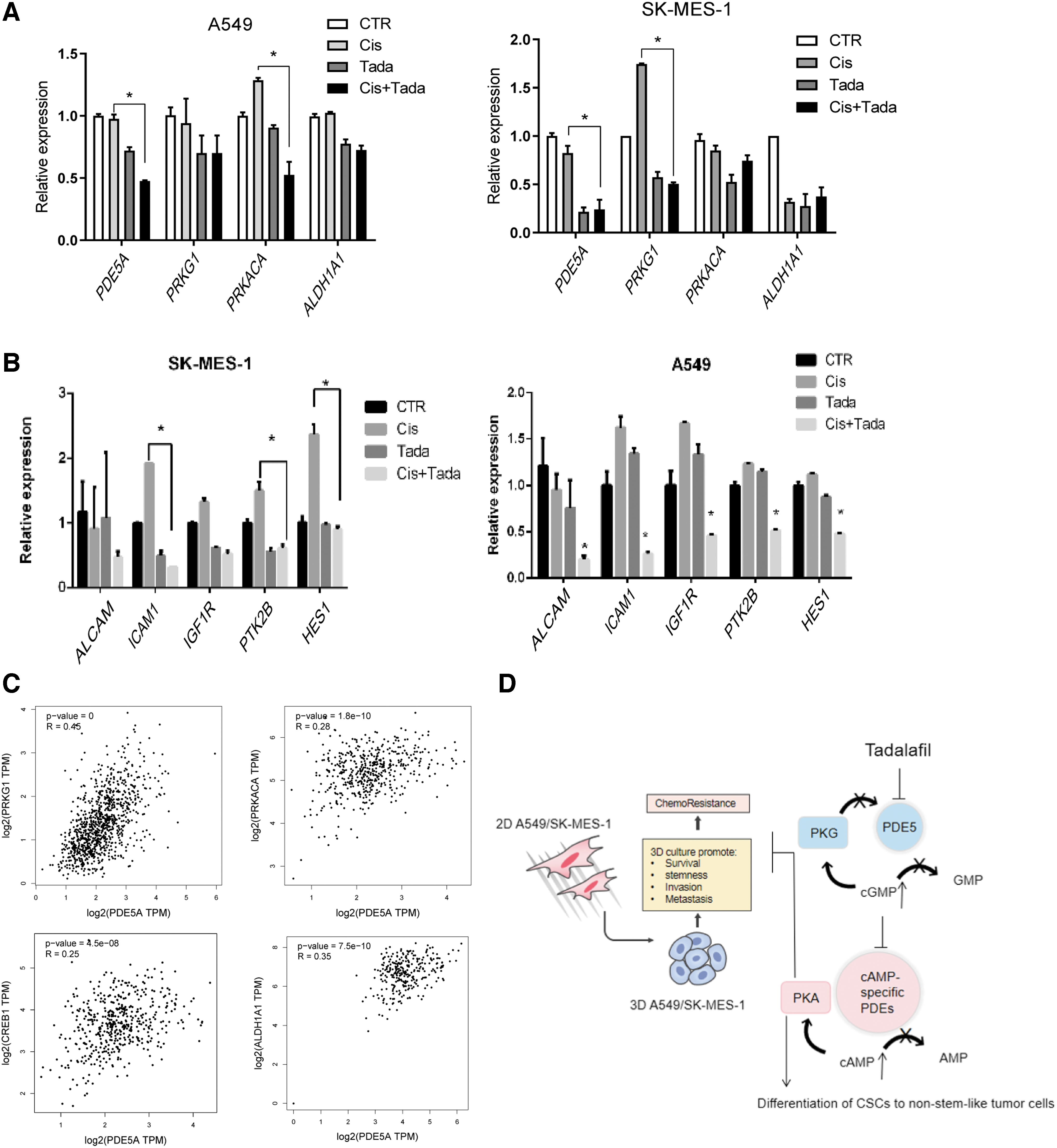
PDE5 inhibitor tadalafil influences PKA signaling.
Discussion
In vitro 3D tumor-ECM models mimic the in vivo tumor morphology in comparison to conventional 2D culture models (Qiu et al., 2020). In the past few years, the advantages of using 3D models for in vitro studies in cancer research have been reported. Since 3D cell culture is highly customizable, it is important to assess the differences between 2D and 3D models to understand the in vitro complexity of the tumor (Duval et al., 2017) and to establish reliable methods for measurement of cell viability for cells growing in spheroids.
In our research, we adopted Liu's 3D culture system to compare differences between 3D and conventional 2D cell culture. It was found that adenocarcinoma cell line A549, and squamous cell carcinoma cell line SK-MES-1 grew well in our 3D model. Although 3D cultured cells had normal proliferation in the experiment, the colony formation ability under cisplatin treatment was significantly enhanced when compared to those of 2D cultured cells. Further functional analyses revealed that 3D culture increased cell migration and invasion capacities. A variety of studies have suggested that EMT endows cancer cells with greater cell motility and may be important for metastasis (Kalluri and Weinberg, 2009; Lee et al., 2020); therefore, we detected typical EMT markers and confirmed the conversion (Dongre and Weinberg, 2019): E markers were decreased and the M markers were increased.
At present, it is recognized that the mobility, stemness, and chemoresistance of cells undergoing EMT conversion are increased (Shibue and Weinberg, 2017) (Kreso and Dick, 2014). We have corroborated it through flow apoptosis experiments showing that 3D culture enhances their antiapoptotic ability. qPCR was also performed to verify the expression of stemness-related markers in lung cancer, and we found it consistent with the previous results. We found that ALDH1A1 and others were significantly upregulated in 3D culture (Liang and Shi, 2012), and with the stemness of CSCs (Huang et al., 2009). In NSCLC, ALDH1A1 was reported to play a role in drug resistance and radioresistance (MacDonagh et al., 2016, 2017). A meta-analysis has shown that the increased expression of ALDH1A1 was associated with poor overall survival and disease-free survival in lung cancer (Wei et al., 2015; Zhou et al., 2016).
The theory of CSCs is now widely accepted (Xiao et al., 2018), and CSC was found in many solid tumors, including NSCLC. It was described to participate in tumor invasion, metastasis, and chemoresistance. CSCs were recognized based on cell surface phenotype, including the high expression of the surface marker CD44 (Herreros-Pomares et al., 2019).
Currently, there are mainly two approaches to the situation when EMT leads to increased stemness. One traditional strategy is to develop drugs that show specific or preferential cytotoxicity to CSCs. For instance, Pattabiraman et al. (2016) have adopted another approach to induce CSCs to exit the mesenchymal tumor initiation state and enter an epithelioid state. They found that this induced differentiation can make the cancer cells more susceptible to conventional cytotoxic treatments and proved that cGMP/PKG and cAMP/PKA signaling pathways were important in this induced transition.
Tadalafil, a selective PDE5 inhibitor, is widely used to treat ED and pulmonary hypertension. In recent years, it was discovered that PDE5 inhibitors may also be effective against cancer (Sponziello et al., 2015). Since it was shown to have an influence on PKA signaling and ALDH1A1 (Klutzny et al., 2018), we assumed tadalafil may reverse stemness in 3D cultured cells.
After confirming that tadalafil alone had weak effects in lung cancer, we combined it with chemotherapy drugs and found that this combination was effective in 3D cultured cells. The results of Operetta CLS showed that tadalafil significantly enhanced the effect of cisplatin on tumor spheroids. To investigate underlying mechanisms, we found that PDE5 inhibitor treatment influenced the PRKACA, PRKG1, and ALDH1A1 levels, which is consistent with previous research results (Klutzny et al., 2018). In this study, we provide insights into the interaction of PDE5A and propose a new mode of action for PDE5A in the maintenance of CSCs in 3D cultured cells (Booth et al., 2015). It was previously reported that the combination of sildenafil with celecoxib was cytotoxic in several types of cancer, including breast cancer, liver cancer, colorectal cancer, glioblastoma, and medulloblastoma cell lines (Webb et al., 2015). Since tadalafil has a low cost and low toxicity with great preclinical potential in NSCLC, it would be helpful if future studies explore additional underlying mechanisms of tadalafil in cancer.
In conclusion, our 3D cultured cell lines allowed the establishment of long-term lung CSC cultures, which showed histological and pathway characteristics similar to tissues in vivo. Therefore, this is a potential platform for research on chemotherapy and drug resistance. In 3D culture, EMT and stemness-related genes were upregulated and influenced cell stemness and drug resistance. We confirmed that tadalafil could reduce the expression of stemness-related genes by PKA signaling and enhance the effect of chemotherapy drugs.
Footnotes
Disclosure Statement
No competing financial interests exist.
Funding Information
This work was supported by Shenzhen Sanming Project (SZSM201612041) and Shenzhen Science, Technology Innovation Commission Project (GJHZ20180754917, ZDSYS201909020 92855097)
Supplementary Material
Supplementary Table S1
Supplementary Table S2
References
Supplementary Material
Please find the following supplemental material available below.
For Open Access articles published under a Creative Commons License, all supplemental material carries the same license as the article it is associated with.
For non-Open Access articles published, all supplemental material carries a non-exclusive license, and permission requests for re-use of supplemental material or any part of supplemental material shall be sent directly to the copyright owner as specified in the copyright notice associated with the article.