
Abstract
Antibiotic resistance genes (ARGs) pose a critical global health threat, driving the emergence of antibiotic-resistant bacteria (ARB) and causing an estimated 7,00,000 deaths annually. While current wastewater treatment technologies significantly reduce bacterial loads, residual ARGs in effluent highlight the need for advanced, targeted solutions. Plasmonic photocatalysts, leveraging localized surface plasmon resonance of nanoparticles provide an innovative solution by generating high-energy charge carriers (“hot” electrons and holes) under visible light. Despite their promise, the application of plasmonic photocatalysts for ARG removal remains underexplored. This study introduces a composite plasmonic photocatalyst comprising silver (Ag) nanoparticles, graphitic carbon nitride (g-C3N4), and polyvinylidene fluoride nanofibers. Characterized by a bandgap energy of 2.89 ± 0.06 eV and optimized for visible light activation, this material addresses limitations of traditional photocatalysts. Real wastewater samples from a primary clarifier effluent at a South Dakota Water Reclamation Facility were analyzed. A custom photostation equipped with chips-on-board light-emitting diode (2,87,000 lux) and a cassette-type photocatalytic mat enclosed in identical Teflon sheets (2 cm × 2 cm window) was utilized. Microbial community analysis and microfluidic-based qPCR revealed the presence of 29 ARGs, 6 heavy metal resistance genes (HMRGs), and 7 integrase-encoding genes in the primary clarifier effluent. Key genes, including ermB, ermF, intl1, intl3, merA, qacF, strB, sul1, and tetX were detected at abundances exceeding 5 log10 gene copies/µL, indicating coselection mechanisms. Plasmonic photocatalyst treatment significantly reduced critical bacterial groups, including Epsilonproteobacteria, Bacteroides, and Leptrichiaceae. Specific ARGs, including vanA and qnrA, showed substantial reductions, with differences in the copy numbers of blaNDM-1, blaSHV, blaimp13, qnrA, tetM, tetL, dfr13, vanA, and mexB. Importantly, the treatment mitigated ARB and ARGs without impacting HMRGs and integrase genes. This photocatalytic approach demonstrates potential as a supplementary treatment strategy for targeting ARGs in wastewater systems, particularly under visible and ambient light conditions. This study introduces an innovative approach to wastewater treatment using light-activated membranes to mitigate ARGs.
Introduction
Antibiotic resistance genes (ARGs) and antibiotic-resistant bacteria (ARB) represent a growing global health crisis, with wastewater treatment plants (WWTPs) serving as critical reservoirs and dissemination pathways for these contaminants. WWTPs also harbor other pollutants including antibiotics and heavy metals, which contribute to the complexity of this issue. Antibiotics such as β-lactams (a class of antibiotics) and vancomycin (a specific antibiotic) target bacterial cell walls, while fluoroquinolones and rifamycin impact DNA/RNA synthesis. Other antibiotics such as trimethoprim and sulfonamide disrupt folate synthesis, whereas daptomycin impacts cell membranes, and tetracycline, linezolid, macrolides, and aminoglycoside target protein synthesis (Pazda et al., 2019; Wright, 2010). This diversity in antibiotic action necessitates the evolution of complex resistance mechanisms in bacteria.
To survive antibiotics, bacteria acquire genetic machinery (Grundmann et al., 2006), including ARGs (Kristensen et al., 2020), heavy metal resistance genes (HMRGs) (Swannell, 2013), and multidrug resistance (MDR) genes (Gullberg et al., 2014). These genes function through mechanisms such as efflux, target modification, immunity, bypass, and enzyme-catalyzed destruction (Wright, 2010). In WWTPs, ARGs often coexist with mobile genetic elements (e.g., integrons, transposons, plasmids, and bacteriophages) (Pazda et al., 2019), facilitating their spread among different bacteria and potentially creating antibiotic-resistant pathogens. The presence of ARGs in wastewater effluent (Tehrani and Gilbride, 2018a, b; Wang et al., 2014) raises concerns about their dissemination in the environment and their potential integration into harmful bacteria (O’neill, 2014). Such integration can worsen the spread of antibiotic resistance, posing significant risks to public health and underming the effectiveness of current antimicrobial treatments. This issue has been linked to nearly 23,000 deaths annually in the United States alone (CDC, 2025).
The combination of slow filtration and low-pressure nanofiltration has shown improved efficiency in removing ARGs and dissolved organic carbon from secondary effluent in WWTPs, outperforming the effectiveness of each method when used individually (Sun et al., 2023). However, membrane biofouling remains a potential challenge for this treatment method (Sun et al., 2023). Although conventional disinfection methods in WWTPs can significantly reduce bacterial loads, their effectiveness against ARGs is limited (Dodd, 2012; Wang and Chen, 2022a, b, c). These methods often require high disinfectant doses, which, while effective for microbial control (Bairán et al., 2020), are not optimized for ARG removal. Photocatalytic oxidation has emerged as a promising alternative for ARG treatment (Loeb et al., 2018; Yuan et al., 2020), leveraging light to eliminate these threats without the use of harsh chemicals.
However, the widespread implementation of photocatalysis in real-word WWTPs faces challenge related to the properties and limitations of existing photocatalyst materials. Conventional photocatalysts, such as copper (Ma et al., 2016) and doped metals such as Mn/Co-TiO2 (manganese/cobalt-titanium dioxide) (Venieri et al., 2017), exhibit sluggish reaction kinetics and instability due to light-induced corrosion and leaching (Ishiguro et al., 2013). In addition, their powdered forms (Adibkia et al., 2012; Seil and Webster, 2012) present practical challenges, including unfavorable morphology (Adibkia et al., 2012; Buzea et al., 2007), recovery difficulties, and reuse inefficiencies in wastewater systems.
Plasmonic photocatalysts offer a promising solution to these limitations. These materials, composed of nanoparticles made from specific metals such as silver (Ag) or gold (Au), leverage localized surface plasmon resonance under visible light to achieve enhanced photocatalytic performance. When irradiated with visible light, plasmonic photocatalysts generate high-energy charge carriers (“hot” electrons and “hot” holes) (Zhang et al., 2013; Zhang et al., 2018), that facilitate degradation of organic pollutants such as ARGs through chemical oxidation and reduction reactions. The strong electromagnetic field near the nanoparticles facilitates the forced polarization of organic species in contact with the catalyst, which further promotes their chemical degradation Reactive oxygen species (ROS) generated by these carriers contribute to pollutant degradation, enhancing efficiency through mechanisms such as chemical interface damping (Zhang et al., 2013; Zhang et al., 2018).
Despite their potential, studies on the application of plasmonic photocatalysts for ARG removal are limited. Existing work has primarily focused on the general mechanisms of bacterial plasmid DNA destruction in the presence of a photocatalysts (Liu and Zhang, 2015; Patra and Gopinath, 2018; Saha et al., 2020), which involve the plasmid transitions from supercoiled form to a nicked-circle and relaxed double-stranded form, ultimately leading to fragmentation. Briefly, the double-stranded plasmid is converted into a single-stranded plasmid because of hydrogen bond cleavage (Liu and Zhang, 2015; Patra and Gopinath, 2018). The generation of hydroxyl radicals during photocatalysis may render the reaction medium partially basic, further facilitating the breakage of hydrogen bonds. Once converted into the single-stranded form, the plasmid DNA undergoes fragmentation or complete degradation in the presence of ROS generated by the photocatalyst (Liu and Zhang, 2015; Patra and Gopinath, 2018; Saha et al., 2020).
This study introduces a novel 3D composite plasmonic photocatalyst consisting of silver nanoparticles (AgNPs), graphitic carbon nitride (g-C3N4), and polyvinylidene fluoride (PVDF) nanofibers, specifically designed to address the limitations of traditional photocatalysts. The material combines common PVDF nanofibers with a light-activated graphitic carbon nitride (g-C3N4) component and AgNPs and is characterized by a bandgap energy of 2.89 ± 0.06 eV, optimized for visible-light activation. Visible light photocatalysts can generate charge carriers by absorbing visible light, unlike UV-based photocatalysts, making them easier to use and eliminating the need for a hazardous UV source. However, the ambient visible light typically lacks sufficient intensity (or photon quantity) to excite the photocatalyst to its optimal performance level. As a result, an additional visible light source is necessary for these photocatalysts to function effectively.
Tested on real wastewater samples (Jawaharraj et al., 2021), this study addresses a critical gap by showcasing the efficacy of plasmonic photocatalysts for ARG mitigation using visible light. This advancement paves the way for scalable integration of these materials into wastewater treatment systems to mitigate ARG dissemination. Through comprehensive microbial community analysis and microfluidic-based qPCR, a range of genetic elements within the microbial consortia was identified, indicating the active involvement of ARGs, HMRGs and integrase-encoding genes in coselection mechanisms. Subsequently, plasmonic treatment was performed to evaluate its capacity to reduce ARB and specific ARGs, such as vanA and qnrA.
Materials and Methods
Fabrication of a 3D nanofiber composite of plasmonic photocatalyst
A 3D nanofiber composite was synthesized following the method described by Grundmann et al. (2006). Briefly, a suspension of AgNPs (displaying dark purple color) was prepared by mixing 2.0 mL of sodium citrate (≥
A 22 kV electric potential was applied between the needle and the collector, which was positioned 10 cm apart. The polymer suspension was electrospun at a flow rate of 15 mL/h. After the electrospinning process, the collector plate was air-dried, and the resulting mat was carefully peeled off for further analysis.
The morphology and composition of the mat were examined using an FEI Quanta 400 environmental scanning electron microscope (Thermo Fisher Scientific, Hillsboro, OR, USA) equipped with energy dispersive X-ray (EDX) capabilities. Observations were made in Z-contrast and backscattered electron modes. In addition, UV-Vis reflectance analysis was conducted using an Agilent Cary 300 Bio UV-Vis spectrophotometer (Agilent Technologies, Santa Clara, CA, USA) to characterize the material’s optical properties. Figure 1 provides an overview of the synthesis of the plasmonic photocatalyst by electrospinning and illustrates its application in the degradation of ARGs within wastewater treatment systems.
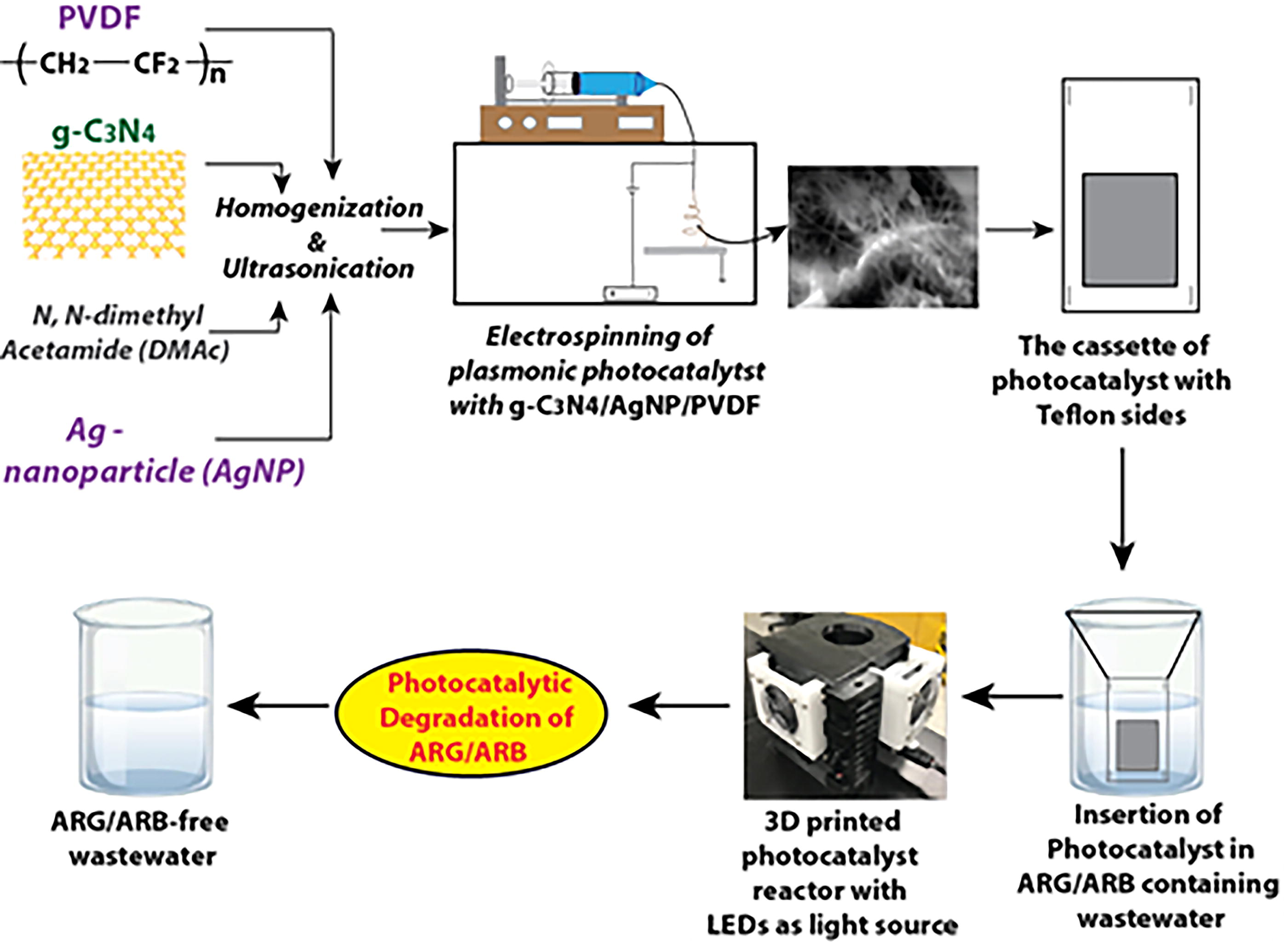
Schematic illustrating the synthesis of plasmonic photocatalyst using the electrospinning technique and its application in the photocatalytic degradation of ARG in wastewater, as evidenced by the reduction in gene copies. PVDF, polyvinylidene fluoride; g-C3N4, graphitic carbon nitride; LED, light-emitting diode.
Wastewater sample collection and photocatalytic treatment
Aliquots of 50 mL were collected in sterile vials from the primary clarifier effluent (Petrovich et al., 2018) at the Rapid City Water Reclamation Facility, SD (for wastewater details, Shrestha et al., 2016). The samples were transported to a wet laboratory for analysis. Upon arrival, the samples were transferred into an autoclaved glass bottle and placed within a specialized photostation, which was equipped with chips-on-board light-emitting diodes (LEDs) emitting visible light at an intensity of 2,87,000 lux (Desipio et al., 2019). The photostation utilizes a cassette-type photocatalytic mat housed between two Teflon sheets, each with a 2 cm × 2 cm window (Saha et al., 2021). Samples were stirred at approximately 150 rpm for 4 h during testing to ensure uniform mixing without vigorous agitation.
The experimental design included three conditions to evaluate the role of the photocatalytic material and light in ARG degradation: a control sample (PE-1) that was not exposed to any light, and test samples subjected to 2-h (PE-2) and 4-h (PE-3) photocatalysis. While specific controls involving visible light without the photocatalytic material were not included, previous studies from Saha et al., 2020. demonstrated that neither visible light alone nor the absence of photocatalytic materials resulted in significant degradation of ARGs. These findings confirm that the degradation observed in this study is primarily driven by the synergistic interaction between the plasmonic photocatalyst and visible light. This conclusion underscores the critical role of photocatalytic processes in ARG removal and aligns with our study’s objectives.
16S rRNA gene sequencing by Illumina platform
Amplicon sequencing of the 16S rRNA gene was conducted to evaluate the impact of photocatalysis on microbial diversity in wastewater samples. Samples were collected at 20-min intervals during the 4-h testing phase. After centrifugation (7,000 rpm, 20 min, 4°C), total genomic DNA was extracted from the bacterial cell pellet using the PureLink™ Microbiome DNA Purification Kit (Thermo Fisher Scientific, Waltham, MA, USA). Illumina 2-step MiSeq sequencing was performed at a commercial facility (RTL Genomics, Lubbock, TX, USA). Briefly, the extracted DNA was amplified using specific primers (Illumina i5 sequencing forward primer and Illumina i7 sequencing reverse primer (Jawaharraj et al., 2020)) and quantified using qPCR. A 25 µL reaction was performed using Qiagen HotStarTaq Master Mix (Qiagen Inc., Valencia, CA, USA), 1 µL of forward and reverse primers (5 mM each), and 1 µL of the DNA template.
PCR amplification was carried out on ABI Veriti thermocyclers (Applied Biosystems, Carlsbad, CA, USA) using the standard thermal program for 35 cycles. The PCR products were purified using Illumina Nextera PCR primers (i5 index forward primer/i7 index reverse primer) for an additional 10 cycles. Amplified libraries were visualized using eGels (Life Technologies, Grand Island, NY, USA) and pooled equimolarly. The pooled libraries underwent size selection using SPRIselect Reagent (0.75 ratio, repeated twice) (Beckman Coulter, Indianapolis, IN, USA) to achieve the targeted fragment size. The size-selected pools were quantified using a Qubit 4 Fluorometer (Life Technologies, Carlsbad, CA, USA) and loaded onto an Illumina MiSeq 2 × 300 flow cell at a loading concentration of 10 pM (Illumina Inc., San Diego, CA, USA). Sequencing results were analyzed to identify bacterial genera by matching sequences against public 16S rRNA gene sequence databases. Three experimental conditions were examined: the unexposed control sample (PE-1), the 2-h photocatalysis exposure sample (PE-2), and the 4-h photocatalysis exposure sample (PE-3).
Quantification of resistance genes by microfluidic qPCR
Microfluidic qPCR (MFQPCR) was employed to quantify the abundance of various ARGs in the DNA samples, following the method described by Sandberg et al., 2018. The bacterial biomass in each sample was measured using 16S rRNA gene detection and expressed as log10 (gene copies per µL). The 16S rRNA gene served as a reference gene for normalization, ensuring accurate comparison across samples.
For the analysis, a BioMark HD System (Fluidigm, South San Francisco, CA, USA) with 96.96 Dynamic Array IFCs (Fluidigm, South San Francisco, CA, USA) was utilized. Serial dilutions (ranging from 2 × 100 to 2 × 10⁶ copies/μL) of a synthetic gBlock DNA mixture containing the sequences of all target genes were included to generate standard curves. Detailed information on the synthetic DNA molecules used as qPCR standards can be found in Sandberg et al. (2018). Specific target amplification (STA) was performed for 14 PCR cycles to enrich target DNA molecules prior to MFQPCR, following established protocols (Ishii et al., 2013). STA was applied to sample DNA, gBlock standards, and no-template controls. Quantitative analyses were carried out using Real-Time PCR Analysis software (version 4.7.1) (Fluidigm, South San Francisco, CA, USA), as described in (Ishii et al., 2013).
Data analysis
ARG data were converted into biom format the method described by McDonald et al., 2012, and analyzed using R with the phyloseq package (McMurdie and Holmes, 2013). Principal coordinates analysis (PCoA) with Bray–Curtis distance matrices were performed to visualize the dissimilarities in ARG profiles across samples. Statistical differences in the ARG profiles were assessed using permutational multivariate analysis of variance (PERMANOVA).
Results and Discussion
Material characteristics of the composite photocatalyst
The composite material exhibits two different morphologies: a fibrous or filamentous structure composed of PVDF and larger nodules consisting of g-C3N4 and AgNPs (see SEM images, Fig. 2a). The selection of g-C3N4 as the catalyst was based on its well-established properties, including its narrow bandgap (∼2.7 eV) that facilitates effective absorption of visible light. Its low cost, thermal stability, and straightforward, nonhazardous synthesis process makes it an ideal candidate for wastewater treatment under ambient light conditions. AgNPs were incorporated due to their strong localized surface plasmon resonance effect, which enhances the photocatalytic efficiency of g-C3N4 by improving light harvesting and facilitating charge separation. Moreover, AgNPs are simple to synthesize, cost-effective, and derived from nontoxic precursors, ensuring scalability for real-world applications.
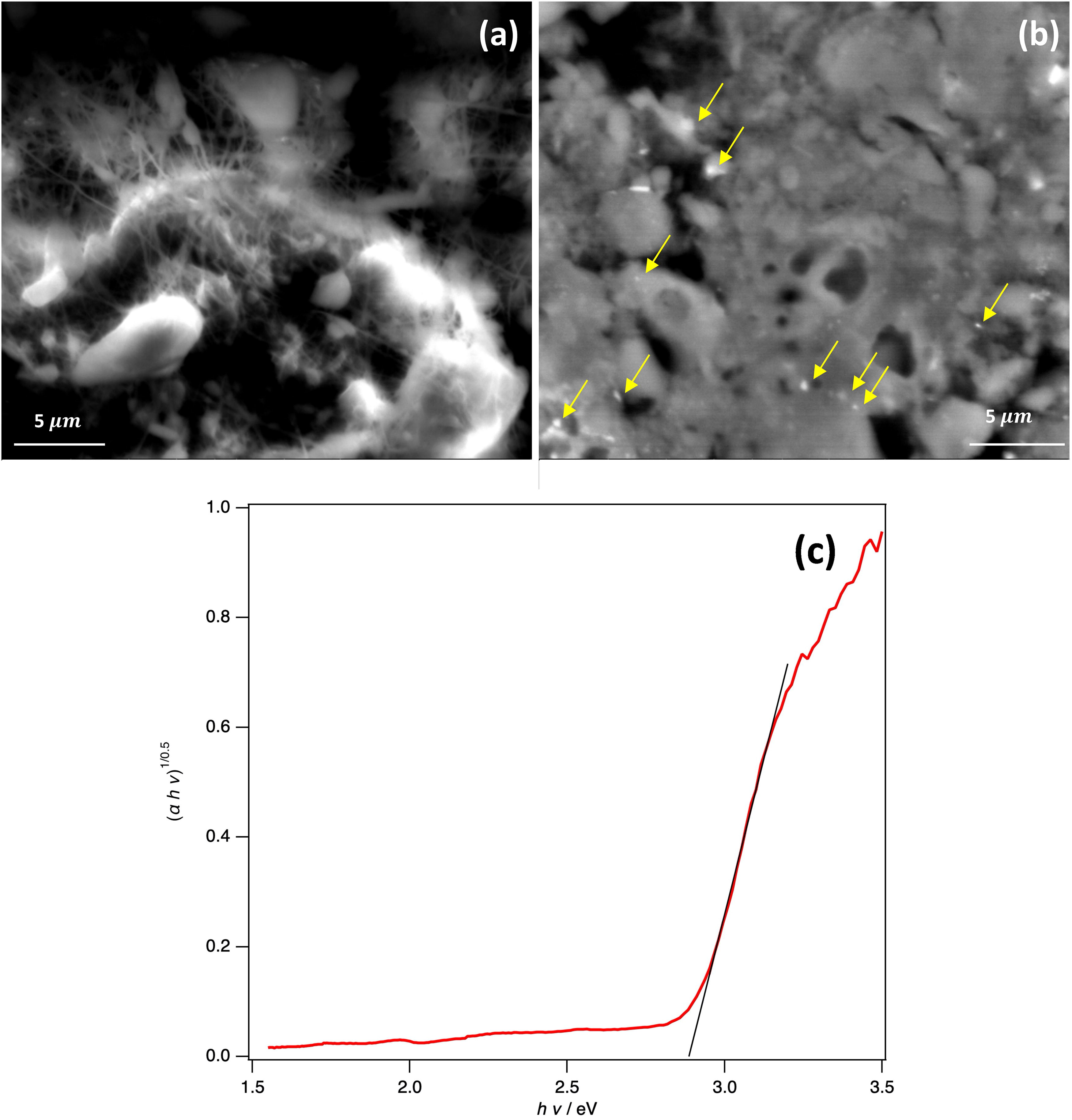
Scanning electron microscopy images of the polyvinylidene fluoride (PVDF) nanofiber composite:
The PVDF nanofibers measure approximately 100–200 nm in width and extend to several micrometers in length. The g-C3N4 particles are irregular in shape, with typical sizes around 5
Differential microbial community analysis
The microbial richness analysis showed that the Shannon alpha diversity metrics were similar across the samples (PE-1: 2.7; PE-2: 2.5; PE-3: 2.7). The Shannon index estimates both the species evenness and species richness, with weight on the species richness factor. In addition, the Shannon richness indices for PE-1, PE-2, and PE-3 were 181, 177, and 179, respectively, indicating minor differences in microbial diversity. However, the diversity richness indicated statistical differences among the tested samples. For instance, sample PE-2, which underwent 2 h of photocatalytic treatment, exhibited a slightly lower Shannon index of 2.5 and a richness index of 177, likely influenced by treatment conditions such as varying pH, temperature, and light intensity.
The 16S rRNA gene libraries identified 50 different bacterial species across the three samples, representing various phyla. Proteobacteria emerged as the predominant phylum in all three samples, with relative abundances of 77.4% in PE-1, 84.0% in PE-2, and 76.0% in PE-3. Other phyla, such as Bacteroides, Leptrichiaceae (family), and Firmicutes were present in minor proportions (Fig. 3). These results represent descriptive trends, as statistical analyses focused on ARG profiles rather than bacterial phyla abundances. At the genus level, the distribution revealed Arcobacter spp., Acinetobacter spp., and Acidovorax spp. as dominant taxa across the three samples (before treatment—PE 1; after 2 h—PE 2; after 4 h—PE 3) (Fig. 4). Notably, the proportion of Epsilonproteobacteria decreased in PE-3, from 33% in PE-1% to 26% in PE-3. In addition, the proportions of Firmicutes, Leptrichiaceae, Bacteroides, and Deltaproteobacteria also decreased across treatment conditions, which reduced in PE 3 by 10.0%, PE 2 by 13.0%, and PE 1 by 22.4%. These shifts in community structure suggest that photocatalytic treatment influenced microbial composition.
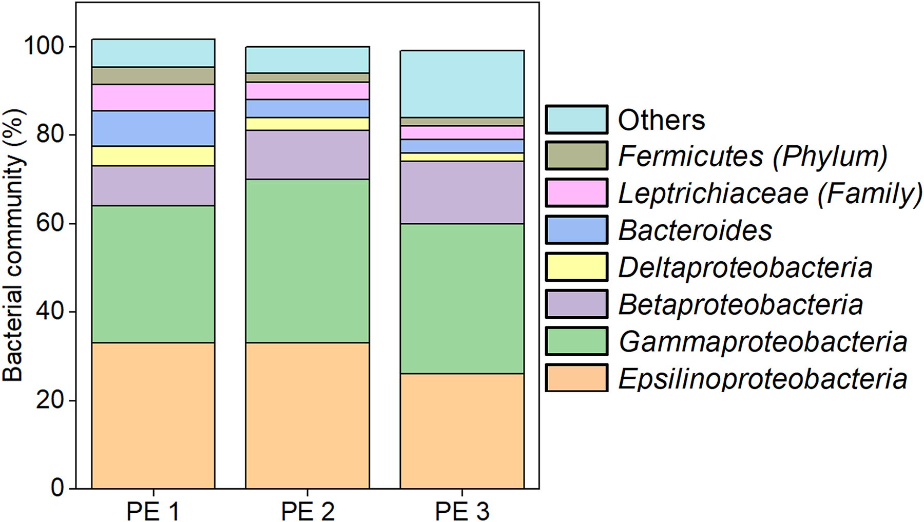
Class-level distribution of bacterial communities in wastewater samples collected before treatment (PE 1), after 2 h of treatment (PE 2), and after 4 h of treatment (PE 3). The percentage distribution represents the bacterial communities at the class level, with “Others” denoting sequences assigned to unclassified bacteria.
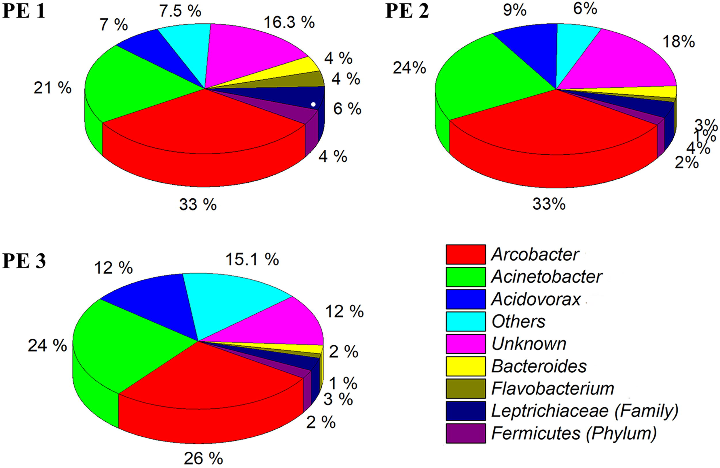
Genus-level distribution of bacterial communities in wastewater samples collected before treatment (PE 1), after 2 h of treatment (PE 2), and after 4 h of treatment (PE 3). The chart shows the relative taxonomic abundance of 16S rRNA gene sequences in the microbial community. Taxonomic assignments were determined by running gene sequences using the USEARCH global alignment algorithm and a Python program. “Others” represent sequences assigned to unclassified bacteria.
Importantly, this study does not conclude that the phyla or genera reported are inherently antibiotic-resistant, as culture-based techniques were not employed to confirm ARB presence. Instead, these results indicate changes in microbial community composition after photocatalytic treatment, with potential implications for wastewater microbiomes. While the genus Arcobacter spp., widely reported in wastewater systems, exhibited a reduction after treatment (e.g., in PE-3), the increases in Acinetobacter spp. and Acidovorax spp., both associated with MDR (Manchanda et al., 2010; Miura et al., 2013), warrant further investigation into how treatment conditions may influence community dynamics. Additional studies are needed to establish the functional significance of these shifts and their potential relevance to ARG mitigation.
DNA biomarker quantification
The gene copies of 16S rRNA genes from wastewater samples, before and after the treatment remained nearly the same, ranging from 6.1 to 7.9 log10 gene copies/µL (Fig. 5 and Supplementary Fig. S2). This consistency aligns with previously reported bacterial biomass values from studies on tubercles (4.4 × 10⁷ gene copies per g tubercle) and biofilms (3.5 × 10⁵ 16S rRNA gene copies per cm2) in water systems (Kimbell et al., 2021). However, the key focus of this study lies in the abundance and removal rates of ARGs, as their mere presence is ubiquitous in wastewater and biofilm systems. Understanding these metrics is crucial for evaluating the efficacy of photocatalytic treatments and their potential scalability in wastewater treatment.
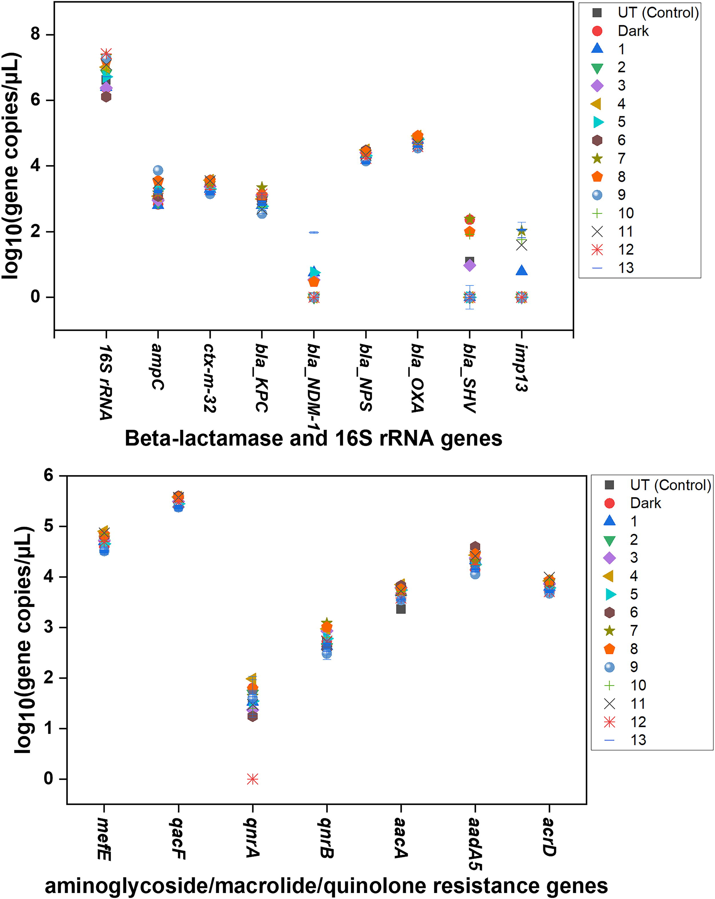
Quantities of 16 distinct ARGs evaluated in this study under various experimental conditions. “UT Control” represents the untreated primary clarifier sample (PE-1); “Dark” indicates samples incubated without light exposure. Time points for light treatment are as follows: 0 h (no treatment), 20 min, 40 min, 60 min, 80 min, 100 min, 120 min (PE-2), 140 min, 160 min, 180 min, 200 min, 220 min, and 240 min (PE-3). ARG, antibiotic resistance gene.
The microfluidic qPCR analysis identified 42 different ARGs in the raw sewage sample, marking this as the first comprehensive study on ARGs in the primary clarifier effluent at the Water Reclamation Facility in Rapid City, SD. The results highlight the abundance of specific ARGs and their resistance mechanisms, with Table 1 listing their copy numbers and roles. Table 1 lists the copy numbers and roles of these ARGs, highlighting key genes such as beta-lactamases (ampC, blaCTX-M-32, blaKPC, blaNPS, blaOXA, blaSHV), aminoglycosides (aadA5, aacA, qacF, acrD), quinolones (qnrA, qnrB), and macrolides (mefE). In addition, tetracycline resistance genes (tetA, tetL, tetM, tetS, tetW, tetX), vancomycin resistance genes (vanA, vanB), and ARGs conferring resistance to rifampicin (arr2), erythromycin (ermB, ermF), streptomycin (strB), trimethoprim (dfr13), and florfenicol–amphenicol (floR) were detected.
List of ARGs, MDRG, MRGs, and Integrons Detected from the Rapid City Wastewater Treatment Plant at Rapid City, South Dakota
Quantities of Gene Copies in log10 (Gene Copies/µL). ARG, antibiotic resistance gene.
HMRGs were also identified, including those conferring resistance to cadmium (cadA), copper (copA), chromium (chrA), mercury (merA), nickel (nikA) and nickel/cadmium/cobalt (rcnA). In addition, integrons conferring resistance to chloramphenicol (class 1 integrons catB8), sulfonamides (sul1, sul2, sul3), and class 1 integrons (intl1), class 2 integrons (intl2), and class 3 integrons (intl3) were detected. These integrons likely play a role in facilitating the spread of ARGs via mobile genetic elements, emphasizing the need for research into their ecological mechanisms and coselection processes to inform the development of more effective treatment strategies (Di Cesare et al., 2016). Moreover, the PCoA illustrated the statistical dimensionality of the datasets, providing additional insights into the relationships among detected ARGs and their resistance mechanisms (Supplementary Fig. S1). The PCoA results offer critical insights into the clustering and variance of ARG profiles across samples, providing a visual representation of how photocatalytic treatments influence microbial and genetic community structures.
Nine crucial genes exceeded 5 log10 gene copies/µL across all photocatalytic treatments, including ermB, ermF, intl1, intl3, merA, strB, sul1, tetX (Supplementary Fig. S2) and qacF (Fig. 5
Among the nine different gene clusters of vancomycin resistance genes, vanA and vanB are predominant and are associated with the transposon sequences of Tn1546 and Tn1549, respectively (Arredondo-Alonso et al., 2021). After 4 h of photocatalytic treatment, varying gene copy levels of blaSHV (Fig. 5) and mexB (Supplementary Fig. S2) were observed, partially confirming that the plasmonic effect of nanofiber mats influences the genetic response of ARGs. Among the variants of β-lactamases genes, blaSHV is particularly crucial due to its broadened substrate specificity against ceftriaxone, ceftazidime, and cefotaxime (Oteo-Iglesias et al., 2006; Pfeifer et al., 2010). In addition, the MexAB-OprM complex, a tripartite protein system, plays a critical role in MDR as a transporter. Within this complex, mexB functions as an inner membrane protein and a key component of the resistance-nodulation division family (Ohene-Agyei et al., 2012).
Interestingly, the blaNDM-1 gene (Fig. 5) was absent in the control sample but detected after photocatalytic treatment, suggesting that the membrane-based photocatalytic treatment may selectively enrich specific ARGs like blaNDM-1 to detectable levels. This ARG, blaNDM-1 (New Delhi metallo-beta-lactamase—Klebsiella), confers resistance to beta-lactam antibiotics, including penicillin, cephalosporin, and carbapenem (Bahr et al., 2021). These findings align with previous studies indicating that heterogeneous photocatalytic treatment can enhance the intl1 gene, which is involved in antibiotic resistance of E. coli. This highlights the need for modifications to photocatalytic membrane-based wastewater treatment methods to achieve complete ARG mitigation.
No significant changes were observed in the gene copies of blaCTX-M-32, blaOXA (Fig. 5), blaNPS, qacF, arr2, and tetA (Supplementary Fig. S2). These results corroborate previous reports suggesting that some ARGs are more resistant to degradation using currently utilized wastewater treatment methods. Moreover, the gene copy values of ARGs in this study align well with previously reported values on the absolute abundance of blaTEM, sul1, qnrA, vanA, and intl1 in biofilms associated with wastewater treatment systems. These concentrations, ranging up to 4.2 log10 copies, are consistent with findings from WWTP studies (Di Cesare et al., 2016; Tehrani and Gilbride, 2018a, b; Wang et al., 2014), providing context for the prevalence and persistence of ARGs in such environments.
Potential pathways used by the consortia for conferring antibiotic resistance
Our gene sequencing analysis identified Arcobacter spp. and Acinetobacter spp. as dominant bacteria in both control and treated samples. These bacteria can develop antibiotic resistance through various mechanisms, including those associated with ROS generated during typical treatment processes. In addition, they play a key role in spreading resistance to other bacteria in wastewater through HGT. Our results demonstrate the use of photocatalytic treatment using a nanocomposite material to degrade ARGs, potentially mitigating the spread of antibiotic resistance in wastewater. These findings are explained in greater detail below.
Arcobacter spp. and Acinetobacter spp. are classified as critical priority pathogens by the World Health Organization due to their potential to cause serious infections and diarrheal illness (Grotiuz et al., 2006; Mansfield and Forsythe, 2000; Ramees et al., 2017; Tacconelli et al., 2018). While Arcobacter spp. is recognized for its significant impact on human health (Skovgaard, 2003), comprehensive information on its genome-wide interactions remains limited. Arcobacter butzleri, known for its high genome diversity and resistome plasticity, highlights its pathogenic potential (Isidro et al., 2020). ROS, generated during chlorine-based disinfection processes (Zhang et al., 2021a, b), can promote antibiotic tolerance and resistance in these bacteria through mechanisms such as mutation, ARG transformation signaling, targeting the TCA cycle to impede cellular respiration, and increasing the production of MDR efflux pump transporters (Li et al., 2021a, b). Studies on Escherichia coli (Wu et al., 2012) and Staphylococcus aureus (Rowe et al., 2020) demonstrate similar ROS-induced antibiotic tolerance. Moreover, whole-genome sequencing of Arcobacter butzleri reveals resistance genes linked to heavy metal exposure, indicating coselection of multiple antibiotic resistances (Fanelli et al., 2019).
In this study, 42 distinct ARGs were identified in the analyzed wastewater samples, highlighting the impact of antibiotic use on the microbial community. This was facilitated by a syntrophic relationship between key bacteria such as Arcobacter spp. and Acinetobacter spp., along with other microorganisms. Mutations and HGT (Wright, 2011), particularly among bacterial consortia is hypothesized to be facilitated by Arcobacter spp. and Acinetobacter spp., conferring resistance to 42 ARGs (Table 1 and Supplementary Figure S2). Acinetobacter baumannii, for instance, has evolved into an MDR bacterium, harboring 45 different ARGs (Fournier et al., 2006). Acinetobacter spp., a predominant bacterium in the consortia, are known to harbor integrons, which are mobile genetic elements in the ARG cassettes. These integrons can integrate their genetic material into plasmids or chromosomes of other bacteria via HGT (Leungtongkam et al., 2018). Detection of seven crucial integrons by MF-qPCR support this hypothesis, aligning with previous findings on wastewater biofilms, where high bacterial biomass and diversity in wastewater biofilms expedite HGT (Zhang et al., 2009).
As outlined in the introduction, photocatalysis is suggested to facilitate the transition of plasmid DNA transfers from supercoiled state to relaxed state, followed by its conversion to a single-helix and subsequent fragmentation into smaller segments. The proposed mechanism is illustrated in Figure 6. Furthermore, the Ag-functionalized PVDF membrane used in visible-light-based photocatalytic treatment demonstrated a significant reduction of critical ARGs, such as qnrA and vanA, which are known to persist in wastewater due to their plasmid-borne nature and resistance to conventional WWT methods. Conventional approaches, such as biological treatment and chlorination, often fail to effectively target ARGs because of their stability and integration into mobile genetic elements (Li et al., 2021a, b; Wang and Chen, 2022a, b, c; Cai et al., 2021). The acquisition of exogenous genetic material allows microbial consortia to develop and transfer ARGs via mobile genetic elements (Lowe, 1982), employing enzymatic mechanisms such as beta-lactamase synthesis and nonenzymatic mechanisms such as efflux pump activation and altered membrane permeability (Vrancianu et al., 2020). The observed photocatalytic degradation of key genes, including qnrA and vanA, highlights the potential of the photocatalytic system to complement existing wastewater treatment processes by targeting ARGs that are otherwise challenging to remove. However, further research is needed to delineate the specific pathways of ARG degradation in wastewater and their interactions with surface-modified membranes. These insights will pave the way for optimizing photocatalytic systems and integrating them effectively into existing wastewater treatment frameworks, as explored further in the Outlook section.
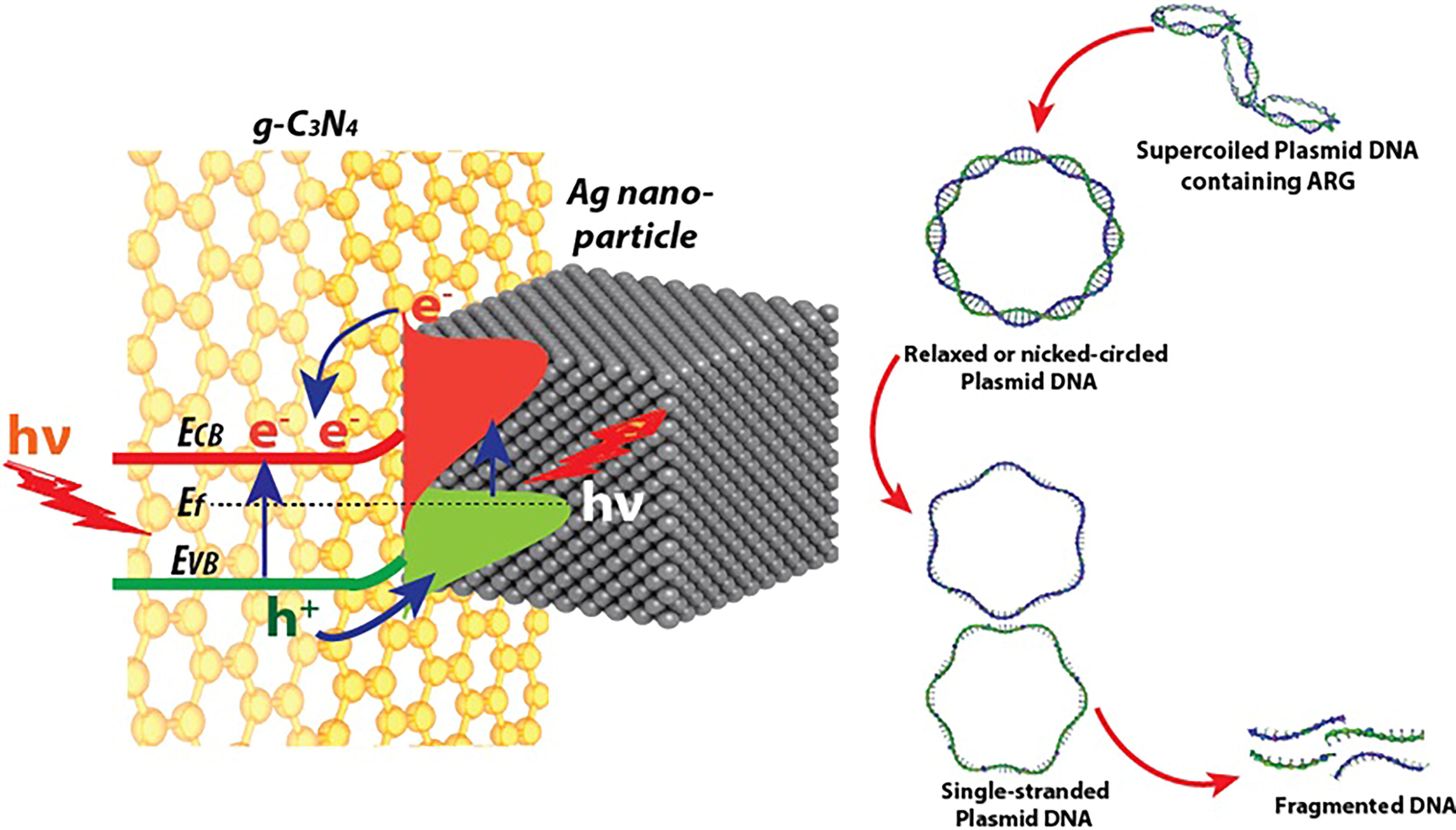
The schematic of proposed mechanism of destruction of plasmic DNA by plasmonic photocatalyst of Ag nanoparticle on g-C3N4 surface.
Outlook
Similar to other photocatalytic reactors, full-scale implementation of the visible-light-based photocatalytic technique will require further research and optimization before adoption in risk-averse industries such as water treatment systems (Loeb et al., 2018). However, the current study shows promise for integration as a supplementary process for ARG removal into existing wastewater treatment systems. Advances in energy-efficient LED technology, electrospun substrates (Loeb et al., 2018), scalable and modular configurations (e.g., the cassette-based module used in this study), and integration with renewable energy sources, as highlighted by Wang et al., 2021 and Alalm et al., 2021, emphasize the feasibility of transitioning from pilot-scale experiments to field-scale applications. The scalability of plasmonic materials, similar to those used in this study, has been demonstrated in studies such as Murphy et al., 2021, further supporting the practical relevance of this technology. Future efforts should focus on ensuring consistent light intensity, maintaining catalyst stability across diverse wastewater conditions, optimizing economic feasibility, refining the system to adapt to varying wastewater conditions, fostering industrial partnerships, and facilitating seamless integration into existing treatment infrastructures.
Key wastewater parameters, such as pH, turbidity, chemical oxygen demand (COD), and nutrient levels (N and P), significantly influence photocatalytic efficiency. Graphitic carbon nitride and AgNP-based photocatalysts perform optimally under near-neutral to slightly acidic conditions (pH 6–7), typical of municipal wastewater. Total suspended solids, regulated at 30 mg/L in the United States, can limit light penetration, but pretreatment steps such as sedimentation or filtration can mitigate this (Wang et al., 2021). High COD levels compete with ARG degradation for reactive species, making photocatalysis more effective as a posttreatment step. Chlorides can quench reactive radicals, while nitrates may enhance photocatalysis. Heavy metals can deactivate catalytic sites, necessitating tailored catalyst coatings. In addition, organic loads and nutrients can indirectly influence ARG dynamics and photocatalytic efficiency (Wang et al., 2021).
The scalability of plasmonic materials, similar to those used in this study, has been demonstrated in studies such as Murphy et al., 2021, further supporting the practical relevance of this technology. EU-funded initiatives such as Photo4Future and PHOTOREACT have already demonstrated the effectiveness of advanced photocatalytic systems in treating recalcitrant pollutants in real-world scenarios (KU LEUVEN, 2024). These findings provide a strong foundation for scaling this technology for targeted ARG removal.
A multifaceted approach is essential to mitigate ARB and ARGs. This includes optimizing photocatalytic processes, reducing antibiotic misuse through regulations and stewardship programs (Foxman et al., 2024), proper pharmaceutical waste disposal (Kümmerer, 2009), enhanced monitoring systems, and public awareness campaigns (Foxman et al., 2024). Promoting safe wastewater reuse can further address ARG spread. These combined strategies will significantly enhance ARG mitigation in wastewater treatment systems and ensure the relevance and impact of advanced technologies such as modified PVDF nanofibers in managing ARG contamination across seasons and diverse wastewater conditions.
Conclusions
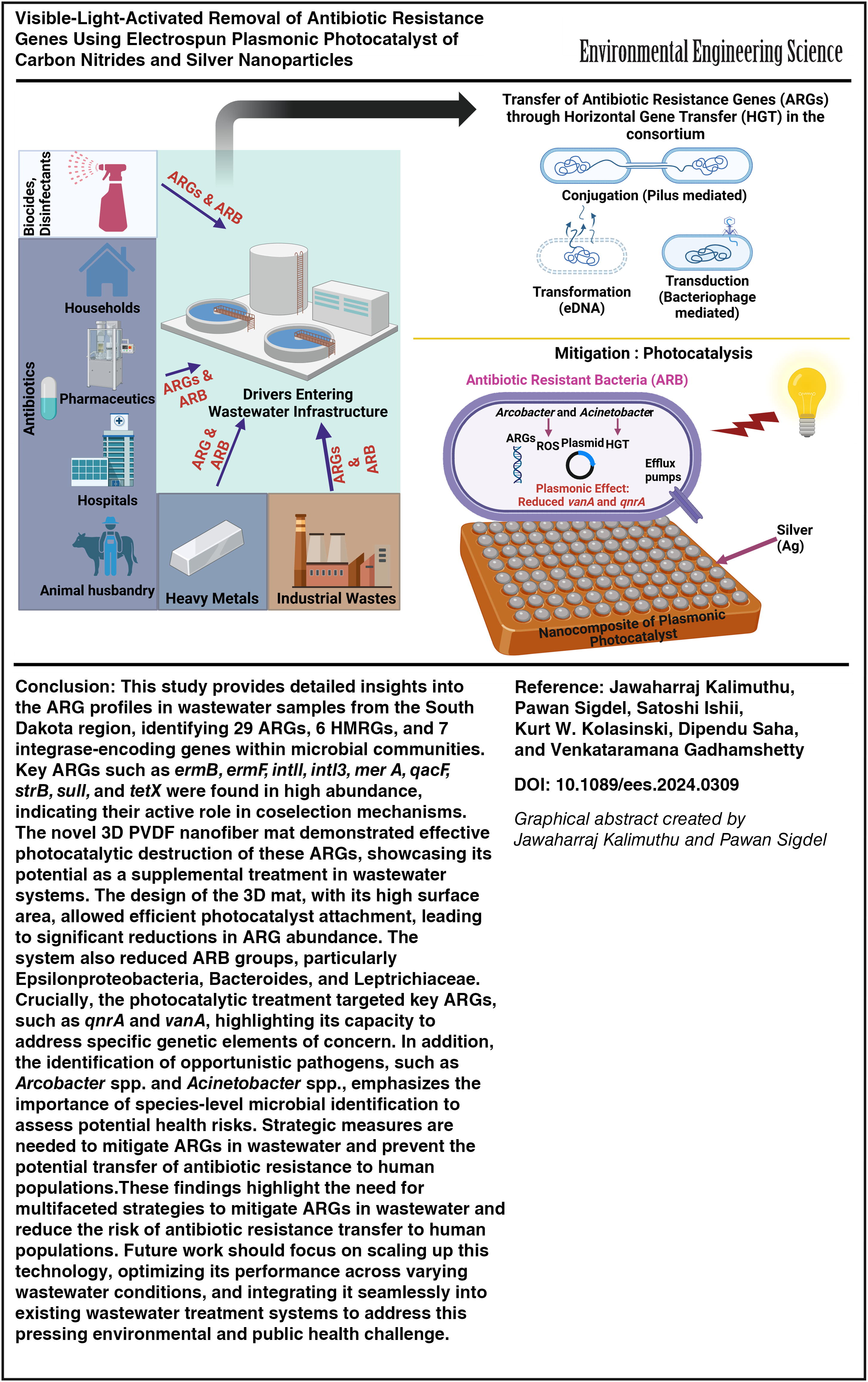
This study provides detailed insights into the ARG profiles in wastewater samples from the South Dakota region, identifying 29 ARGs, 6 HMRGs, and 7 integrase-encoding genes within microbial communities. Key ARGs such as ermB, ermF, intl1, intl3, merA, qacF, strB, sul1, and tetX were found in high abundance, indicating their active role in coselection mechanisms. The novel 3D PVDF nanofiber mat demonstrated effective photocatalytic destruction of these ARGs, showcasing its potential as a supplemental treatment in wastewater systems. The design of the 3D mat, with its high surface area, allowed efficient photocatalyst attachment, leading to significant reductions in ARG abundance. The system also reduced ARB groups, particularly Epsilonproteobacteria, Bacteroides, and Leptrichiaceae. Crucially, the photocatalytic treatment targeted key ARGs, such as qnrA and vanA, highlighting its capacity to address specific genetic elements of concern. In addition, the identification of opportunistic pathogens, such as Arcobacter spp. and Acinetobacter spp., emphasizes the importance of species-level microbial identification to assess potential health risks. Strategic measures are needed to mitigate ARGs in wastewater and prevent the potential transfer of antibiotic resistance to human populations. These findings highlight the need for multifaceted strategies to mitigate ARGs in wastewater and reduce the risk of antibiotic resistance transfer to human populations. Future work should focus on scaling up this technology, optimizing its performance across varying wastewater conditions, and integrating it seamlessly into existing wastewater treatment systems to address this pressing environmental and public health challenge.
Footnotes
Authors’ Contributions
K.J.: Resources, conceptualization, visualization, and validation. P.S.: Editing and validation. S.I.: Resources. K.W.K.: Resources and validation. D.S.: Resources, conceptualization, validation. V.G.: Conceptualization, editing and validation, resources, supervision, validation, project administration, funding acquisition.
Author Disclosure Statement
The authors declare that they have no known competing financial interests or personal relationships that could have appeared to influence the work reported in this paper.
Funding Information
We acknowledge the funding support from National Science Foundation RII FEC awards (#2418752, #1849206, #1920954). Research support from the Department of Civil and Environmental Engineering at the South Dakota School of Mines and Technology is gratefully acknowledged. D.S. acknowledges the funding support from 2018 Water Resources Research projects USGS 104B, Pennsylvania.
References
Supplementary Material
Please find the following supplemental material available below.
For Open Access articles published under a Creative Commons License, all supplemental material carries the same license as the article it is associated with.
For non-Open Access articles published, all supplemental material carries a non-exclusive license, and permission requests for re-use of supplemental material or any part of supplemental material shall be sent directly to the copyright owner as specified in the copyright notice associated with the article.