
Abstract

Abstracts
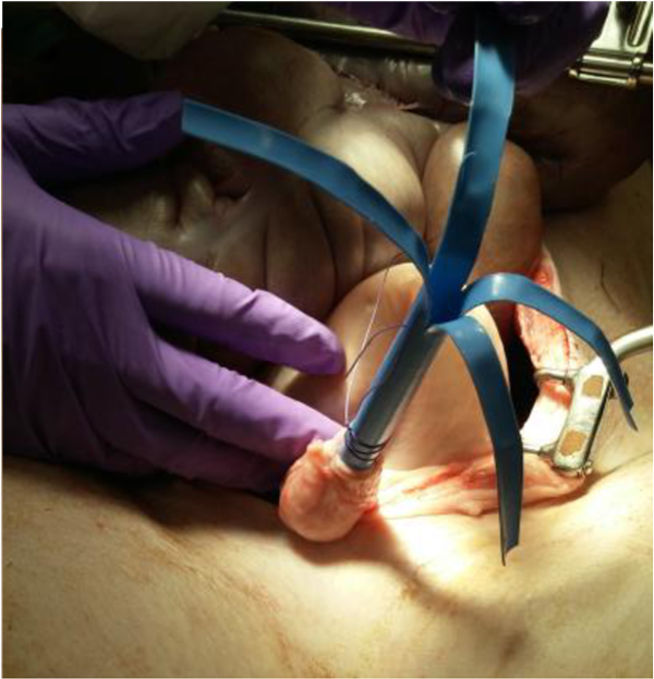
APPLYING UROLITHIASIS TO BILIARY STONES: PERCUTANEOUS TRANSHEPATIC LITHOTRIPSY
University of Wisconsin Department of Urology
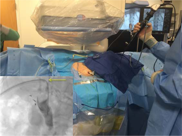
Patient is supine with a ureteral access catheter inserted percutaneously into the common bile duct. The radiograph shows the location of the sheath (yellow arrow) and the ureteroscope in place.
OFFICE-BASED ULTRASOUND GUIDED PERCUTANEOUS RENAL MASS BIOPSY
Department of Urology, University of California, Irvine
Section of Urology, Department of Surgery, University of Manitoba
TOP 10 ABSTRACT
Robotics Laboratory, Urology Department, Johns Hopkins University, Baltimore, MD
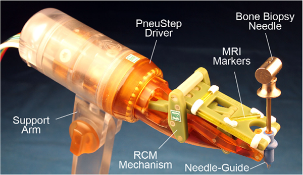
MRI-Safe RCM Robot.
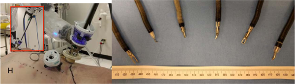
The robot is electricity free, uses air pressure for actuation, light for the position sensors, and is entirely made of nonconductive, non-metallic, and nonmagnetic materials. Accordingly, the robot is MRI-Safe according to ASTM F2052, F2213, and F2182 based on the scientific rationale. The needle-guide, which comes in direct contact with the patient, is built of certified biocompatible material (ISO-10993). The bore of the needle-guide can be made to accommodate various needles. The figure shows the MRI-Conditional Invivo 15100 bone biopsy needle (Invivo, Philips Healthcare, The Netherlands).
The robot includes high contrast MRI markers for registration (filled with MR-Spots contrast, Beekley, Bristol, CT). A custom image-to-model registration algorithm and image-guided control software was developed. Initial tests were conducted in a Siemens MAGNETOM Tim4G scanner. Images of a gelatin mock-up were acquired together with the robot. These initial tests showed no apparent image artifacts or problems in operating the robot within the MRI.
ROBOTIC VERSUS CONVENTIONAL FLEXIBLE URETEROSCOPY IN RENAL STONES: EXPERIENCE OF 132 CASES
Sanador Hospital, Bucharest, Romania
Johns Hopkins University School of Medicine, Baltimore, MD, USA
As a precursor to a clinical system, this study presents preliminary results with an adaptive, pre-clinical system based on the integration of the Philips Sonalleve MR-HIFU software platform, a pre-clinical-specific transducer, and an MR-Safe robot.
The system was evaluated for accuracy in a tissue-mimicking phantom by sonicating in a 15 mm grid with different powers and durations (4–32 W, 30–90 s, continuous wave) to assess the accuracy of the combined robot transducer system. Second, sonications were performed in a 10 mm grid on a fresh, excised pig leg (16 W, 120 s) to evaluate the targeting accuracy and the ability of the system to couple to the leg properly. The MRI SNR was also evaluated with and without the system in place.
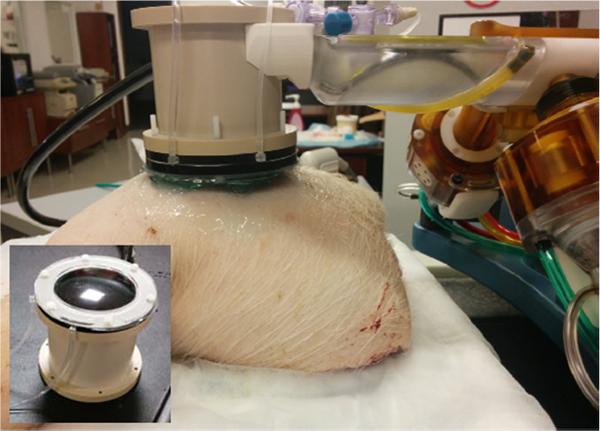
The transducer coupled to an excised pig leg, mounted to the MR-Safe robot. The top of the transducer is shown, inset.
TOP 10 ABSTRACT
Urology Service, Department of Surgery, Memorial Sloan Kettering Cancer Center, New York, NY
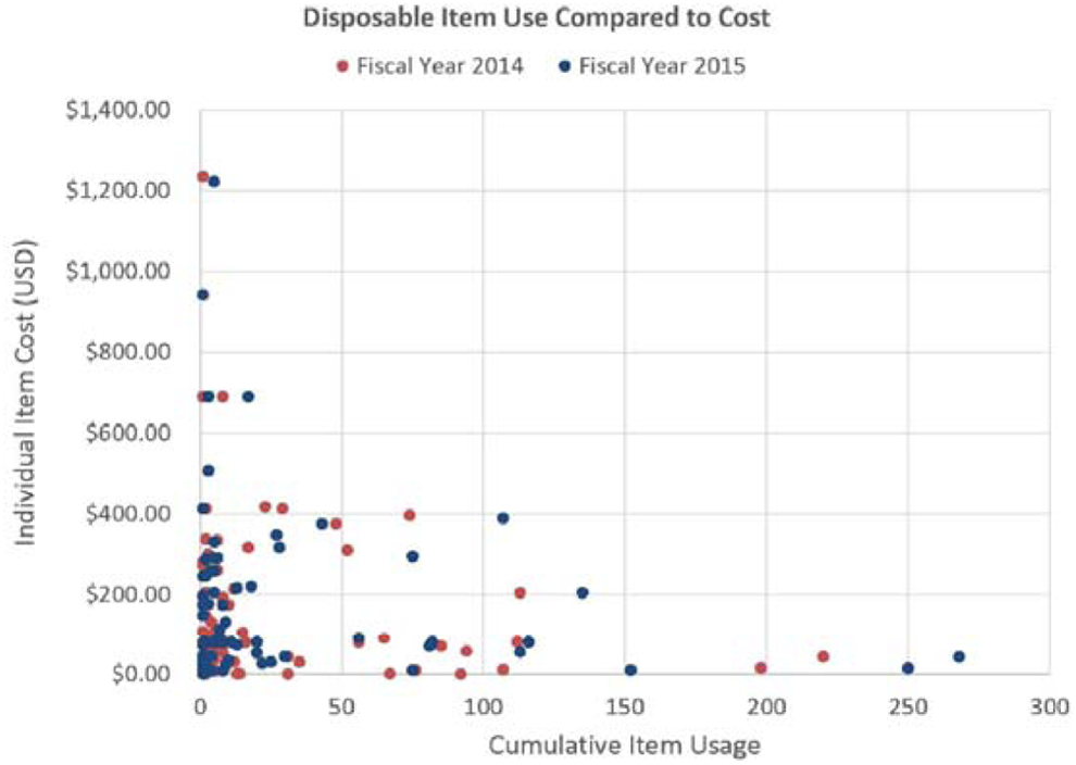
Relationship between item usage and cost.
TOP 10 ABSTRACT
A DEVICE FOR RARE CELL ISOLATION AND CHARACTERIZATION
The James Buchanan Brady Urological Institute, Johns Hopkins School of Medicine, Baltimore, MD, USA
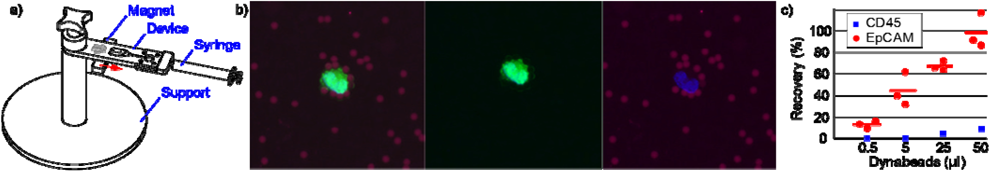
a) Schematic diagram of the device, b) LNCaP prostate cancer cell captured and visualized on membrane. Red = magnetic beads, green = cytosol of the cell, and blue = nucleus. c) Percent recovery of LNCaP prostate cancer cells using different volumes of anti-EpCAM Dynabeads (n = 3 per concentration)), as well as with a negative control using anti-CD45 Dynabeads (n = 1 per concentration).
Department of Urology, University of Illinois at Chicago College of Medicine, Chicago, IL
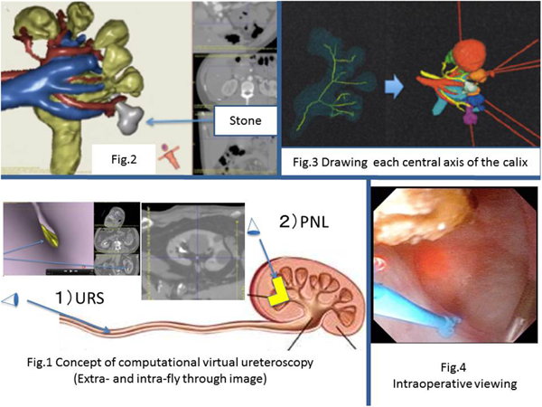
Nagoya University Graduate School of Medicine, Department of Urology, Nagoya, Japan
Centre of Uronephrology and Renal Transplantation, Fundeni Clinical Institute, Bucharest, Romania
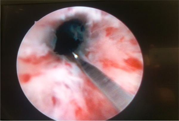
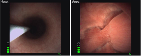
Cold knife incision of vesico-urethral anastomotic stricture
NEW GENERATION SINGLE-PORT ROBOTIC PLATFORM: FEASIBILITY ASSESSMENT IN A CADAVERIC MODEL
(left): Subcostal single-port robotic access for retroperitoneal radical nephrectomy.
TOP 10 ABSTRACT
Dept. of Pathology and Lab Medicine & Centre for Blood Research, University of British Columbia, Vancouver, BC, Canada
Dept. of Pathology and Lab Medicine & Centre for Blood Research, University of British Columbia, Vancouver, BC, Canada
Ctr. for Industrial and Medical Ultrasound, Applied Physics Lab., Univ. of Washington, Seattle, WA
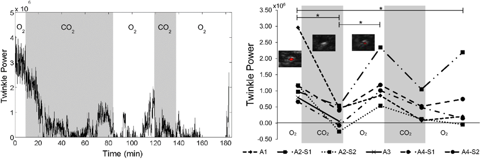
(Left) Plot of the twinkle power, or the magnitude of the Doppler signal, versus time for one animal. Twinkling begins to decrease immediately after the exposure to CO2 and begins to increase near the end of the O2 exposures. (Right) Summary plot of the mean twinkle power for 8-minute intervals at the end of each O2 or CO2 exposure for all four animals. Twinkling was found to be reduced by exposure to CO2 and increased by exposures to O2.
Work supported by the National Space Biomedical Research Institute through NASA NCC 9-58 and NIH NIDDK grants DK043881 and DK092197; this material is the result of work supported by resources from the VA Puget Sound Health Care System, Seattle, WA.
BEST PAPER AWARD
INCREASED CONTRAST OF STONE SPECIFIC ULTRASOUND IMAGING IN HUMAN SUBJECTS
Center for Industrial and Medical Ultrasound, Applied Physics Laboratory, University of Washington, Moscow 119991, Russia
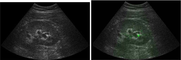
ultrasound image (12 cm depth) showing the B-mode (left) and S-mode (right).
University of California Los Angeles, Department of Bioengineering
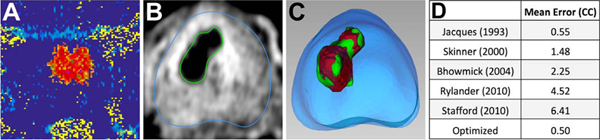
(left), MRI thermometry during treatment; Fig. B (middle-left), DCE MRI with contoured non-perfused tissue (green); Fig. C (middle-right), non-perfused tissue model (green) registered to Arrhenius damage estimate (red); Fig. D (right), mean error of damage volume estimated using Arrhenius values from 5 publications, and using coefficients optimized to our dataset.
University of California Los Angeles, Department of Bioengineering
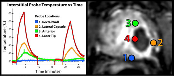
Left: Interstitial probe temperatures during FLA treatment. Right: Post-treatment dynamic contrast-enhancement image showing treated region as non-perfused.
Kesem Health, Melbourne, Australia
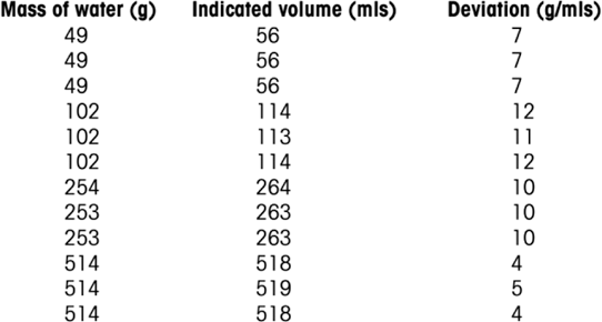
iUFlow (indicated volume) repeatability and accuracy testing.
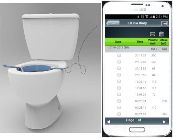
iUFlow Fully Automated Voiding Diary Digital Health Solution.
University of California Los Angeles, Department of Bioengineering
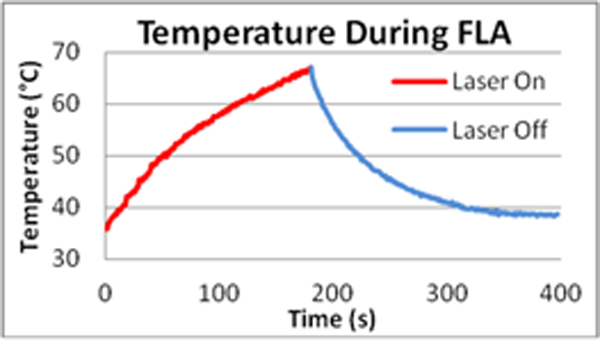
Interstitial probe temperature vs time for patient B.
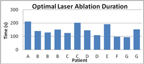
Optimal laser ablation duration for selected patients.
Biomedical Engineering, University of Michigan, Ann Arbor, MI, USA.
Hemorrhage and hematoma induced during treatment appeared as dark pixels on the T2-weighted, MR scans. An image segmentation algorithm was able to separate hemorrhage from nominal parenchyma. The volume of hemorrhage was computed by summing the number of image pixels in a sample that had been identified as hemorrhage and then multiplying by the volume represented by an image pixel.
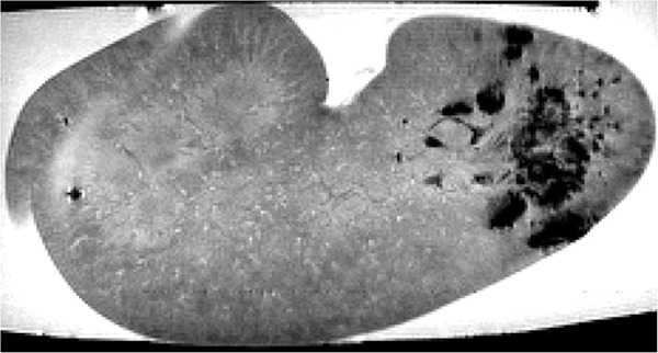
Example MR image slice of a kidney treated with SWL. Hemorrhage causes a readily apparent loss in image signal (right half of image).
THE APPLICATION OF AN INNOVATIVE SURFACE ELECTRODE SYSTEM IN EXTERNAL URETHRAL SPHINCTER ELECTROMYOGRAPHY TESTING IN RATS
Dept of BME, Lerner Research Institute, Cleveland, OH 44106
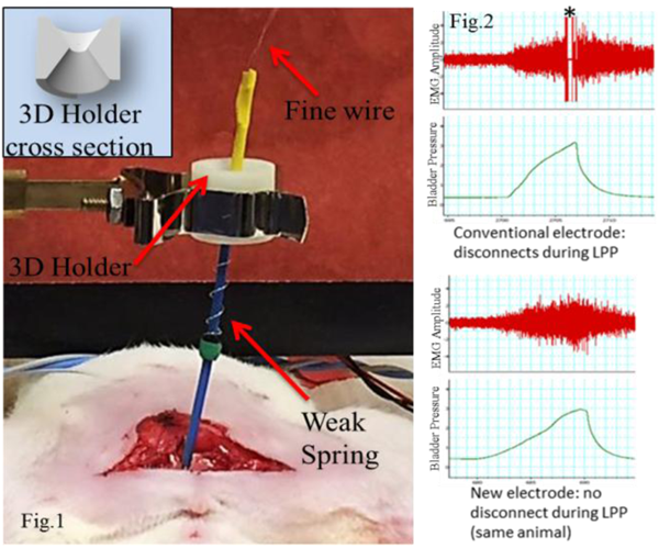
Bioengineering Program, School of Engineering and Applied Science, Hofstra University
Johns Hopkins Medical Institutions
TOP 10 ABSTRACT
University of Nagoya, Department of Urology
Department of Genitourinary Oncology, Moffitt Cancer Center, Tampa, FL
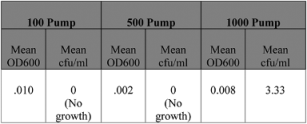
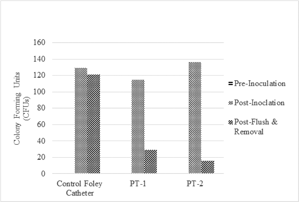
IN VITRO MICROBIOLOGICAL EVALUATION OF INSUFFLATION FILTER PERFORMANCE
Spectrophotometer and LB-Agar Plate Growth data
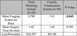
Fogging Events, Surgery Time, Cost;
*
Omits One-Time Purchase of Fluid Warming System
Department of Urology, University of California, Irvine
University of Illinois at Chicago – Department of Urology
Department of Surgery, Division of Urology, Memorial Sloan Kettering Cancer Center, New York, NY
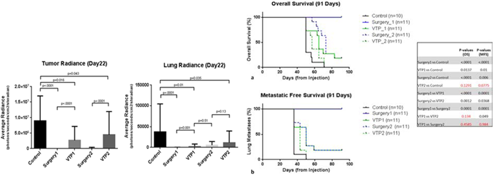
Urothelial carcinoma (UC) is a common malignancy of the bladder or upper urinary tract which usually presents in non-muscle-invasive form at diagnosis. Yet we lack conservative treatment options for patients for whom bacillus Calmette-Guerin (BCG) immunotherapy has failed or is not indicated. To address this lack, we investigated the effectiveness of combination treatment using vascular targeted photodynamic therapy (VTP) and anti-CTLA-4 immunotherapy in a mouse model of UC. C57BL/6 mice injected with murine bladder 49 (MB-49) cell line were allocated into 4 treatment groups: VTP only, anti-CTLA-4 only, combination therapy, and control. We monitored tumor growth and development of lung metastases using bioluminescent imaging. Tumor cell population was studied with flow cytometry, and survival was evaluated with Kaplan-Meier curves. The combination treatment group had significantly lower tumor signal than the other three groups (p < 0.0001), as well as decreased lung signal uptake compared to the control (p < 0.0001), VTP only (p < 0.0001), and anti-CTLA-4 only (p = 0.002) groups. Combination therapy provided prolonged survival (p < 0.0001). We also rechallenged tumors in previously treated mice and compared tumor growth to that of a group of naïve mice, finding that mice previously treated with VTP only or combination therapy did not present tumor growth after rechallenge. The combination of VTP with anti-CTLA-4 was shown to be an effective therapy in an UC syngeneic mouse model. Our results suggest this therapy as a potential treatment option for both bladder and upper tract tumors in future clinical trials.
TOP 10 ABSTRACT
Johns Hopkins Hospital, Baltimore, MD
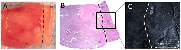
Tumor to left of dotted line, margin to right. A) Gross sample with 2 mm margin. B) H E of same sample. C) OCT en face image of region within box.
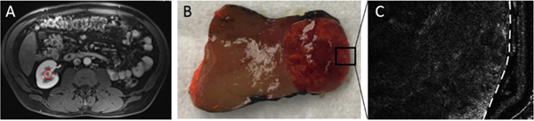
A) Contrast-enhanced MRI, with mas circled. B) Gross image of enucleated tumor. C) OCT en face image of region within box, thin margin to left of line.
Department of Urology and Urological Science Institute, Yonsei University College of Medicine, Seoul
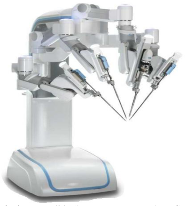
REVO-I robotic surgical system (2011); Meere Company (Inc. Seongnam, Republic ofKorea).
Rutgers New Jersey Medical School, Department of Urology, Newark, NJ
1. Single-use, flexible 9FR articulating tip ureteroscope 2. LED light source/video processor with monitor
University of Washington Department of Urology
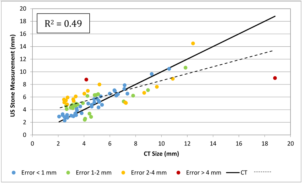
Correlation of stone size measurement between US and CT. Each measurement is color-coded based on the absolute difference (error), with the CT size used as the reference measurement. The solid black line represents a 1:1 correspondence between the US and CT measurements. The dashed line represents the correlation between the US and CT measurements.
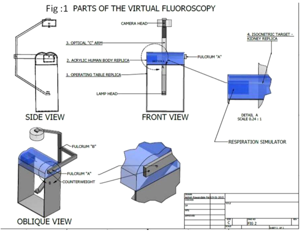
CAD drawing.
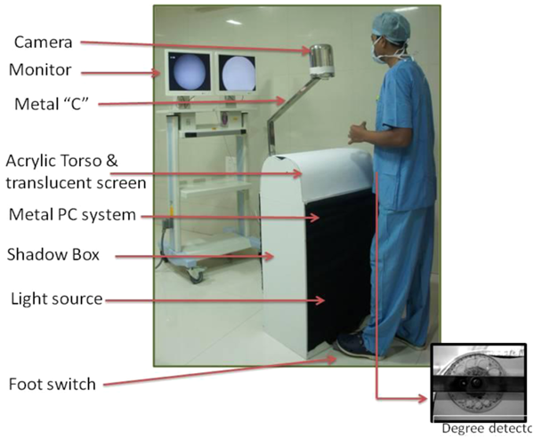
The First Prototype.
Department of Urology, University of Illinois at Chicago College of Medicine, Chicago, IL
Department of Urology, Stanford University Hospital, Stanford, CA, USA
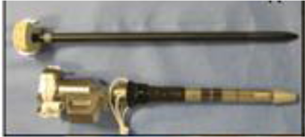
Sensorized cannula with obturator.
Department of Urology, University of Texas Southwestern Medical Center, Dallas, TX, USA
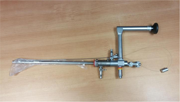
Nephroscope fitted with PercSac.
30Fr Amplatz sheath placed in porcine bladder for cystolitholapaxy.
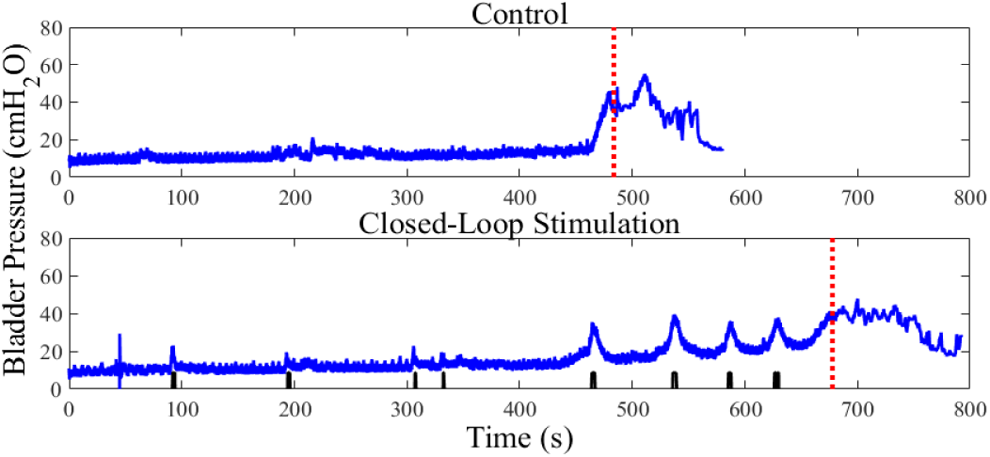
Dept of ECE, University of Florida, Gainesville, FL 32608
Autonomous, closed-loop GNS inhibited bladder activity and increased bladder capacity. (A) A hyper-reflexic bladder contraction occurred at 480 seconds after 400 mL of saline was infused into the bladder, resulting in urine leakage (long vertical dashed line). (B) Automatic conditional GNS triggered by CAT (short vertical lines) increased bladder capacity by 40% after inhibiting 4 unwanted contractions in this example.
BEST PAPER AWARD
The James Buchanan Brady Urological Institute and Department of Urology
The James Buchanan Brady Urological Institute and Department of Urology, Johns Hopkins University School of Medicine, Baltimore, MD, USA
Urology Department, Kaplan Medical Center, Rehovot, Israel
Portela Soni Medical LLC
TOP 10 ABSTRACT
Center for Industrial and Medical Ultrasound, Applied Physics Laboratory, University of Washington
Work support by NIH NIDDK grants DK043881, DK107094, and DK092197, and the National Space Biomedical Research Institute through NASA NCC 9-58. This material is the result of work supported by resources from the VA Puget Sound Health Care System.
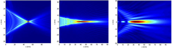
Simulated beam widths of the three methods.
TOP 10 ABSTRACT
Department of Urology, College of Medicine, University of Florida
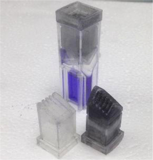
Single-faceted (left) and double-faceted (right) passive flow column designs in front of an assembled, 3D printed oxalometer with high oxalate-containing urinary sample (violet).
University of California, Los Angeles Department of Chemistry
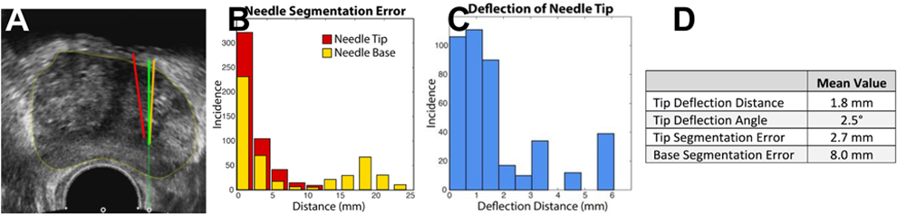
US image with intended needle path (green), segmentation (red), and true location (orange); Fig B, histogram of segmentation errors for needle tip (red) and base (yellow); Fig C, histogram of deflection errors for needle tip; Fig D, mean deflection and segmentation errors relative to true core location
Biomedical Engineering, University of Michigan, Ann Arbor, USA
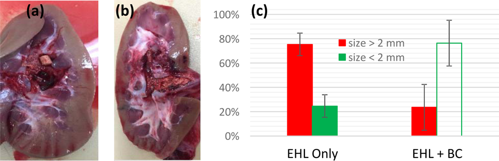
(a) Image of a kidney and remnant fragments treated with SWL only
(b) Image of a kidney and remnant fragments treated with SWL and bubble coalescence
(c) Remnant stone fragment size mass distributions normalized to initial stone mass
Department of Mechanical Engineering, University of Minnesota, Minneapolis, MN
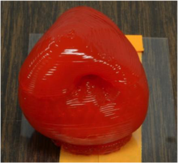
Preliminary 3D Printed Prostate Model
Center for Industrial and Medical Ultrasound, Applied Physics Laboratory, University of Washington, Moscow, Russia

Left: 2D ultrasound. Right:Twinkling power vs transmit voltage for different frequencies.
Work support by NIH NIDDK grants DK043881 and DK092197, and the National Space Biomedical Research Institute through NASA NCC 9-58.
Dept. of Bioengineering, University of Illinois at Chicago
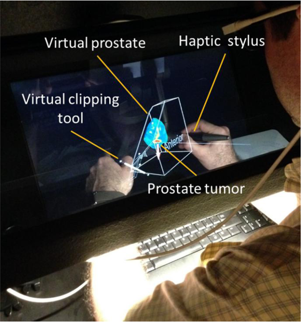
Augmented reality and haptic interface for exploration of holographic prostates using MRE. Arrow indicates the direction and magnitude of the force feedback while touching the cancerous tissue with the virtual needle.
Department of Urology, University of Washington
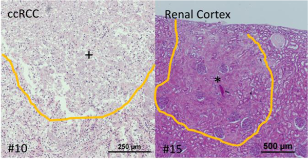
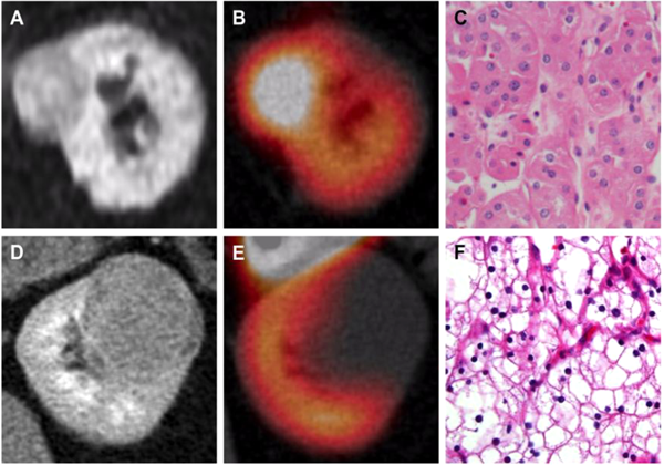
Histologic appearance of nearly completely homogenized (+) clear cell RCC treated with 10 BH pulses and sparsely homogenized (*) renal cortex with intact glomeruli and tubules treated with 15 pulses.
Biomedical Optics & Laser Laboratory, Biomedical Engineering, University of Miami, Coral Gables, FL
Department of Urology, Tokai University Hachioji Hospital, Japan
(right): Instrumentation for the SP1098 da Vinci SP platform.