
Abstract
Hepatitis E virus (HEV) is an emerging foodborne pathogen with domestic and wild pigs (and likely other species such as deer or rabbits) recognized as reservoir. Pathogenesis in pigs usually leads to an asymptomatic course of disease. Since there is no enzyme-linked immunosorbent assay (ELISA) kit for the detection of anti-HEV antibodies in pigs commercially available, the objective of this study was to assess the seroprevalence in fattening pigs at slaughter and at herd level using a newly developed ELISA based on genotype (GT) 1 and GT 3 in Bavaria, Germany. Based on 516 serum and 198 meat juice samples collected from different herds at four different Bavarian slaughterhouses, the overall seroprevalence of anti-HEV IgG in serum and meat juice samples was 68.6% and 67.6%, respectively. Analyzing the serum for the presence of anti-HEV IgM, 36/516 (7%) were positive for anti-HEV IgM. At herd level, most of the herds were seropositive for anti-HEV antibodies. The present study shows that HEV is widespread among the Bavarian pig population and that some pigs might test positive for anti-HEV IgM even at the age of slaughter. Also, meat juice serves as an equivalent matrix to serum to test for anti-HEV antibodies in pigs.
Introduction
H
Several methods have been available for the diagnosis of hepatitis E in humans, including immune electron microscopy, fluorescent antibody blocking assay, polymerase chain reaction, ELISA, and a recently developed immunochromatographic assay (Zhang et al., 2011). Amongst these methods, ELISA detecting anti-HEV IgM and/or anti-HEV IgG is most convenient (Panda et al., 2007). Due to high cross-reactivity of the different genotypes, most ELISAs are based on recombinant antigens of GT 1. Since there is no similar kit for serological diagnosis of HEV in pigs commercially available (Rose et al., 2010; Zhang et al., 2011), the objective of this study was to develop an ELISA for the detection of anti-HEV IgG and IgM in porcine serum and meat juice samples, and to assess the seroprevalence in fattening pigs at slaughter and at herd level in Bavaria, Germany.
Methods
Sampling and sample preparation
From August 2009 to February 2010, 516 serum samples and 198 corresponding meat-juice samples from pigs at slaughter were collected at four Bavarian slaughterhouses (A–D). In total, 516 pigs belonging to 41 different fattening farms were sampled. Blood samples (20–30 mL) were obtained during exsanguination. Sampled pigs were marked, and an approximately 9 cm2 sized sample of the diaphragm was removed during the slaughtering process. The serial number assigned to the pig at slaughter was noted in order to obtain information about age, weight, and origin for each pig sampled.
The samples were immediately transported to the Institute of Food Science, Ludwig-Maximilians-University, under refrigeration. To obtain serum, the blood samples were processed in a centrifuge (model 5810R; Eppendorf, Hamburg, Germany) until serum and cellular particles were clearly separated. The serum was then transferred to a sterile 2-mL safe-lock tube (Eppendorf). The muscle samples were stored at −20°C overnight and thawed at room temperature the next day to collect meat juice. The obtained volume of meat juice differed from sample to sample in a range of 20–1000 μL. All sera and meat juice samples were stored at −20°C until processing.
Development of ELISA
The polystyrene microwell plate was coated with recombinant antigens of GT 1 and 3 containing the antigens of the complete ORF3 protein and the C- and N-terminal part of ORF2 protein (O2C and O2N, respectively) (Table 1). Table 1 shows the coating in comparison to the coating used for the commercially available recomLine HEV (Mikrogen, Neuried, Germany), which was used as a reference adapted to pig samples using anti-swine IgG conjugate. The conjugate used was a horseradish-peroxidase–labeled conjugate for the detection of porcine IgG and IgM, respectively. All other solutions such as dilution and washing buffer, substrate, and stop solution are standardized for all commercially available diagnostic tests produced and sold by Mikrogen.
GT, genotype.
ELISA testing
Serum samples were diluted 1:100 according to standard manuals and meat juice samples were diluted 1:5 and 1:10 to obtain the optimal dilution testing for reactivity in correspondence to serum. The procedure was according to standard ELISA protocols. Shortly, 100 μL of the diluted sample (muscle sample 1:100, meat juice 1:10) were transferred onto the polystyrene microwell plate. A positive, negative, and cut-off control were included in each run. The plate was sealed and incubated at 37°C for 60 min. The cavities were washed four times with 300 μL of washing buffer using the 96 Plate Washer™ (Tecan, Männedorf, Switzerland), and 100 μL of horseradish-peroxidase–labeled conjugate was added to each well. The plate was sealed and incubated at 37°C for 30 min, followed by washing the cavities. Afterwards 100 μL of substrate were added to each well, and the plate was incubated for another 30 min at room temperature. Adding stop solution terminated the reaction, and the extinction was measured at 450 nm using Sunrise™ microplate reader (Tecan).
Reference methods
recomLine HEV (Mikrogen), which is commercially available for HEV diagnostic in humans and has been adapted for porcine samples using anti-swine Ig conjugate, was used as reference method. This line assay contains the antigens of O2N and O2C of GT 1 and 3, the antigens of the middle part of ORF2 protein (O2M) of GT 1 and the antigen of the complete ORF3 protein of GT 1 and 3. To compare the results obtained in the newly developed ELISA assay the commercially available HEV Ab ELISA (Axiom Diagnostic, Worms, Germany) was used. This assay is based on recombinant HEV antigens corresponding to structural proteins of ORF2 of the native virus of GT 1 enabling the detection of anti-HEV antibodies in both human and animal samples. Both tests were performed according to manufacturers' instructions.
Test evaluation
The ELISA developed in this study includes cut-off controls in each run. The cut-off is calculated multiplying the mean value of the extinctions of the cut-off controls by 1.2. Samples with extinction above this value were considered as positive and samples with extinction below the extinction of the cut-off control were considered as negative. Samples with extinction in between these two values were considered as questionable. The test evaluation of HEV Ab ELISA (Axiom) was done according to the instructor's manual, and the line assay was evaluated using recomScan software (Mikrogen).
Seroprevalence
Anti-HEV seroprevalence was calculated separately for all three test systems—newly developed ELISA assay, HEV Ab ELISA (Axiom), and recomLine HEV (Mikrogen)—using serum as sample material. Afterwards, an overall seroprevalence was calculated. Thus, an animal was considered seropositive if at least one of the three assays (newly developed ELISA assay with IgM or IgG, and HEV Ab ELISA [Axiom]) showed antibody reactivity, and this has further been confirmed using recomLine HEV (Mikrogen). Furthermore, 198 meat juice samples were analyzed in comparison to their corresponding serum samples using the newly developed assay for the detection of IgG.
Statistical analysis
Statistical analysis was performed using SPSS version 18. Sensitivity and specificity of the newly developed assay were calculated in comparison to the line assay for serum samples and meat juice samples using IgG. Furthermore, the negative (NPV) and positive predictive value (PPV) was calculated. Also, linearity was assessed for the different sample types and immunoglobulin classes. The validation of the test method was done according to the certification basis for diagnostic tests. Furthermore, logistic regression was performed to estimate the effect of fattening farm on seropositivity for anti-HEV in the pigs sampled.
Results
Statistical analysis
The newly developed ELISA assay using serum with IgG showed a sensitivity of 91.0% and a specificity of 94.0% in reference to recomLine HEV (Mikrogen) (Table 2). The PPV and NPV was 97.4% and 81.1%, respectively.
The reference was the assay developed in this study using corresponding serum samples.
n.d., not done.
The meat juice testing showed a sensitivity of 97.6% and a specificity of 94.4% in reference to the newly developed assay for serum (Table 2). This was chosen as reference to avoid the comparison of two different test systems and two different kinds of samples. The PPV and NPV for the meat juice assay was 99.2% and 71.4%, respectively. The correlation coefficient was 0.94 to the corresponding serum sample (Fig. 1).
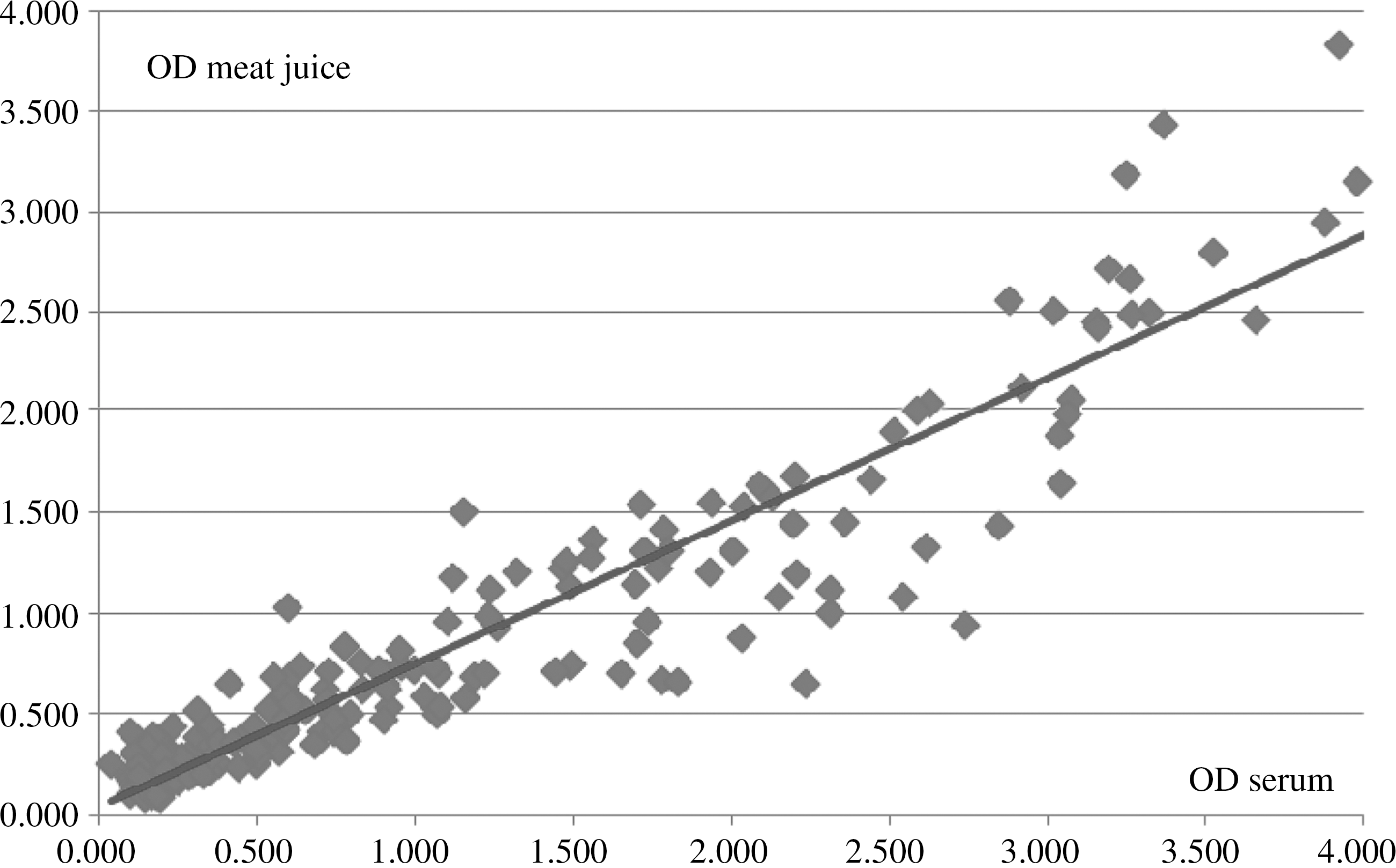
Correlation of optical densities (OD) at 450 nm of meat juice samples and their corresponding serum from 198 pigs at slaughter.
Statistical analysis of linearity gave R2=0.9951 and R2=0.9781 for serum samples with IgG and IgM, respectively, and using meat juice, R2 was 0.9962.
Seroprevalence study
The overall seroprevalence of anti-HEV IgG in 516 serum samples from fattening pigs at four different Bavarian slaughterhouses was 68.6%. Using the newly developed ELISA assay, the seroprevalence of anti-HEV IgG was 67.8%, and using HEV Ab ELISA (Axiom) and recomLine HEV (Mikrogen), the seroprevalence was 73.3% and 71.3%, respectively. Analyzing the anti-HEV IgM seroprevalence using the newly developed ELISA gave 36/516 (7%) positive serum samples. Three of these samples were IgG negative, whereas the other samples showed both IgG and IgM reactivity. Assigning the seroprevalence to the fattening farms that the pigs belonged to and excluding those farms that had fewer than 10 animals sampled showed that, for most of the fattening farms, a seroprevalence of at least 70% was obtained. The pigs of one farm had a seroprevalence of 46.2%, whereas the fatteners of another farm had a seroprevalence of 6.9%; in one farm, all sampled pigs were seronegative for anti-HEV IgG and IgM (Fig. 2). Assigning the seroprevalence to the slaughterhouses that the pigs were sampled at gave a seroprevalence of 71.0%, 91.1%, 34.3%, and 71.3% for slaughterhouses A–D, respectively.
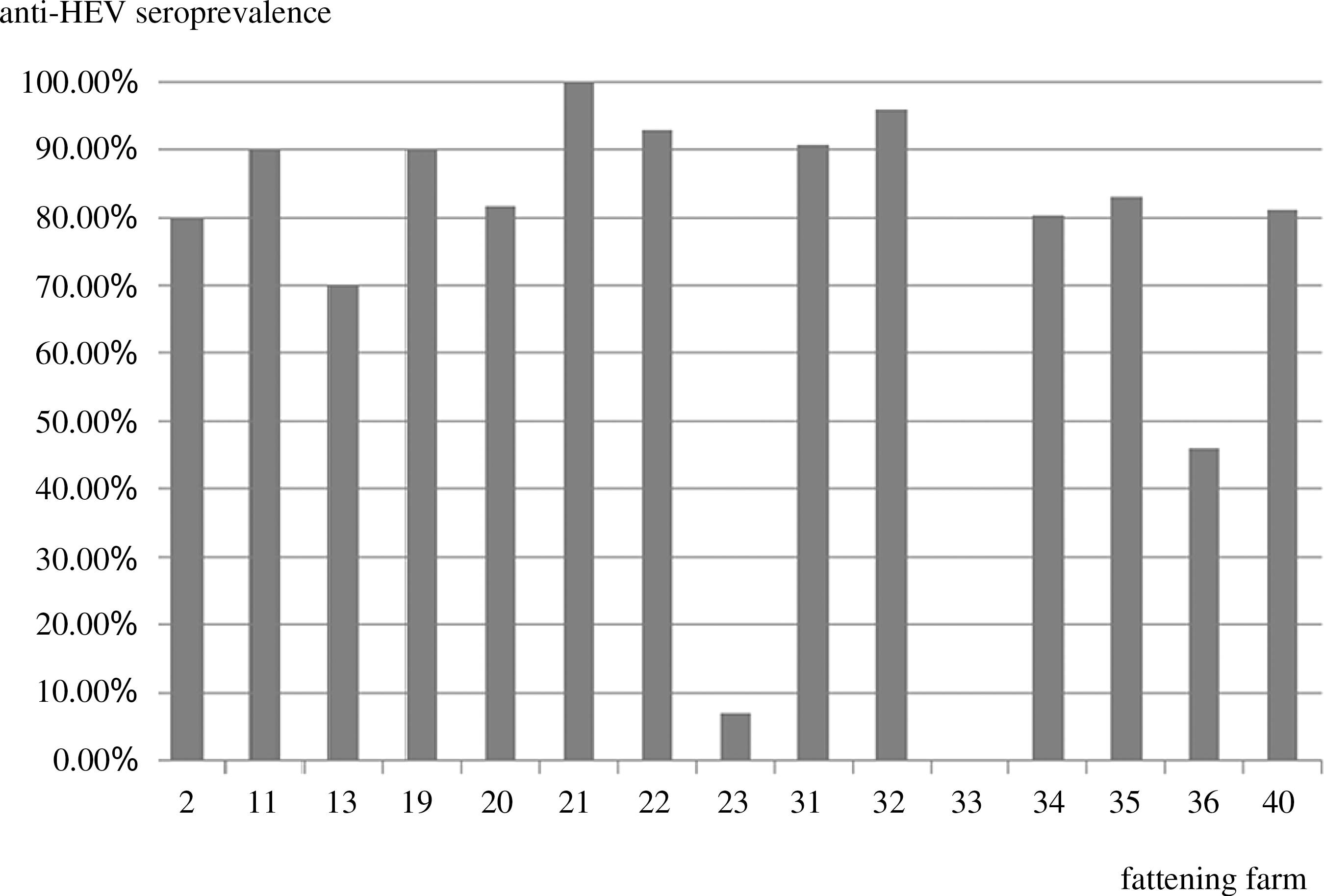
Anti-HEV seroprevalence in the fattening farms with more than 10 animals sampled using the assay developed in this study with IgG.
Of the 198 meat juice samples, 67.6% were positive for anti-HEV IgG using the newly developed ELISA assay.
To estimate the effect of fattening farms on seropositivity for anti-HEV, logistic regression was performed. In reference to farm 41, nine farms showed a statistically significant effect (α=0.05) on the dependent variable seropositivity. Farms 32, 22, and 31 showed enormously higher odds to be seropositive for anti-HEV in reference to farm 41 with ß=72.0, ß=39.0, and ß=30.0, respectively. Also, farms 11, 19, and 40 showed much higher odds with ß=27.0, ß=27.0, and ß=21.0, respectively, in reference to farm no. 41.
Discussion
Domestic pigs are considered as a reservoir for HEV. Yet, little is known on the seroprevalence of HEV in the domestic pig population in Germany. Thus, our objective was to determine the seroprevalence of anti-HEV antibodies in pigs at slaughter using different assays. Furthermore, this study represents the first report of the detection of IgM anti-HEV in pigs at slaughter, and it is the first study describing the use of meat juice to test for anti-HEV antibodies in pigs in Germany.
The overall seroprevalence of anti-HEV IgG was 68.6% in the present study. This is in concordance with a previous study in Germany which found 70.7% of pig sera collected at farms in Bavaria to test positive for anti-HEV IgG using an in-house ELISA based on a synthetic peptide of a genotype 1 strain (Baechlein et al., 2010). Of the three assays used in the present study, HEV Ab ELISA (Axiom Diagnostics) was more sensitive in comparison to the line assay than the newly developed ELISA although it was less specific (data not shown). This is due to the difference in immunoglobulin detection: while HEV Ab ELISA (Axiom) and recomLine HEV (Mikrogen) detect all immunoglobulin classes the newly developed assay detects IgG only. Another explanation might be the differing genotypes used for coating of the polystyrene microwell titer plate. While the HEV Ab ELISA (Axiom) is coated with antigens of HEV GT 1 only, the newly developed assay is coated with antigens of GT 1 and 3. It has recently been shown that using an ELISA coated with antigens of GT 1 and 3 is more specific for the detection of anti-HEV antibodies in humans (S. Dorn, personal communication). Also, coating of the microplate well with antigens of HEV GT 1 and 3 gives reliable results on the absence and presence of anti-HEV antibodies in pigs as has been described previously (Zhang et al., 2011).
The detection of IgM anti-HEV in pigs has been described recently in Spain (Casas et al., 2011a; Seminati et al., 2008). The prevalence of IgM anti-HEV in the present study was clearly less than those reported in Spain. This might be due to the different study design: while the present study was cross-sectional collecting samples at four different Bavarian slaughterhouses, the study by Casas et al. (2011) was a longitudinal study in six farrow-to-finish swine herds. Nevertheless, assuming that IgM is related to viraemia (de Deus et al., 2008), the occurrence of IgM anti-HEV in pigs at slaughter indicates that these animals must have had recent contact to the virus. This might be due to a decline in the force of infection elevating the age of infection (Satou and Nishiura, 2007), since most of the domestic pigs get infected with 2–3 months of age (Meng, 2010a). These data indicate that HEV is widespread among the domestic pig population in Bavaria, Germany, as has been previously shown for other European countries (Breum et al., 2010; Di Bartolo et al., 2011; Jimenez de Oya et al., 2011; Kaba et al., 2009; Martinelli et al., 2011; Meader et al., 2010; Rose et al., 2011; Seminati et al., 2008). Furthermore, the detection of farms with a lower seroprevalence and one farm which was seronegative for anti-HEV IgG and IgM demonstrates that even though the animals are in contact with the virus during their life span they can be seronegative at slaughtering age which has been reported previously in a longitudinal study in Spanish farrow-to-finish herds (Casas et al., 2011a). Also, in Bavaria, Germany, most of the pigs are held in farms with a relative small animal number per herd, which might reduce the infection pressure and probability of infection in some herds. Another possible explanation might be a different herd management as suggested by Rose et al. (2011). They found that pigs from finishing farms were more likely to be HEV positive, suggesting that a rearing management plan might reduce the overall seroprevalence among the pig population (Rose et al., 2011). For the present study, there was no information on the herd management available. In reference to the slaughterhouses, the difference of the seroprevalence could be explained by the different fatteners, including the different seroprevalence status of anti-HEV antibodies of the animals delivered to the abattoirs.
It has been described before that meat juice serves as a suitable sample matrix for testing for antibodies against salmonellosis in pigs (Nielsen et al., 1998) and recently to test for antibodies against HEV (Casas et al., 2011b). We were able to demonstrate that serum and its corresponding meat juice had a correlation coefficient of 0.94 in the newly developed ELISA assay supporting this finding.
To conclude, we were able to show that HEV is widespread in the domestic pig population in Bavaria, Germany, even though some farms showed a low or absent seroprevalence of anti-HEV antibodies, and that some pigs might test IgM positive even at the age of slaughter. Furthermore, we showed that meat juice is an equivalent matrix to serum to test for anti-HEV antibodies in pigs. This newly developed assay is now commercially available for the detection of anti-HEV antibodies in serum and meat juice samples from pigs (PrioCHECK® HEV Ab porcine; Prionics, Schlieren-Zurich, Switzerland), enabling us to compare results of veterinary surveillance across country borders.
Footnotes
Acknowledgments
We would like to thank Jutta Koerber for technical assistance.
Disclosure Statement
No competing financial interests exist.