
Abstract
Cronobacter sakazakii is an emerging pathogen associated with the ingestion of contaminated reconstituted formula, which causes necrotizing enterocolitis, sepsis, and meningitis in low-birth-weight preterm neonatal infants. Sensitive and specific detection methods are needed to better control C. sakazakii infections. This study aims to develop a highly specific and sensitive loop-mediated isothermal amplification (LAMP) assay for detecting C. sakazakii in powdered infant formula (PIF). A set of four LAMP primers were designed based on the published C. sakazakii ompA gene sequence. Specificity of the assay was evaluated using a panel of 22 C. sakazakii, 27 Enterobacteriaceae family except C. sakazakii, and 25 other strains. Assay sensitivity was determined using serial dilutions of C. sakazakii American Type Culture Collection 51329 culture ranging from 106 colony-forming units (CFU)/mL to extinction. The assay was also tested in experimentally inoculated PIF samples. The ompA-based LAMP assay was able to detect specifically all of the 22 C. sakazakii strains without amplification from 52 non–C. sakazakii strains. The detection limit was 101 CFU/mL in pure culture, up to 10-fold more sensitive than that of the ompA–polymerase chain reaction (PCR). When applied to PIF, sensitivity was 102 CFU/mL, up to 10-fold that of the ompA-PCR. The ompA-based LAMP assay developed in this study was sensitive, specific, and low cost with great potential for future field detection of C. sakazakii in PIF.
Introduction
Detection of C. sakazakii using conventional culture- and biochemical-based assays is time-consuming and laborious, requiring more than 7 days (
LAMP is a nucleic acid amplification method that synthesizes large amounts of DNA in a short period of time with high specificity (Notomi et al., 2000; Mori et al., 2001). The strand displacement activity of Bst DNA polymerase impels auto-cyclic DNA synthesis with loop-forming primers to yield long-stem loop products under isothermal conditions: 60–65°C for about 60 min (Nagamine et al., 2002). The LAMP reaction requires four or six primers that target six or eight separate DNA sequences on the target and give the assay very high specificity (Notomi et al., 2000; Nagamine et al., 2002). LAMP amplification products can be detected by gel electrophoresis, by real-time monitoring of turbidity with a turbidimeter (Mori et al., 2001), or by the naked eye. Visual detection can be accomplished using different methods such as detection of a white precipitate (magnesium pyrophosphate) or use of an intercalating DNA dye such SYBR Green I gel strain (Soliman et al., 2005). LAMP assays have been developed for the detection of various pathogens, including Vibrio parahaemolyticus (Wataru et al., 2008; Prompamorn et al., 2011), Yersinia ruckeri (Mona et al., 2008), Escherichia coli (Joshua et al., 2008), Mycobacterium tuberculosis (Tomotada et al., 2003), and Norovirus (Shinji et al., 2008). This study aims to develop a sensitive and specific LAMP-based method in detecting C. sakazakii and to compare its results with a conventional PCR assay of PIF samples.
Materials and Methods
Bacterial isolates, culture conditions, and DNA extraction
A total of 22 isolates of C. sakazakii, including strain Cronobacter muytjensii American Type Culture Collection (ATCC) 51329, C. sakazakii ATCC 29544, and 20 wild isolates from food (powdered milk and PIF), were obtained from the Guangdong Provincial Key Laboratory of Microbial Culture Collection and Application. An additional 27 strains of Enterobacteriaceae family and 25 non-Enterobacteriaceae strains were used to evaluate assay specificity. The bacterial strains used in this study are listed in Table 1. All bacterial strains were cultured in Luria-Bertani broth (Huankai, China) at 37°C overnight. DNA was extracted using a DNA Gel extraction kit (Sangon Ltd., Shanghai, China). The DNA samples were stored at−20°C for later use.
LAMP, loop-mediated isothermal amplification; SYBR, PCR, polymerase chain reaction; ATCC, American Type Culture Collection; GIM, China Inspection and Quarantine in Guangdong; SMU, Southern Medical University; CMCC, China Microbiological Culture Collection.
LAMP primers and reaction conditions
The C. sakazakii LAMP assay required two sets of primers (outer and inner), which were designed by analyzing a conserved region of the C. sakazakii ompA gene (GenBank accession no. DQ000206) (Manoj et al., 2006; Nair et al., 2009; Zhou et al., 2008; Zaid et al., 2009) with Primer Explorer version 4.0 software (
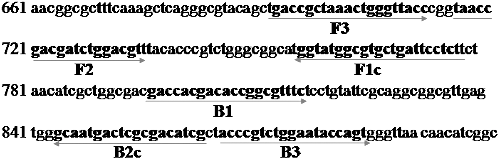
Names and locations of target sequences used as primers for loop-mediated isothermal amplification (LAMP). The sequence of the second half of the Cronobacter muytjensii American Type Culture Collection (ATCC) 51329 ompA gene, together with the primer name (in boldface) and the location of each target sequence (in boldface and underlined in red) are shown.
The LAMP reaction was conducted as described previously(Wataru et al., 2008; Prompamorn et al., 2011; Mona et al., 2008; Joshua et al., 2008; Tomotada et al., 2003; Shinji et al., 2008). Each reaction mixture (total volume of 25 μL) contained 2.5 μL of 10×Bst ThermoPol buffer [20 mM Tris-HCl, 10 mM KCl, 8 mM MgSO4, 10 mM (NH4)2SO4, 0.1% Triton X-100], 600 μM of each deoxynucleoside triphosphate, 1 μL (8 units) Bst DNA polymerase (New England Biolabs, Inc., Ipswich, MA), 2 mM MgCl2, 1.6 M Betaine (Sigma-Aldrich, St. Louis, MO), 20 pmol each of FIP and BIP primers, 5 pmol each of the F3 and B3 primers, and 2 μL of DNA sample. Samples were incubated at 60°C for 60 min and then heated at 80°C for 3 min.
PCR
For comparison, C. sakazakii PCR reactions were performed; LAMP outer primers (F3 and B3) would be the PCR primers. The PCR reaction, which contains 2.5 μL 10×PCR buffer (500 mM KCl, 100 mM Tris-HCl, pH 8.3, 15 mM MgCl2, and 0.01% gelatin), 200 μM of each deoxynucleoside triphosphate, 200 nM of each primer (F3 and B3), 1 U of Taq polymerase, and 2 μL of sample DNA in a total volume of 25 μL, yields a product with 191 bp. The reaction mixture was denatured by incubating at 94°C for 5 min. The mixture was then subjected to 30 cycles at 94°C for 40 s, annealed at 60°C for 40 s, and extension at 72°C for 1 min, followed by a final extension at 72°C for 7 min.
Analysis of LAMP product
LAMP amplicons in the reaction tube were directly detected with the naked eye by monitoring the presence of magnesium pyrophosphate, a white precipitate generated as a byproduct during the reaction, of by visual observation of fluorescence of DNA binding SYBR green I (1 μL of 10,000×concentrate in dimethyl sulfoxide [DMSO]) under ultraviolet (UV) light. For further confirmation, some of the amplified products were also detected by agarose gel electrophoresis stained with ethidium bromide; the positive result can get a special ladder strap, while it did not appear without amplification. PCR monitoring using agarose gel electrophoresis stained with ethidium bromide was utilized.
Sensitivity of the C. sakazakii LAMP assay
To determine the detection limit of the LAMP assay, serial 10-fold dilutions of C. sakazakii ATCC 51329 were made and 1-mL volumes were added into 9.0 mL of reconstituted infant formulas to obtain a final concentration of C. sakazakii that ranged from 106 colony-forming units (CFU)/mL to extinction. DNA templates were then prepared from each dilution by the method described above, and aliquots (2 μL) were subjected to both LAMP and PCR amplifications. Sensitivity tests were repeated three times, and the lower limits of detection (CFU/mL) were reported.
Specificity of the C. sakazakii LAMP assay
A total of 74 bacterial strains were used to determine LAMP specificity. DNA templates were made from fresh overnight bacterial cultures, and aliquots (2 μL) were subjected to both LAMP and PCR amplifications. Specificity tests were repeated twice.
LAMP testing in experimentally inoculated PIF samples
Five commercially available brands of PIF from supermarkets in China were used in this study. All the PIF were determined to be C. sakazakii negative by the U.S. Food and Drug Administration (FDA, 2002; Gurtler et al., 2005) and traditional methods. The traditional method was reconstituted by mixing 1.0-g aliquots in 8 mL of sterile distilled water in 15-mL tubes. A serial 10-fold dilution of C. sakazakii was made, and 1-mL volumes were added into 9.0 mL of reconstituted infant formulas to obtain a final concentration of Enterobacter sakazakii (ATCC 51329 and one isolate) that ranged from 107 to 100 CFU/mL. The extraction of DNA template from reconstituted samples was described by Nair and Venkitanarayanan (2006). The LAMP and PCR amplification was previously described. The PIF sensitivity tests were repeated three times, and the lower limits of detection (CFU/g) were reported.
LAMP testing in commercial PIF samples
The enrichment procedure for 54 infant formulas is described by Ye et al. (2008). DNA samples were extracted from 1.5-mL samples of mLST vancomycin enrichments (Huankai, Guangzhou, China) using the DNA Gel Extraction Kit (Sangon Ltd., Shanghai, China). The LAMP, PCR assay, and electrophoresis were performed as described above. All experiments were performed in triplicate. DNA from Cronobacter muytjensii ATCC 51329 and the sterile water were used as the positive and negative controls, respectively.
Results
Detection and confirmation of LAMP product
The LAMP-positive results (C. muytjensii ATCC 51329 and C. sakazakii ATCC 29544) were observed three ways. Figure 2A is the result of white precipitate (magnesium pyrophosphate) of C. sakazakii LAMP products observed by naked-eye inspection. Figure 2B is the result of visual observation of green fluorescence of DNA binding SYBR green I under UV light. Figure 2C shows the typical ladder-like bands on agarose gels. After amplifying the DNA products by using PCR, an expected 191-bp fragment was obtained (Fig. 2D) and verified by ABI PRISM 377, which gave 100% homology.
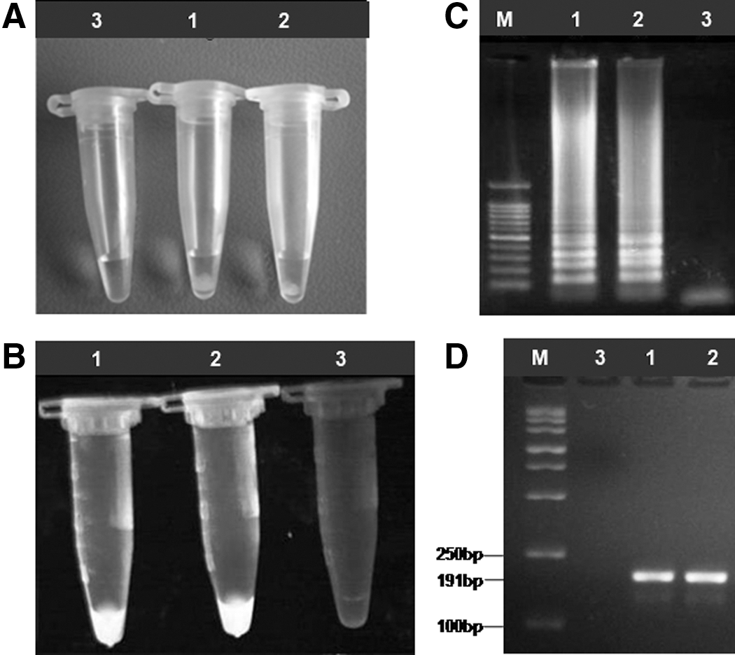
Results of loop-mediated isothermal amplification (LAMP) and polymerase chain reaction (PCR) detection of different Cronobacter sakazakii samples. M, marker. (1) American Type Culture Collection (ATCC) 51329; (2) ATCC 25944; (3) negative control.
Specificity of the LAMP assay
LAMP was tested for its specificity using 74 bacterial strain (22 Enterobacter sakazakii strains and 52 non–C. sakazakii strains) DNA samples as controls. Only the target C. sakazakii DNA was amplified. No LAMP products were detected in the reactions performed with DNA from other strains (Table 1). Both LAMP and PCR specifically amplified C. sakazakii DNA, whereas the negative-control DNA was not amplified (Table 1). Thus, the results indicate that the C. sakazakii LAMP can detect C. sakazakii with high specificity, in agreement with previous studies showing extremely high specificity of LAMP assays in the detection of V. parahaemolyticus, Y. ruckeri, E. coli, M. tuberculosis, Norovirus, and C. sakazakii (Wataru et al., 2008; Prompamorn et al., 2011; Mona et al., 2008; Joshua et al., 2008; Tomotada et al., 2003; Shinji et al., 2008; Liu et al., 2012).
Sensitivity of the LAMP assay
To determine the detection limit of the LAMP assay, about 1×10⁶ C. sakazakii through plate counts from ATCC 51329 was diluted 1:10 serially sevenfold to a minimum concentration equivalent to 101 bacterium. DNA was then extracted from each C. sakazakii dilution. The LAMP procedure amplified the DNA of C. sakazakii diluted to a minimum concentration equivalent to 1×101 CFU/mL (Fig. 3A). A positive LAMP reaction can be easily detected with the naked eye by observing fluorescence in the solution (Fig. 3B). In comparison, PCR amplified the same DNA only to a minimum concentration equivalent to that of 1×10² CFU/mL C. sakazakii (Fig. 3C), indicating that LAMP was 10 times more sensitive than the conventional PCR assay. No amplification from the water used as the negative control was observed with either method.
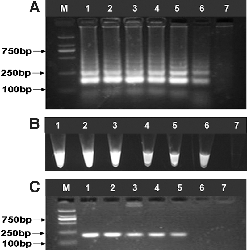
Sensitivity of the Cronobacter sakazakii loop-mediated isothermal amplification (LAMP) versus that of polymerase chain reaction (PCR). M, marker. The C. sakazakii LAMP and PCR were carried out at the following concentrations: (1) 1×10⁶ colony-forming units (CFU)/mL; (2) 1×10⁵ CFU/mL; (3) 1×10⁴ CFU/mL; (4) 1×10³ CFU/mL; (5) 1×10² CFU/mL; (6) 1×101 CFU/mL; (7) negative control.
Detection of C. sakazakii in PIF samples
The sensitivity of detecting C. muytjensii ATCC 51329 cells in PIF samples is shown in Table 3. In five independent sample experiments, the ompA-based LAMP assay using the two platforms consistently detected down to 102 CFU/g in the infant formula samples without enrichment. However, for the two PCR assays using F3/B3 primer, the lowest detection limit achieved was 103 to 104 CFU/g, which was up to 10–100-fold less sensitive than that of the ompA-based LAMP assay.
CFU, colony-forming units.
As for the real PIF samples, the targeted ompA gene was amplified from 49 formula samples, whereas five other samples showed a different result in which there did not appear to be any growth of bacteria after two enrichment steps (the first step is to add 100 g of infant formula milk in 900 mL of BPW incubation at 37°C for 16 h; the second step is to transfer 1.0 mL of BPW culture into 9 mL of mLST/vancomycin medium incubation at 44°C for 20 h) (Ye et al., 2008) by LAMP and PCR assay; this indicated that the DNA quality from commercial formula samples was reliable. The results for detection of C. sakazakii by LAMP and PCR are shown in Figure 4. There were two positive samples from the 6– 12-month age group. For further confirmation, the two positive infant formulas were determined to be C. sakazakii positive by FDA methods (FDA, 2002).
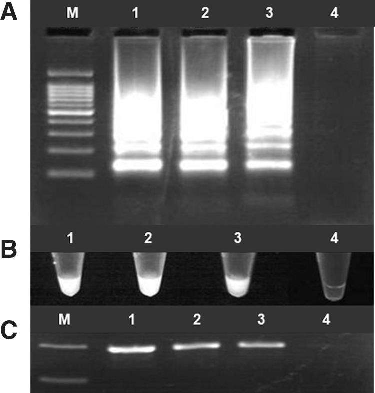
Detection of Cronobacter sakazakii in powdered infant formula (PIF): (1) positive control (American Type Culture Collection [ATCC] 51329); (2) positive sample 1; (3) positive sample 2; (4) negative control.
Discussion
In this study, we designed a set of four LAMP primers to target specifically the C. sakazakii ompA gene, a gene previously shown to possess better specificity for C. sakazakii detection by PCR (Manoj et al., 2006, Nair et al., 2009; Zhou et al., 2008; Zaid et al., 2009). We also developed LAMP assays and PCR assays to detect C. sakazakii in pure cultures and PIF samples. This is the first report demonstrating the efficacy of an ompA-based LAMP assay for detecting C. sakazakii in PIF. LAMP assays for specific Cronobacter strains have been reported (Hu et al., 2009; Liu et al., 2012), and these two studies selected 16S-23S rRNA to use as target sequences. In the Zaid et al. (2009) study, they used eight sets of Cronobacter spp.–specific PCR primers (α-GluA, α-GluB, SG, SI, Saka, OmpA, Zpx, BAM), which confirmed as non–Cronobacter spp. By 16S rRNA sequence analysis, the results showed that all primers except OmpA appeared false positive. So, the OmpA gene and 16S rRNA can be used as ideal primer target sequences for LAMP detection.
The LAMP primers were selected from regions of the C. sakazakii ompA gene coding sequence that are highly specific to C. sakazakii (Nair et al., 2009; Zaid et al., 2009). The four primers (F3, B3, FIP, and BIP) targeted six regions of C. sakazakii ompA (Fig. 1), providing additional levels of specificity compared to PCR primers (with two target regions). Among a total of 22 C. sakazakii and 52 non–C. sakazakii strains, the ompA-based LAMP assay obtained 100% inclusivity and 100% exclusivity. This level of specificity was the same as that of the two ompA-based PCR assays evaluated simultaneously in this study and that of an oligo-1,6-glucosidase gene–based PCR assay developed by Ye et al. (2009).
The ompA-LAMP assay was able to detect 101 CFU/mL in contrast to 102 CFU/mL for the ompA-PCR; our data was consistent with that of Liu et al. (2012). Similarly, the LAMP assays for other bacteria were reported to be 10–100-fold more sensitive than PCR, with a detection limit of 2 CFU per reaction for LAMP (Han et al., 2008, 2010; Nemoto et al., 2009; Xu et al., 2010; Chen et al., 2011).
In the PIF samples, we found the detection limit of the ompA-based LAMP assay to be 102 to 103 CFU/g; our data was consistent with that of Hu et al. (2009). The FDA requires that all post-harvest processed PIF be analyzed for the presence of C. sakazakii (FDA, 2002). This indicates that, without enrichment, DNA amplification assays such as LAMP lack the needed sensitivity when applied to food samples. Therefore, combining most probable number overnight enrichment (Lversen et al., 2004) or pre-enrichment for 6 h with LAMP or other DNA amplification assays is a desirable approach to achieve the needed sensitivity. In real PIF samples, two infant formula were positive for C. sakazakii by using LAMP, which is consistent with the FDA method. This suggested the LAMP could be applied to directly detect PIF samples effectively.
Combined with 6-h enrichment, the complete LAMP detection system was markedly faster than PCR and conventional methods. LAMP does not require sophisticated equipment; only a water bath or heat block is required to accomplish the whole reaction process, which is of great value for application in poorly equipped laboratories or in large-scale epidemiological studies conducted in isolated areas. The most practical characteristic of LAMP is the visual detection of amplification through the addition of fluorescent dyes such as SYBR Green I (Poon et al., 2006). Based on these advantages, we conclude that LAMP is a useful and practical tool for the early diagnosis of C. sakazakii. The LAMP assay established in this study is cost-effective, easy to perform, and readily adaptable for field diagnosis and disease surveillance in PIF samples.
Footnotes
Acknowledgments
The research was supported by the Science and Technology project of Guangzhou, China (no. 2010U1-E00611), School of Public Health and Tropical Medicine of Southern Medical University, China (no. GW201202), and Doctoral Fund of Ministy of Education of China (no. 20104433120015).
Disclosure Statement
No competing financial interests exist.