
Abstract
Salmonella, a highly virulent food-borne pathogen transmitted through food, can cause severe infectious diseases in a large number of people through a single outbreak, due to its low infective doses. In this study, a flow cytometry (FCM)-based method was developed for the rapid detection of single viable Salmonella cells with dual staining of fluorescein isothiocyanate (FITC)-labeled anti-Salmonella antibody and propidium iodide (PI) dyes. The FCM-based method includes 6 h of pre-enrichment, 40 min of target cell isolation, and 20 min of dual staining and FCM analysis. The developed method demonstrated high specificity for the detection of 23 Salmonella strains and 22 food-borne pathogenic non-Salmonella strains. Furthermore, the analyses of 30 samples of milk powder artificially contaminated with single Salmonella cells, 123 samples of retail milk powder, and 6 samples of Salmonella-positive milk powder were performed by the FCM-based as well as traditional plate-based methods for testing the efficiency of the methods. The two methods yielded similar results for the detection of pathogens in all milk powder samples. In conclusion, the developed FCM-based method was found to be efficient in detecting single viable Salmonella cells in milk powder within 7 h. The proposed dual-color FITC assay combined with pre-enrichment offers a great potential for the rapid and sensitive detection of other pathogens in dairy products.
Introduction
Salmonella is a common bacterial pathogen found in humans and animals, causing diseases such as animal typhoid and cholera, and human gastroenteritis and septicemia (WHO, 2015; CDC, 2016). Salmonella includes more than 2600 serotypes (Gal-Mor et al., 2014), the majority of which are disease-causing agents. As Salmonella is found in many food-based products such as milk, eggs, meat (poultry and beef), vegetables, and fresh fruits (Almeida et al., 2013; Arguello et al., 2018), it is the main cause of food-borne diseases among almost all other food-related pathogens (Crim et al., 2018; Srisa-Art et al., 2018). The high prevalence of Salmonella in milk and dairy products has received considerable attention worldwide (Nisar et al., 2017; Marder et al., 2018). The largest outbreak of Salmonella reported in 1985 in America was from a dairy firm (Harris et al., 1990); recently, serious contamination of Salmonella in nearly 7000 tons of milk powder was reported in France (Jourdan-da Silva et al., 2018). Milk powder is deemed to be a high-risk food product that can be easily contaminated by Salmonella (Yan et al., 2010). Therefore, the quality control of milk powder is crucial to the food department, dairy factories, wholesalers, and customers.
Salmonella causes food poisoning at very low infective doses with an estimation of <10 cells/mL (Blaser et al., 1982; Todd et al., 2008). European Commission (EC) regulations require zero Salmonella cells in all kinds of food products, including milk powder (EC, 2007). Therefore, for a more accurate and effective detection method, isolation and separation of a single Salmonella cell are desirable.
Previously published methods of detection or identification of Salmonella have relied mainly on culture-based approaches (Kokkinos et al., 2013), standardized by the International Organization for Standardization (ISO) (Mooijman et al., 2018). Although these methods are used for single-cell detection, they are time-consuming, requiring almost 18 to 24 h (Koyama et al., 2016). For the detection of Salmonella, many rapid and sensitive methods have been used, such as polymerase chain reaction (PCR) (Rodriguez-Lazaro et al., 2014; Bai et al., 2019), integrated passive micromixer-magnetic separation-capillary electrophoresis (PMMS-CE) microdevices (Jung et al., 2011), and enzyme-linked immunomagnetic electrochemical (ELIME) assays (Volpe et al., 2016). However, it is difficult to distinguish between viable lethal cells and their dead innocuous counterparts by integrated PMMS-CE microdevices. On the other hand, the PCR-based method and ELIME assays offer an acceptable detection limit, but these methods still require more than 10 h.
Several flow cytometry (FCM)-based methods have also been used for the detection of pathogenic microbes in food, as these methods can efficiently distinguish viable cells from nonviable cells rapidly and sensitively (Comas-Riu and Rius, 2009; Morishige et al., 2015; Lang et al., 2018). Recently, the application of FCM in combination with an enrichment process was successful in detecting single Escherichia coli O157:H7 cells in 25 g of sample (Williams et al., 2017). As for Salmonella, the previous methods with the detection limit of >10 cells/mL in milk are invalid for the detection of a single cell (McClelland and Pinder, 1994; Pinder and McClelland, 1994). Hence, from the above-stated descriptions, the development of an efficient method for the detection of single Salmonella cells in milk powder is crucial and demanding.
In this study, dual staining combined with an additional enrichment procedure was used to develop an FCM-based method for the rapid detection of single viable Salmonella cells in milk powder. The method was comprehensively evaluated by analyzing a total of 30 samples of milk powder artificially contaminated with single Salmonella cells, 123 samples of retail milk powder, and 6 samples of Salmonella-positive milk powder for proficiency testing.
Materials and Methods
Bacterial strains
All bacterial strains listed in Table 1 were purchased from the China Center of Industrial Culture Collection (CICC, Beijing, China) and American Type Culture Collection (ATCC).
Specificity of the Flow Cytometry-Based Method
Serogroup, O-antigen serogroup of the Salmonella strain.
The gating of flow cytometer was determined empirically from multiple experiments.
Symbol “+,” positive, means that the strain can react with FITC-labeled anti-Salmonella antibody.
The symbol “/” means that the bacterial strain is non-Salmonella strain.
Symbol “−,” negative, means that the strain cannot react with FITC-labeled anti-Salmonella antibody.
FITC, fluorescein isothiocyanate.
A total of 23 Salmonella strains were selected for specific detection, whose serogroups cover the most common O-antigen serogroups (A, B, C1, C2, D, and E) of Salmonella (Popoff and Le Minor, 1997; Popoff et al., 2000). In this study, Salmonella Typhimurium (ATCC 13311) was used as the target strain, as described in the previous research (Biswas et al., 2010; Edilu et al., 2015). In addition, a total number of 22 non-Salmonella strains were used for the evaluation of specificity.
All strains were collected during the logarithmic growth phase at 37°C in a nutrient broth (NB; Oxoid, Basingstoke, United Kingdom). Bacteria-containing pellets were obtained by centrifugation at 10,000 × g for 10 min and were resuspended immediately in 1 mL of phosphate-buffered saline (PBS; pH 7.2). All solutions were prepared using ultrapure water obtained from a Milli-Q water purification system (Millipore, Bedford, MA).
FCM and dual staining of Salmonella
The FCM used was of A50-Micro model (Apogee, Hemel Hempstead, United Kingdom). With the capability of low- and high-angle light scattering channels, the A50-Micro achieves an optical resolution of 80 nm and is particularly useful for detecting bacteria. The excitation source was a solid-state 50-mW laser (488 nm). For FCM, fluorescence emission was detected at standard wavelengths: FL1 = 525 λ, FL2 = 575 λ, and FL3 = 680 λ. Fluorescein isothiocyanate (FITC)-labeled anti-Salmonella antibody was purchased from SeraCare Life Sciences (Milford, MA), which was a polyclonal goat antibody against anti-Salmonella common structural antigens (CSA) labeled with FITC. The anti-Salmonella antibody should theoretically be capable of recognizing any strain that belongs to the genus Salmonella (Li et al., 2010). Propidium iodide (PI) purchased from Molecular Probes (Eugene, OR), is a red fluorescent nucleic acid stain, which can be used to distinguish viable cells from dead cells.
The final concentration of Salmonella suspension of ∼107 cells/mL was obtained by mixing the antibody (final concentration: 1 μg/mL) to PI (final concentration: 1 μg/mL). The reaction was allowed to proceed at room temperature for ∼15 min in the dark. After incubation, the suspensions were determined by FCM measured on FL1 and FL3 fluorescence channels. The FL1 and FL3 fluorescence channels were used to detect Salmonella cells stained with FITC and PI, respectively. According to principle of the dual labeling with FITC and PI, enumeration of viable cells is usually performed by counting events that scatter the incident blue light at the expected intensities and emit green, but not red fluorescence in the resulting dot plot of FL1 versus FL3.
The specificity of the FCM-based method
To determine whether the presence of non-Salmonella strains affected the identification and detection of Salmonella strains, the 23 Salmonella strains and the 22 non-Salmonella strains were stained with FITC-labeled anti-Salmonella antibody, and then analyzed by the flow cytometer, respectively.
Analysis of FCM method for viable Salmonella cells
A suspension of viable Salmonella cells at a final concentration of ∼107 cells/mL was prepared as described in the section Bacterial Strains. In this study, isopropyl alcohol (IPA), which can be used as a disinfectant (Hendry et al., 2012), was used for the preparation of dead cells. The suspension containing viable cells was centrifuged at 10,000 × g for 10 min to collect the pellets. After collection, the pellets were resuspended in 300 μL of PBS and 700 μL of IPA for 30 min. Finally, the suspensions of dead cells were obtained by centrifugation at 10,000 × g for 10 min, immediately followed by resuspension in 1 mL of PBS solution. Our previous experiment revealed that no distinct bacterial colonies were observed when viable Salmonella cells on treatment with IPA were examined on xylose lysine desoxycholate (XLD; Land Bridge, Beijing, China) agar plates at 37°C for 18 h.
To evaluate the quantitative analysis of FCM for viable Salmonella cells, viable and dead cells were mixed at different ratios of concentration (0/10, 1/9, 5/5, 9/1, and 10/0), and then detected by FCM after incubation with the antibody and PI.
Preparation of single Salmonella cells
Single Salmonella cells were sorted using a flow cytometric cell sorter (FACSAria III; Becton-Dickinson Biosciences, New York) for the preparation of milk powder samples with single bacterial cells (Fig. 1). The cell sorter was flushed twice with ultrapure water to ensure the cleanliness of the instrument before each use. The single cell of a droplet was sorted in an Eppendorf tube containing 50 μL of PBS, and then transferred to a milk powder sample of 25 g using a pipette (Preparation of Artificially Contaminated Milk Powder Samples section). To confirm that the cell sorter could isolate a single cell, 50 samples with nominally single cells and 10 blank samples with no cells were prepared by the cell sorter and then examined on XLD plates incubated at 37°C for 18 h.
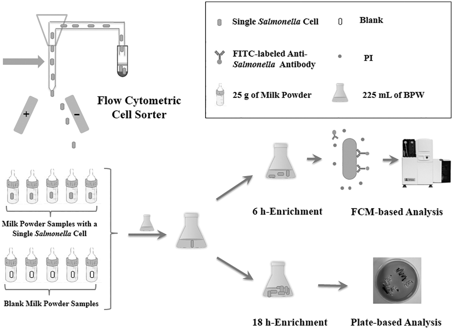
Schematic illustration for the preparation and detection of milk powder samples artificially contaminated with single Salmonella cells. The flow cytometry cell sorter will charge the target cells and the single charged cells of a droplet would be deflected into an Eppendorf tube containing 50 μL of PBS by the charge plates, and then transferred to a milk powder sample of 25 g using a pipette. The artificially contaminated samples were added into 225 mL of sterilized BPW at 37°C for pre-enrichment, and then analyzed by FCM-based method as well as plate-based method. BPW, buffered peptone water; FCM, flow cytometry; FITC, fluorescein isothiocyanate; PBS, phosphate-buffered saline; PI, propidium iodide.
Preparation of artificially contaminated milk powder samples
Milk powder (fat, 26.4%; protein, 11.5%) was purchased from Friesland Campina Domo B.V. (Frisolac, Friesland, Netherlands). In the initial tests, no microbes were detected through surface plating (data not shown). A total of 91 samples of artificially contaminated milk powder (A01–A55 and B01–B36) were prepared with single Salmonella cells (Fig. 1) and other nonrelated bacteria such as E. coli CMCC 44102 and Staphylococcus aureus ATCC 6538P. Detailed information is given in Table 2.
Preparation of Artificially Contaminated Milk Powder Samples
Each sample contains 25 g of the milk powder. The series A samples (A01–A55) were used for the optimization of the pre-enrichment time of the FCM-based method. The series B samples (B01–B36) were used to evaluate the FCM-based method for the detection of single Salmonella cells with a huge background of nonrelated bacteria.
“Nominally Positive” sample contains one Salmonella cell sorted by the cell sorter. Single Salmonella cells were sorted by a flow cytometric cell sorter and then transferred into a milk powder sample of 25 g by a pipette.
“Negative” sample contains no Salmonella cells.
E. coli CMCC 44102 cells were obtained from the bacterial suspensions by the dilution method and then transferred into a milk powder sample of 25 g by a pipette. The population of E. coli cells was confirmed using a plate-based method by inoculating the samples onto plate count agar plates.
S. aureus ATCC 6538P cells were obtained from the bacterial suspensions by the dilution method, and then transferred into a milk powder sample of 25 g by a pipette. The population of S. aureus cells was confirmed using a plate-based method by inoculating the samples onto plate count agar plates.
N, “Negative” sample.
The symbol “/” means that there is no E. coli cells and S. aureus cells in the sample.
P, “Nominally Positive” sample.
CFU, colony-forming unit; FCM, flow cytometry.
The series-A samples (A01–A55) were used for optimizing the pre-enrichment time for the FCM-based method (Optimization of the Pre-enrichment Time Section). Samples A06 to A55 were artificially contaminated with a single Salmonella cell (Preparation of Single Salmonella Cells section), whereas samples A01 to A05 were not contained with Salmonella cells.
Samples B01 to B12 were prepared as follows: a suspension of E. coli at a final concentration of (3.8 ± 0.3) × 103 colony-forming unit (CFU)/mL and a suspension of S. aureus at a final concentration of (5.1 ± 1.0) × 103 CFU/mL were prepared by the dilution method. The populations of the E. coli suspension and the S. aureus suspension were confirmed using a plate-based method by inoculating the samples onto plate count agar (Land Bridge) plates (n = 3). Then, 7.5 μL of the E. coli suspension and 10 μL of the S. aureus suspension were transferred to the milk powder sample using a pipette, of which each sample contained ∼29 cells of E. coli and 51 cells of S. aureus. In addition, samples B01 to B10 were artificially contaminated with a single Salmonella cell (Preparation of Single Salmonella Cells section), while samples B11 and B12 were not contained with Salmonella cells.
Samples B13 to B24 were prepared as follows: 10 μL of E. coli suspension ([2.3 ± 0.3] × 104 CFU/mL) and 6.5 μL of S. aureus suspension ([9.6 ± 1.0] × 104 CFU/mL) were transferred to the milk powder sample, of which each sample contained ∼230 cells of E. coli and 624 cells of S. aureus. In addition, samples B13 to B22 were artificially contaminated with a single Salmonella cell, while samples B23 and B24 were not contained with Salmonella cells.
Samples B25 to B36 were prepared as follows: 22 μL of E. coli suspension ([2.4 ± 0.3] × 105 CFU/mL) and 35 μL of S. aureus suspension ([1.5 ± 0.2] × 105 CFU/mL) were transferred to the milk powder sample, of which each sample contained ∼5280 cells of E. coli and 5250 cells of S. aureus. In addition, samples B25 to B34 were artificially contaminated with a single Salmonella cell, while samples B35 and B36 were not contained with Salmonella cells.
The series-B samples (B01–B36) were used to evaluate the FCM-based method for the detection of single Salmonella cells with a huge background of nonrelated bacteria. These 36 samples were detected by the FCM-based method (Analysis of Sample by FCM-Based Method section) and the plate-based method (Analysis of Sample by Plate-Based Method section).
Optimization of the pre-enrichment time
A total of 50 samples of milk powder (A06–A55) were prepared as the experimental group, while 5 samples (A01–A05) with no Salmonella cells were prepared as the blank control group (Table 2). Each sample (25 g) from the artificially contaminated milk powder was added to 225 mL of sterilized buffered peptone water (BPW) and incubated at 37°C. After 5, 6, and 7 h of pre-enrichment, 1.5 mL of the mixture was removed immediately from each of the well-mixed suspensions, respectively. The removed aliquot mixture was processed and analyzed by the FCM-based method (Analysis of Sample by FCM-Based Method section). The remnant of each mixture was further analyzed by a plate-based method (Analysis of Sample by Plate-Based Method section).
Analysis of sample by FCM-based method
Initially, the milk powder sample (25 g) was added to 225 mL of BPW and incubated at 37°C for the pre-enrichment of Salmonella. After pre-enrichment, 1.5 mL of the mixture was used for the isolation of Salmonella cells. As described in the previous studies, for the isolation of bacteria cells (Gunasekera et al., 2000), 2.5 mg of proteinase K, which was purchased from Molecular Probes, and 500 μL of 0.1% Triton X-100, which was purchased from Andy Bio (Itasca, MN), were added to the 1.5 mL of the mixture containing milk powder. Notably, the treated samples were incubated at 37°C for 30 min, as the specified temperature and time were conducive to enzyme activity in the purification solution. After incubation, 1.5 mL of PBS was added to the sample and mixed thoroughly by inverting the tube 100 times for the reaction to complete. The mixtures were centrifuged at 12,000 × g for 5 min to remove lipid components, and the pellets containing Salmonella cells were collected. Finally, the pellets were resuspended in 150 μL of PBS.
After isolation, the suspension of Salmonella cells was mixed with the antibody and PI at room temperature for ∼15 min in the dark, as described in the FCM and Dual Staining of Salmonella section. After staining, the bacterial cells were detected by FCM (Fig. 1). A sample volume of 100 μL was set to be analyzed based on the Apogee A50-Micro model; however, the actual volume analyzed was only 47.2 μL until the end of the pre-experiment. The final number of cells reached by enrichment was determined by distinct events within the counting gate in 47.2 μL of the sample. A distinct final number indicated that the result obtained is positive.
The results revealed that by increasing the setting of the sample volume, the final number of cells shown by the FCM also increased, which might allow a lower population of Salmonella cells on enrichment to be detected, so that the enrichment time would be reduced. However, according to our previous experiments, there was still a mass of background particles in the pretreated milk powder sample, which may lead the Apogee A50-Micro model not to work efficiently if a larger sample volume was set. Hence, this study revealed that 100 μL is the optimized sample volume for single Salmonella cell detection.
Analysis of sample by plate-based method
In this study, each sample that was split after enrichment was analyzed by the FCM-based and the plate-based methods (Fig. 1). Each artificially contaminated milk powder sample (25 g) was added to 225 mL of BPW and incubated at 37°C. After pre-enrichment, 1.5 mL of the mixture was used for the FCM-based analysis as described in the Analysis of Sample by FCM-Based Method section. The remnant of the mixture was further incubated for a total of 18 h and then analyzed by a plate-based method as described by the ISO (2017). Distinct bacterial colonies on the XLD plate were indicative of a positive result; otherwise, the result was considered to be negative.
Detection of Salmonella retail milk powder samples and milk powder samples for proficiency testing
A total of 123 retail milk powder samples with different brands or different lot numbers were purchased from local markets. In addition, six Salmonella-positive milk powder samples for proficiency testing (No. NIFDC-PT-098) were obtained as a gift from Dr. Shenghui Cui (National Institutes for Food and Drug Control, P.R. China). All the 123 retail samples and 6 Salmonella-positive samples were detected by the FCM-based method (Analysis of Sample by FCM-Based Method section) and the plate-based method (Analysis of Sample by Plate-Based Method section).
Results
Dual staining of Salmonella
In this study, the FITC-labeled anti-Salmonella antibody was used to specifically label the target bacteria over nontarget bacteria. PI was used to distinguish viable Salmonella cells from dead Salmonella cells (Stiefel et al., 2015). Viable cells emitted only green fluorescence, while dead cells emitted red and green dual-color fluorescence. The results showed that the FL1 and FL3 fluorescence channels used for FCM worked efficiently (Fig. 2).
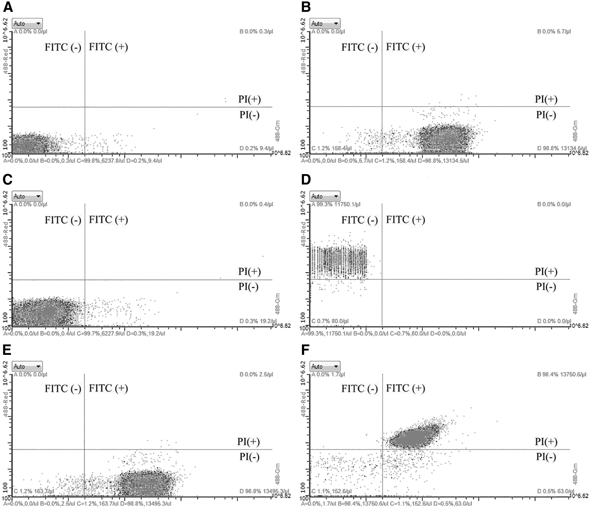
Staining of Salmonella cells.
Specificity of the FCM-based method
There are currently >2600 serotypes of Salmonella (Gal-Mor et al., 2014). An FCM-based method specific toward all Salmonella serotypes can only be considered effective. In this study, the 45 bacterial strains listed in Table 1 were allowed to react with the FITC-labeled anti-Salmonella antibody to evaluate the specificity of the FCM-based method. The results showed that all the selected Salmonella serotypes were labeled with FITC antibody, while 22 non-Salmonella strains were not labeled with FITC. Therefore, the results revealed that the FCM-based method exhibited high specificity toward Salmonella spp.
Evaluation of the FCM-based method analysis for viable Salmonella cells
Viable and dead cells were mixed at different ratios of concentration to evaluate the quantitative analysis of the FCM-based method for viable Salmonella cells. The results indicated that viable cells can be distinguished from dead bacterial cells by this method. The obtained values for the ratio of viable to dead cells were similar to previously observed values (Fig. 3).
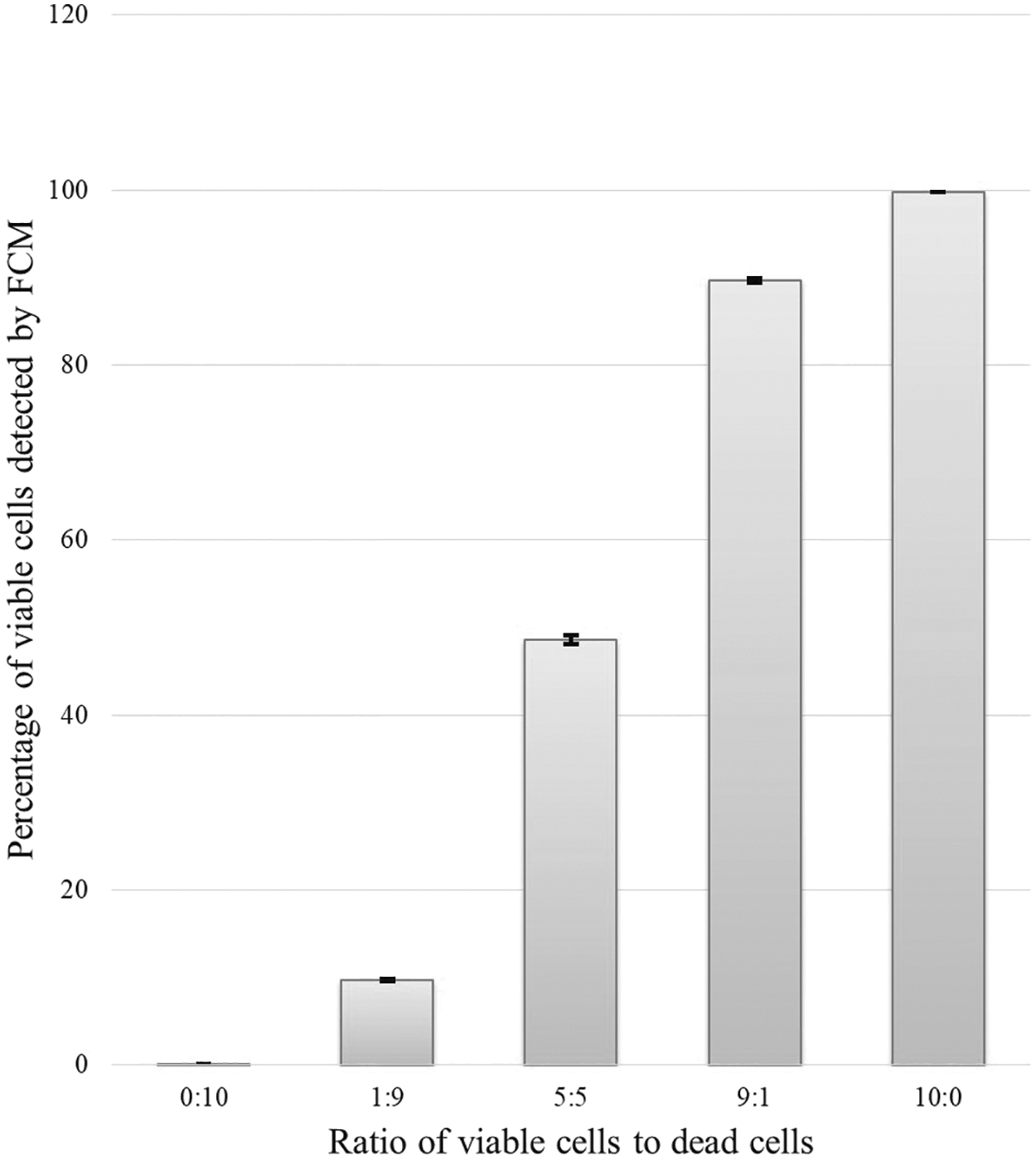
Percentage of viable Salmonella cells with different concentration ratios (n = 3). Viable Salmonella cells and dead Salmonella cells were mixed with different ratios of concentration (0/10, 1/9, 5/5, 9/1, and 10/0), and then detected by FCM after incubation with the FITC-labeled antibody and PI. FCM, flow cytometry. FITC, fluorescein isothiocyanate; PI, propidium iodide.
Validation of single-cell isolation by the flow cytometric cell sorter
To confirm that the flow cytometric cell sorter could isolate single cells, 50 samples containing nominally single cells and 10 blank samples were directly tested on XLD plates. The number of positive samples that contained only one colony per plate was 24, and the number of negative samples that contained no colony was 26, whereas all the 10 negative control samples exhibited no colony formation. This result showed that the sorting ratio of the flow cytometric cell sorter for single viable target cells was 48%, with 24 positive samples obtained from 50 nominally positive samples.
Optimization of the pre-enrichment time
A total of 55 artificially contaminated milk powder samples (A01–A55) were prepared and analyzed by the FCM-based method with 5, 6, and 7 h of pre-enrichment, respectively, and by the plate-based method with 18 h of enrichment. The results are shown in Table 2. For the five blank control samples (A01–A05), no positive results were obtained with the two methods. For the 50 nominally positive samples analyzed by the FCM-based method (A06–A55), the number of positive results by enrichment of 5 h was 14, while the results by enrichment of 6 and 7 h were identical, thereby yielding 30 positive results. In total, 16 of the nominally positive samples were deemed to be negative by the FCM-based method with 5 h of enrichment; however, these 16 samples were all determined to be positive by the FCM-based method with 6 and 7 h of enrichment. Therefore, 6 h was used as the optimized enrichment time for the subsequent experiment.
Furthermore, there were 30 positive results and 20 negative results for the 50 nominally positive samples determined by the FCM-based method with 6 h of enrichment and by the plate-based method with 18 h of enrichment. For each sample, both the FCM-based method and plate-based method yielded the same results.
Detection of single Salmonella cells in artificially contaminated milk powder samples
A total of 36 artificially contaminated samples (B01–B36) were prepared and analyzed by the FCM-based method and the plate-based method. The results are shown in Table 4. For the six negative control samples (B11, B12, B23, B24, B35, and B36), no positive results were obtained with the two methods. For the 30 nominally positive samples (B01–B10, B13–B22, and B25–B34) with different populations of the nonrelated bacteria, both FCM-based method and plate-based method yielded 18 positive results and 12 negative results, respectively.
Detection of Salmonella in retail milk powder samples and milk powder samples for proficiency testing
To further confirm the FCM-based method, a total of 123 retail samples and 6 Salmonella-positive samples for proficiency testing were detected by the FCM-based method and the plate-based method. As a result, both methods reported 123 negative results for all the 123 retail milk powder samples (Table 5). As for six milk powder samples for proficiency testing, there were six positive results and zero negative result according to the two methods (Table 5). For each sample, both methods, the FCM based and plate based, yielded the same results. The results provided by the FCM-based method for all the six Salmonella-positive samples were the same as the results provided by the National Institutes for Food and Drug Control, P.R. China.
Discussion
Milk powder and dairy products have the greatest risk of Salmonella contamination, which can be harmful to humans (Lang et al., 2018), especially for children. Hence, zero detection of Salmonella is necessary for milk powder. The rapid detection of single Salmonella cells in milk powder is important for monitoring and controlling Salmonella pathogens.
The preparation of milk powder samples containing single cells is an important prerequisite for the detection of Salmonella at the single-cell level (Chen et al., 2018). In this study, a flow-cytometric cell sorter was used instead of a dilution-based method to obtain single Salmonella cells. The dilution-based methods, with complicated procedures, might result in the detection of more than one Salmonella cell per sample. To confirm that the cell sorter could isolate single cells, 50 samples of nominally single cells were prepared by the cell sorter and then tested by a plate-based method. The result showed that the single-cell sorting ratio of the cell sorter was 48% (24 out of 50), thereby demonstrating absence of single dead Salmonella cells or no cells in 52% of milk powder samples because single cells are typically subjected to physical stress during sorting such as fluidic pressure, laser beams, and collisions with container surfaces. Nevertheless, in this study, the single-cell sorting ratio is acceptable, as 48% of the tested samples contained only one single Salmonella cell, not two or more. This study revealed that the sorting ratio for single viable Salmonella cells may not be an exact number, and instead, the sorting ratio may be within a range of percentage because the precise isolation of single cells by a cell sorter is still a difficult work (Svensson et al., 2018). In this study, although 80 samples with nominally single Salmonella cells (A06–A55, B01–B10, B13–B22, B25–B34) were prepared (Table 2), the results indicated the actual presence of 48 positive samples (Tables 3 and 4). The percentage of positive results obtained from nominally single-cell samples by using the FCM-based method was 60% (48 out of 80), which was close to the sorting ratio of 48% obtained with the cell sorter for single cells.
Results of Single Salmonella Cell Detection in Milk Powder with Different Enrichment Time
“Nominally Positive” sample contains one Salmonella cell sorted by the cell sorter. Single Salmonella cells were sorted by a flow cytometric cell sorter and then transferred into a milk powder sample of 25 g by a pipette.
“Negative” sample contains no Salmonella cells.
Milk powder samples were analyzed by FCM after pre-enrichment. The final number of Salmonella cells reached by enrichment was determined by distinct events within the counting gate in 47.2 μL of each sample.
N, “Negative” sample.
The symbol “−” means negative samples—that there is no distinct bacterial colony on the XLD plate.
P, “Nominally Positive” sample.
The symbol “+” means positive samples—that there are distinct bacterial colonies on the XLD plate.
FCM, flow cytometry; XLD, xylose lysine desoxycholate.
Detection of Single Salmonella Cells in Artificially Contaminated Milk Powder Samples
“Nominally Positive” sample contains one Salmonella cell sorted by the cell sorter. Single Salmonella cells were sorted by a flow cytometric cell sorter and then transferred into a milk powder sample of 25 g by a pipette.
“Negative” sample contains no Salmonella cells.
The population of the nonrelated bacteria was the sum of Escherichia coli cells and Staphylococcus aureus cells, which were the confirmed using the plate-based method by inoculating the samples onto plate count agar plates, respectively.
The nonrelated bacteria were E. coli CMCC 44102 and S. aureus ATCC 6538P. E. coli cells and S. aureus cells were obtained from the bacterial suspensions by the dilution method, respectively, and then transferred into the milk powder sample.
The sample was analyzed by the FCM-based method. The symbol “+” means that the sample was Salmonella positive, confirmed by the FCM-based method; the symbol “−” means that the sample was Salmonella negative, confirmed by the FCM-based method.
The sample was analyzed by the plate-based method. The symbol “+” means that the sample was Salmonella positive, confirmed by the plate-based method; the symbol “−” means that the sample was Salmonella negative, confirmed by the plate-based method.
P, “Nominally Positive” sample.
N, “Negative” sample.
CFU, colony-forming unit; FCM, flow cytometry.
Every single cell has different cultivability, which may lead to different final bacterial populations after enrichment. Optimization of enrichment time was required to ensure the detection of any single viable Salmonella cells by the FCM. In this study, 50 samples with nominally single Salmonella cells were analyzed by the FCM-based method with 5, 6, and 7 h of enrichment. The results indicated that just 14 out of 30 positive samples confirmed by the plate-based method were detected after 5 h of enrichment, while all the 30 positive samples were detected after 6 and 7 h of enrichment (Table 3). Therefore, the pre-enrichment time for the FCM-based method was determined to be 6 h.
In general, a certain amount of other nonrelated bacteria exists in the milk powder. Hence, the real diagnostic challenge for Salmonella would be testing the presence of Salmonella with a huge background of other nonrelated bacteria. Thus, in this study, 30 nominally positive samples with 3 different populations of nonrelated bacteria were prepared to confirm the FCM-based method, to determine its diagnostic efficiency. The results showed that FCM-based method was able to detect one Salmonella cell in the milk powder among almost 104 cells of nonrelated bacteria (Table 4). In addition, in this study, a total of 123 retail samples and 6 Salmonella-positive samples were used to further confirm the FCM-based method (Table 5).
Detection of Retail Milk Powder Samples and Milk Powder Samples for Proficiency Testing
For each milk powder sample, both the FCM-based method and plate-based method yielded the same results.
The 123 retail milk powder samples with different brands or different lot numbers were purchased from local markets.
The six Salmonella-positive milk powder samples for proficiency testing were provided by the National Institutes for Food and Drug Control, P.R. China.
FCM, flow cytometry.
Therefore, it can be inferred that the FCM-based method is able to detect one viable Salmonella cell in 25 g of milk powder. This method has a lower detection limit than the previous study (McClelland and Pinder, 1994). The reason may be due to the relatively high sensitivity of the A50-Micro FCM used in this study, and the isolation procedure that increased the bacterial concentration by 10 times, as the mixture of Salmonella cells in 1.5 mL was initially resuspended in a final volume of 150 μL.
The FCM-based method requires ∼7 h in total, including 6 h of enrichment, 40 min of isolation, and 20 min of staining and analysis. The time required to obtain the results by the FCM-based method was much shorter compared with the plate-based method. Furthermore, the plate-based method may also lead to the underestimation of the results due to the inability of bacteria to absorb nutrient components from the medium, sublethal environmental injury, and other physiological factors that reduce its cultivability (Smith et al., 1994; Pyle et al., 1999; Trevors, 2011). In addition, there are a variety of technologies available for the detection of single Salmonella cells, such as PCR-based methods (Rodriguez-Lazaro et al., 2014), integrated PMMS-CE microdevices (Jung et al., 2011), and ELIME assays (Volpe et al., 2016). Unfortunately, PCR-based methods and integrated PMMS-CE microdevices are invalid for the detection of viable cells (Jung et al., 2011; Rodriguez-Lazaro et al., 2014), unlike the FCM-based method. Both the PCR-based methods and ELIME assays have slightly longer operation time than the FCM-based method. ELIME assays require ∼12 h in total (10 h for enrichment, 1 h for the immunoassay, and 1 h for electrochemical measurement) (Volpe et al., 2016) and PCR-based methods require nearly 20 h (18 h for enrichment, 1 h for DNA extraction, and 1 h for real-time PCR).
At present, the FCM-based method used in this study may be only available in large industries or developed countries, and may not be feasible in remote areas with the underdeveloped testing facility as it requires sophisticated equipment with relatively high cost. However, the FCM-based method could offer great potential for the rapid and sensitive detection of Salmonella; this method might be more popularized and the cost would be reduced with further development of the FCM. In addition, Salmonella could also be found in the environmental samples, such as feces or water samples (with a large number of other nonrelated bacteria and background particles), which might be of great diagnostic challenge to the FCM-based method. However, it is believed that with further optimization of the sample pretreatments, the FCM-based method might be applied in the environmental samples and provides a wide range of potential applications in the assay of pathogenic microorganisms.
Conclusion
A method based on flow cytometric analysis was successfully developed for the rapid detection of single viable Salmonella cells in milk powder. By applying the three subsequent steps of pre-enrichment, isolation, and FCM analysis in 7 h of duration, the FCM-based method can detect a single viable Salmonella cell in 25 g of milk powder. This method provides an effective platform for the detection of Salmonella contamination in milk powder, such as the contamination that occurred in France in 2017. As a proof-of-concept study, this proposed assay can be used for other pathogenic microorganisms by rational screening of specific antibody and optimization of enrichment. Furthermore, it has a wide range of potential applications for the rapid and sensitive detection of pathogens in the food industry.
Footnotes
Acknowledgments
The authors thank Dr. Jiayi Yang and Ms. Tingye Zhuo from Center for Advanced Measurement Science of National Institute of metrology, P.R. China, for the contribution on the partial data analysis and suggestions for drawing figure. They also thank Dr. Shenghui Cui from the National Institutes for Food and Drug Control, P.R. China, for providing the six Salmonella-positive milk powder samples for proficiency testing.
Disclosure Statement
No competing financial interests exist.
Funding Information
This study was supported by grant 2017YFF0204602 from the National Key Research and Development Program of China, grant ANL1812 from Project for Improvement Quality Capability, grant AKY1958 from the National Institute of Metrology, P.R. China, grant AKY1818 from the National Institute of Metrology, P.R. China, and grant 201603D21108-03 from the Key Research and Development Program of Shanxi Province.