
Abstract
Objective: The aim of this study was to investigate the roles of cyclin D1, CDK4, and p53 in knee osteoarthritis (KOA). Methods: A total of 76 healthy controls and 154 KOA cases (grades ranging from II to IV) were recruited. Protein expression of cyclin D1, CDK4, and p53 were detected by immunohistochemistry, and mRNA expression levels of the cyclin D1, the CDK4, and the p53 genes were measured by reverse transcription-polymerase chain reaction. Results: Both protein and mRNA expression levels of cyclin D1 and CDK4 were significantly lower in KOA cases than those in healthy controls, while protein and mRNA expression of p53 was significantly higher in KOA cases than that in healthy controls (all p < 0.05). As the grades of KOA increased, Cyclin D1 and CDK4 mRNA expressions decreased, whereas p53 mRNA expression increased (all p < 0.05). In KOA cases, mRNA expression of Cyclin D1 was positively correlated to CDK4 mRNA levels (r = 0.386, p < 0.001), while negatively correlated with p53 mRNA levels (r = −0.227, p = 0.005). Conclusions: Expression of the Cyclin D1, CDK4, and p53 genes are correlated with the disease grades of KOA.
Introduction
O
Cyclin D1 is a cytokine that plays an important role in cell cycle regulation, especially in the G1/S phase of the cell cycle (Baldin et al., 1993). Cyclin-dependent kinase 4 (CDK4) interacts with cyclin D1 and together form a complex that can directly lead to G1/S conversion (Zhong et al., 2010). P53 is a tumor suppressor gene that has been extensively studied in cancer research (Kim et al., 2015). The principal role of p53 activation is to arrest the cell cycle at G1 or S phase so that the damaged cells could be repaired (Dolezalova et al., 2012). Many studies have found that cyclin D1 and CDK4 could promote the progression of the cell cycle and thus shorten the time of each cycle. This leads to an increase in cell proliferation in many cancer cells, while p53 inhibits the proliferation of cancerous cells, such as in breast cancer (Dukelow et al., 2015), lung cancer (Baharuddin et al., 2016), and liver cancer (Huang et al., 2013).
Chondrocytes are the resident cells in the cartilage. Recent studies have suggested that chondrocyte apoptosis and proliferation are the main reasons for OA progression and chondrocytes are the target cells for OA therapy (Hwang and Kim, 2015; Huang et al., 2016). On this ground, we hypothesized that cyclin D1, i, and p53 might be involved in the chondrocyte apoptosis or proliferation through controlling the cell cycle; therefore cyclin D1, CDK4, and p53. In the present study, we compared the protein and mRNA expressions of cyclin D1, CDK4, and p53 in KOA patients and healthy controls, aiming to investigate the roles of cyclin D1, CDK4, and p53 in the development of KOA.
Materials and Methods
Study population and sample source
A total of 154 hospitalized patients and outpatients with KOA were included as the KOA group. All KOA patients received replacement of total knee and total hip joint or half hip between January 2012 and March 2014 in The First Affiliated Hospital of Wenzhou Medical University. The patients' KOA grade varied from II to IV according to the Kellgren-Lawrence (K-L) grading scale. The K-L grading scale was as follows: grade 0, no radiographic features of osteoarthritis; grade I, possible joint space narrowing and osteophyte formation; grade II, definite osteophyte formation with possible joint space narrowing; grade III, multiple osteophytes, definite joint space narrowing, sclerosis, and possible bony deformity; grade IV, large osteophytes, marked joint space narrowing, severe sclerosis, and definite bony deformity. Criteria of inclusion: positive clinical symptom of KOA and K-L grade II to IV as determined by X-ray scans. Patients with metabolic bone disease, neuropathic arthropathy, acute trauma, articular bodies, acute synovitis, hemophilic arthritis, psoriatic arthritis, severe joint deformity, cardiovascular, hematopoietic, endocrine system, or other primary diseases were excluded from the current study. Femoral neck fracture patients (n = 76) who received replacement of total knee and total hip joint or half hip in The First Affiliated Hospital of Wenzhou Medical University were allocated into the control group. All of these patients had no history of knee pain and its complications and no radiographic features of osteoarthritis.
From the excised joints of the KOA group, three types of cartilage tissues were collected. The three cartilages included surface cartilages on the hip and knee joint, deep cartilages from the weight-bearing zone in the tibia side of the knee joint, and also deep cartilages from the weight-bearing zone in the femoral head of the hip joint. From the control group, deep cartilages were collected from the weight-bearing zone in the femoral head of the hip joint. The collected tissues were sampled as 0.5 × 0.5 × 0.5 cm pieces and immediately dropped into liquid nitrogen for preservation. This study was discussed and approved by the Ethics Committee of The First Affiliated Hospital of Wenzhou Medical University, and all patients signed written informed consent.
Immunohistochemistry for detection of Cyclin D1, CDK4, and p53 protein
Tissues were dehydrated in ascending series of ethanol, cleared in xylene, embedded in paraffin, then sliced, and mounted on slides (the slides were precoated with polylysine). The paraffin sections were dewaxed in xylene, hydrated in descending series of ethanol, washed in PBS, and then went through the process of antigen retrieval in the pressure cooker. After that, the tissues were added one drop of peroxidase blocking the solution in each section, incubated for 10 min at room temperature, then washed with PBS. Next, normal nonimmune serum was added, followed by another 10 min of incubation. Then, the sections were incubated in primary antibodies (cyclin D1, CDK4, p52 monoclonal antibody, PBS as blank control) overnight. After this, the samples were washed with PBS, incubated with biotin-tagged secondary antibodies for 10 min, followed by washing in PBS again, and incubation with streptavidin enzyme solution for 10 min. Finally, the section was washed again with PBS, then further incubated with DAB solution, washed with PBS, restained with hematoxylin, dehydrated in ascending series of ethanol, cleared in xylene, and mounted with neutral resin. The sections were photographed using microscopy under observation, and then the proportion of positive cells was calculated. The positive expression of cyclin D1 and CDK4 localized in the nucleus, shown as brown granules; the positive expression of p53 located in the nucleus, shown as brown to brownish yellow granules.
Reverse transcription polymerase chain reaction for detection of cyclin D1, CDK4, and p53 mRNA
Sequences of all primers are shown in Table 1. Total RNA was extracted from the cartilage frozen tissue samples using the Bio-Rad kit, according to the cesium chloride purification principle. Polymerase chain reaction (PCR) was performed in a 20 μL reaction containing the 5 μL RNA sample, primer oligo (dT), Rnase-free ddH2O, 5× reaction buffer, 20 U/μL Rnase inhibitor, 10 mM dNTP mix and M-MuLV reverse transcriptase. The samples were bathed at 70°C for 5 min, then at 37°C for 5 min +60 min, and then incubated at 70°C for 10 min. PCR conditions were as follows: 30 s at 94°C, 40 s at 58°C, 40 s at 72°C, and final extension for 40 s at 72°C. β-actin was used as the internal standard. Ten microliters of amplification products was used for electrophoresis on a 1% agarose gel, stained with ethidium bromide, and observed under UV light. Mean gray value was quantified by computer software and the relative mRNA expression of each amplicon was compared with that of β-actin.
Statistical analysis
SPSS 20.0 was used for data analysis. Quantitative data are expressed as mean ± standard deviation (
Results
General characteristics analysis
There were 76 healthy controls, with an age range of 40-75 and 154 KOA cases, with an age range of 32-74. The duration of KOA ranged from 0.3 to 5.0 years. Among these 154 KOA cases, 55 were grade II, 63 were grade III, and 36 were grade IV. Information regarding the average age, number of males and females, BMI, and disease duration was shown in Table 2. Baseline characteristics of each group were comparable (all p > 0.05).
KOA, knee osteoarthritis; BMI, body mass index.
Protein and mRNA expressions of cyclin D1
The results of reverse transcription (RT)-PCR showed that cyclin D1 mRNA band intensity gradually decreased in the control group, KOA II group, KOA III group, and KOA IV group (Fig. 1). When compared with healthy controls, both protein and mRNA expressions of cyclin D1 were downregulated in KOA cases (both p < 0.05). The protein and mRNA expressions of cyclin D1 were gradually downregulated as the grade of the disease increased. There was no significant difference of protein expressions of cyclin D1 between cases with different grades (all p > 0.05), while mRNA expression was significantly different between cases with different grades (all p < 0.05), as shown in Table 3.
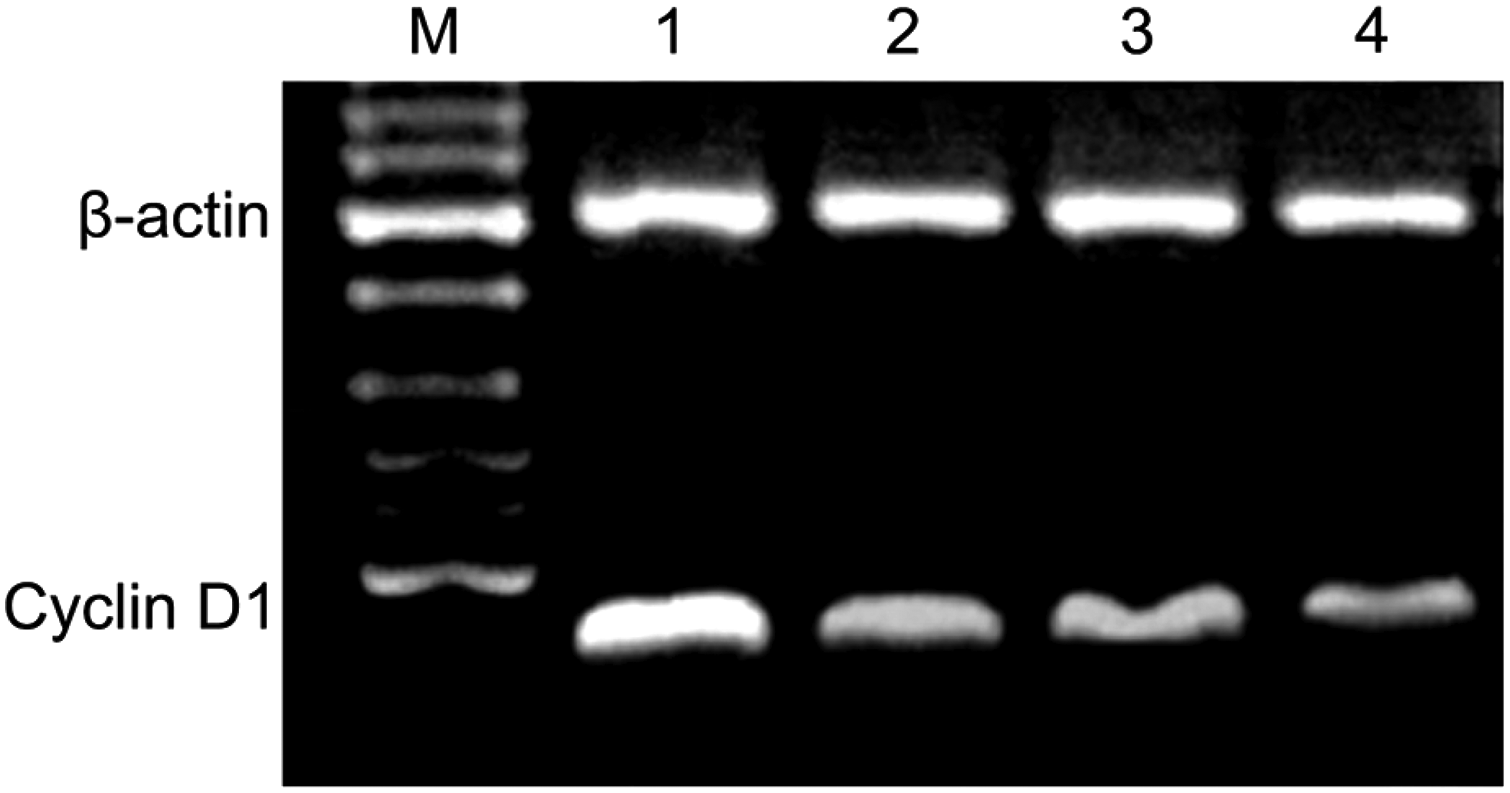
Results of cyclin D1 RT-PCR electrophoretogram. M represents Marker, 1 represents the control group, 2 represents the KOA patients at grade II, 3 represents the KOA patients at grade III, 4 represents the KOA patients at grade IV. KOA, knee osteoarthritis; RT-PCR, reverse transcription-polymerase chain reaction.
p < 0.05 compared with controls after adjustment of such covariants as age, gender, and body mass index.
p < 0.05 compared between groups.
Protein and mRNA expressions of CDK4
The results of RT-PCR showed that CDK4 mRNA band intensity gradually decreased in the control group, KOA II group, KOA III group, and KOA IV group (Fig. 2). When compared with healthy controls, both protein and mRNA expressions of CDK4 were downregulated in KOA cases (both p < 0.05). The protein and mRNA expressions of CDK4 were gradually downregulated as the grade of the disease increased. There was no significant difference of protein expression of CDK4 between cases with different grades (all p > 0.05), while mRNA expression was significantly different between cases with different grades (all p < 0.05), as shown in Table 4.
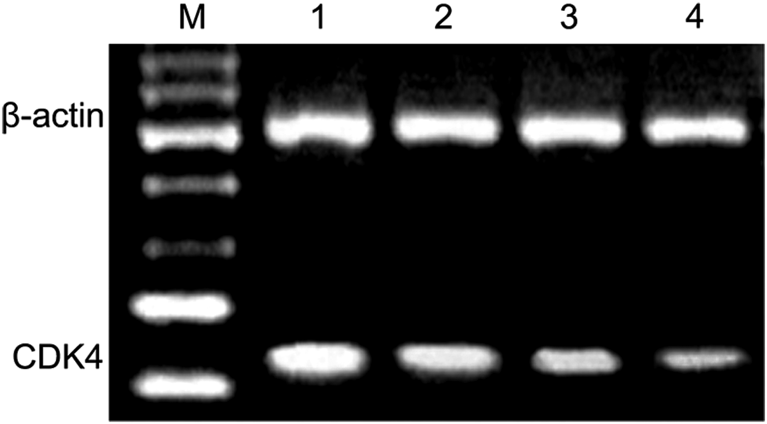
Results of CDK4 RT-PCR electrophoretogram. M represents Marker, 1 represents the control group, 2 represents the KOA patients at grade II, 3 represents the KOA patients at grade III, 4 represents the KOA patients at grade IV.
p < 0.05 compared with controls after adjustment of such covariants as age, gender, and body mass index.
p < 0.05 compared between groups.
Protein and mRNA expression of p53
RT-PCR results showed that p53 mRNA band intensity gradually deepened in the control group, KOA II group, KOA III group, and KOA IV group (Fig. 3). When compared with healthy controls, both protein and mRNA expressions of p53 were upregulated in KOA cases (both p < 0.05). The protein and mRNA expressions of p53 were gradually upregulated as the grade of the disease increased. There was no significant difference of protein expression of p53 between cases with different grades (all p > 0.05), while mRNA expression was significantly different between cases with different grades (all p < 0.05), as shown in Table 5.
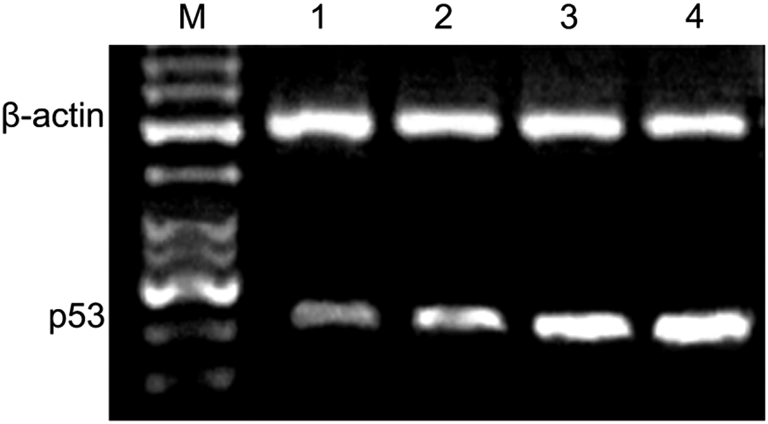
Results of p53 RT-PCR electrophoretogram. M represents Marker, 1 represents the control group, 2 represents the KOA patients at grade II, 3 represents the KOA patients at grade III, 4 represents the KOA patients at grade IV.
p < 0.05 compared with controls after adjustment of such covariants as age, gender, and body mass index.
p < 0.05 compared between groups.
Correlation analysis
No significant correlation of cyclin D1 and p53 protein expressions with CDK4 expression in bone and joint cartilage among three grades in KOA group was detected (all p > 0.05). There was no significant difference between mRNA expressions of p53 and CDK4 in bone and joint cartilage tissue (p > 0.05). In bone and joint cartilage tissue, mRNA expression of cyclin D1 was positively correlated with that of CDK4 (r = 0.386, p < 0.001), while mRNA expression of cyclin D1 was negatively correlated with that of p53 (r = −0.227, p = 0.005) (Fig. 4).
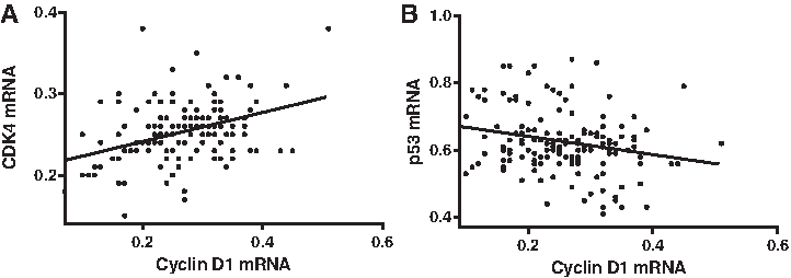
Correlation analysis.
Discussion
In recent years, the cross development of molecular biology in different disciplines and research has entered a rapidly developing stage. As the elderly population has steadily increased, more concerns have aroused regarding diseases affecting the health of this population; KOA is one of these diseases. In the present study, we found that both the protein and mRNA expressions of cyclin D1 and CDK4 were significantly decreased in KOA cases compared with that in healthy controls, suggesting that both decreased cyclin D1 and CDK4 may be useful biomarkers for KOA. Cyclin D1 in cells entering the cell cycle is induced by extracellular signals and subsequently binds to and activates CDK4 (Musgrove et al., 2011). The pathway cyclin D1/CKD4 functions is a central role in the G1/S phase transition (Gibson et al., 2003). Before our study, it was reported that increased cyclin D1 and CDK4 could promote interleukin-1β-induced chondrocyte proliferation through acceleration of G1/S transition (Wu et al., 2013, 2014). In addition, mRNA expressions of cyclin D1 and CDK4 decreased as the grades of KOA increased. We assumed that cyclin D1/CKD4 inhibition-induced G1/S defect in chondrocytes may promote chondrocyte apoptosis and block cartilage formation, therefore contributing KOA exacerbation (Aszodi et al., 2003).In the present study, we also observed that both protein and mRNA expressions of P53 were significantly upregulated in KOA patients, compared with healthy controls and the level of p53 increased as the grade of the disease increased. These might suggest that upregulated p53 may also be involved in KOA pathogenesis. A mechanic review on chondrocyte senescence reported that upregulation of p53 directly induces chondrocyte senescence (Ashraf et al., 2016). Fei et al. (2016) reported that upregulation of such cell cycle regulators as p53 and p21 could lead to G1 cell cycle arrest. Wang et al. reported that p53 activation has robust capacity to inhibit cell proliferation and induce G0/G1 arrest (Wang et al., 2016). Another research found that p53 could bind with Bcl-2 family proteins and thus participate in the regulation of cell apoptosis (Cheok et al., 2011). Evidence supported that p53 could regulate PUMA and MCL-1, which would promote the progress of cell apoptosis (Nakano and Vousden, 2001; Yu et al., 2001; Ekoff et al., 2007). We believed that p53 upregulation may promote chondrocyte apoptosis in KOA progression.
We found that in KOA cases, protein and mRNA expressions of cyclin D1 and CDK4 were upregulated, while protein and mRNA expressions of p53 were downregulated. We also observed that mRNA expression of cyclin D1 was positively correlated with that of CDK4, while mRNA expression of p53 was negatively correlated with that of CDK4. Bigi et al. (2016) suggested that cyclophilin D is p53 dependent. Concordantly, Fei et al. (2016) also reported that upregulation of p53 could downregulate the expression of cyclin D1 and CDK4. Taken together, we believe that the combination of upregulated p53 and suppressed cyclin D1/CDK4 causes KOA development.
However, the limitations in our study are also noteworthy. First, the included sample is relative small. For this, the reliability of our results may be impaired for the possibility of overgeneralization. Second, our study is preliminary. Although we provided evidence that p53, cyclin D1, and CDK4D are closely associated with KOA progression, direct evidence of the effect of p53, cyclin D1, and CDK4D in chondrocyte apoptosis is missing. In the future study, we may focus on the effect of p53, cyclin D1, and CDK4D in chondrocyte apoptosis and verify the association between p53 and cyclin D1/CDK4 in KOA progression.
In summary, cyclin D1, CDK4, and p53 were associated with the severity of KOA and were involved in the development of osteoarthritis through the regulation of cell apoptosis. However, the present study did not demonstrate a direct involvement of p53 in the interaction process of cyclin D1 and CDK4, which can lead to the aggravation of the disease. Therefore, our further studies will aim to evaluate the direct or indirect interaction of p53 with cyclin D1 and CDK4. This will be conducted by measuring the changes of cyclin D1 and CDK4 mRNA and protein expressions after the overexpression and knockout of p53 gene by viral transfection in various KOA-related cells.
Footnotes
Acknowledgment
We acknowledge the helpful comments on this article received from our reviewers.
Author Disclosure Statement
No competing financial interests exist.