
Abstract
Aims:
The aim of this study was to investigate the association of PI3K expression and PIK3CA mutations with various clinical features in Chinese patients diagnosed with esophageal squamous cell carcinoma (ESCC).
Methods:
The study included 112 patients diagnosed with ESCC from Jan 2013 to Dec 2015. Immunohistochemistry was used to determine the expression of PI3K. PIK3CA mutations were determined by sequencing. Statistical analysis was done using SPSS 19.0 software.
Results:
PI3K protein was expressed in 81.3% (91/112) of all ESCC samples, whereas it was found in only 4.9% (5/56) of adjacent normal cells. This rate of expression of PI3K was significantly higher in ESCC tissues (p < 0.001). PI3K protein expression was highly correlated with age, lymph node metastasis, and clinical stage (p < 0.05), but not with gender, location, tobacco use, alcohol use, or degree of differentiation. PIK3CA gene mutations were highly correlated with age, tobacco use, and clinical stage (p < 0.05), but not with gender, location, alcohol use, lymph node metastasis, or degree of differentiation. PI3K protein expression was not statistically correlated with PIK3CA gene mutations.
Conclusion:
PI3K overexpression and PIK3CA mutations are associated with age, tumor staging, and other clinical characteristics in Chinese patients with ESCC and thus can be further exploited as biomarkers and therapeutic targets in esophageal cancer.
Introduction
E
The phosphatidylinositol-3-kinase (PI3K)/Akt signaling pathway plays an important role in the development of various malignancies (Yuan and Cantley, 2008). It is associated with cell cycle progression, proliferation, angiogenesis, and antiapoptosis, as well as tumor invasion and metastasis (Engelman et al., 2006; Manning and Cantley, 2007). PIK3CA is located on chromosome 3q26 and is one of the most commonly mutated oncogenes in human cancers. The PIK3CA gene encodes the p110 catalytic subunit of PI3K (PI3Kp110α) (Hiles et al., 1992). When PIK3CA is mutated, methylated, or amplified, PI3K kinase is constitutively activated, leading to upregulation of the PI3K/Akt signaling pathway and subsequent tumorigenesis (Samuels and Velculescu, 2004). Previous studies have shown that PIK3CA gene mutations were found in various solid tumors, including gastric cancer, intestinal cancer, breast cancer, and lung cancer (Samuels and Velculescu, 2004). Novel therapies targeting PIK3CA mutations are under development (Gustin et al., 2008). Although PIK3CA has been studied extensively in other cancers, its importance in ESCC remains to be carefully examined. Thus, it is important to investigate whether PIK3CA can be an effective biomarker for early diagnosis and a target for therapeutic intervention in ESCC.
In this article, we retrospectively analyzed 112 esophageal cancer patients who were enrolled in the First Affiliated Hospital of Fujian Medical University from January 2013 to December 2015. We investigated the expression of PI3K and the incidence of PIK3CA gene mutations. We also evaluated the associations of PI3K expression or PIK3CA mutations with various clinical features of these enrolled patients. Our study should provide useful insights into the development of using PI3K expression and PIK3CA mutations as diagnostic markers in esophageal cancer.
Materials and Methods
Study subjects
Retrospectively analyzed profiles of 112 patients diagnosed with ESCC in our hospital from Jan 2013 to Dec 2015. The patients consisted of 73 males and 39 females in the age range from 39 to 80 (average age 58.7 ± 9.9). Forty-six patients used tobacco and 43 patients used alcohol. Eight patients had tumors located in the upper chest, 73 cases in the middle chest, and 31 cases in the lower chest. Forty-seven patients had lymph node metastasis; 65 did not. Based on WHO (2000) criteria, 14 cases were highly differentiated, 62 cases were mildly differentiated, and 36 cases were poorly differentiated. Based on the AJCC (2009) TNM stage classification system, 10 cases were at stage I, 54 cases were at stage II, 35 cases were at stage III, and 13 cases were at stage IV.
All enrolled patients were diagnosed with ESCC through histopathologic evaluation of gastroscopic biopsies or surgical tissue specimens. None of these patients had any previous chemotherapies, radiotherapies, or other anticancer treatments. Any patient who did not meet the above criteria or those who had other tumors or severe heart, brain, kidney, or liver diseases were excluded from this study.
Patient samples
A total of 112 tumor samples and 56 adjacent normal tissues as controls were collected within 15 min after open or endoscopic surgery, including thoracoscopy and laparoscopy. For each sample, half of the tissue was snap frozen in liquid nitrogen and then transferred to a −80°C freezer for storage, while the other half was preserved in formalin-fixed paraffin wax-embedded (FFPE) tissue blocks. Acquisitions of all samples were conducted with patient consent and approved by the ethics committee of our hospital.
Immunohistochemistry
Expression of PI3K protein in 112 ESCC tumor tissues and 56 adjacent normal tissues was assessed by immunohistochemistry (IHC) on FFPE tissue sections (4 μm) using a rabbit monoclonal anti-human PI3K antibody (1:1000 dilution, clone: C73F8; Cell Signaling Technology, Inc.) as per vendor's instructions. Five random fields in which the cell number was 200 or more at high magnification were selected. PI3K protein is expressed in the cytosol of cells. The intensity of PI3K expression was scored using a protocol modified from previously published studies (Zhu et al., 2012; Wang et al., 2014). In brief, the intensity score of PI3K expression was based on the following: no stain (0), light stain (1), medium stain (2), and dark stain (3). The rate of positive PI3K expression was scored as follows: <30% (1), 30-70% (2), >70% (3). Total scores were obtained by multiplying the above two scores. A total score of 0-1 was considered negative and a score of ≥2 was considered positive. Correlations between PI3K expression level and gender, age, location of tumor, tobacco use, alcohol use, lymph node metastasis, degree of differentiation, and clinical stage were then analyzed.
Sequencing of the PIK3CA gene
Genomic DNA was extracted from frozen tissue samples with the QIAamp DNA kit (Qiagen, Inc.) according to the vendor's instructions. Mutations on Exon 9 and 20 in the PIK3CA gene were detected by fluorescent real-time quantitative PCR with the Human PIK3CA Mutation Detection Kit (Shanghai RightOn Biotechnology Co., Ltd.) as per the manufacturer's instructions. The PCR conditions were as follows: 42°C 5 min, 94°C 5 min; 40 cycles of 94°C 30 s, 58°C 30 s, and 72°C 1 min; followed by 72°C 5 min, using the 7500 Real-Time PCR System (Applied Biosystems; Thermo Fisher Scientific, Inc.). Mutations were verified through direct DNA sequencing using an Applied Biosystems® 3500 (Applied Biosystems). Correlations between PIK3CA gene mutations and various clinical characteristics were then determined.
Statistical analyses
All statistical analyses were done using SPSS 19.0 software. Comparisons of PI3K protein expression between tumor cells and adjacent normal cells or between PIK3CA gene mutations, or PI3K expression, and various clinical features were done using either a chi-square test or Fisher's exact test. Correlations between PI3K protein expression and PIK3CA gene mutations were done using Spearman correlation analysis. p < 0.05 was considered statistically significant.
Results
Expression of PI3K in ESCC and adjacent normal cells
To determine the expression level of PI3K, we performed IHC on all 112 ESCC samples. We scored the expression of PI3K as described in the Materials and Methods section (Fig. 1). Analysis showed PI3K expression in 81.3% (91/112) of all ESCC samples, but only in 4.9% (5/56) of adjacent normal tissue samples. This rate of PI3K expression was significantly higher in ESCC tissues (p < 0.001), suggesting that aberrant PI3K expression in ESCC cells might contribute to the development and progression of ESCC (Table 1).
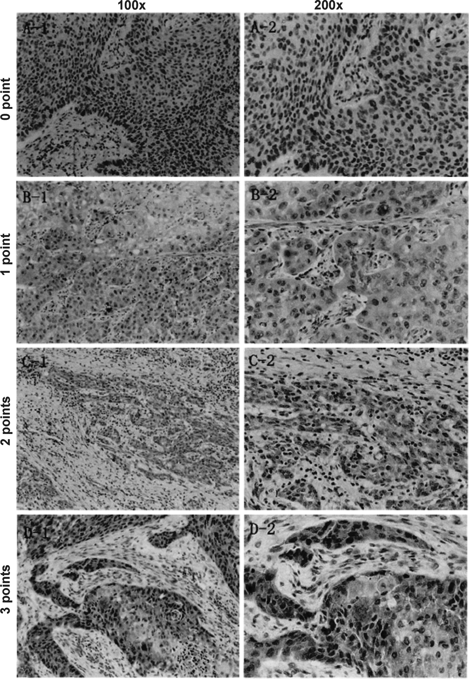
Representative images of immunohistochemistry using an antibody against PI3K on FFPE tissue sections from patients with ESCC. The intensity score of PI3K expression (Materials and Methods section) is indicated on the left. The magnification of images is indicated on top. Tissue sections were counter stained with H&E. ESCC, esophageal squamous cell carcinoma.
ESCC, esophageal squamous cell carcinoma.
Correlation between PI3K expression and various clinical features
Expression of oncogenes is often associated with certain clinical features. Thus, we performed statistical analyses to determine any correlations between PI3K expression and various clinical features. Male and female patients had comparable rates of PI3K positivity (p = 0.801). Older patients (age > 60) had significantly higher rates of PI3K expression (p < 0.014). Lymph node metastasis was more common in PI3K-positive patents (91.5%) than PI3K-negative patients (73.8%) (p < 0.026). Moreover, most of the ESCC patients at advanced stages (III and IV) had positive PI3K expression (97.9%), whereas the PI3K positivity rate was much lower in ESCC patients at earlier stages (64.3%). PI3K expression was otherwise not associated with gender, location, tobacco use, alcohol use, or degree of differentiation (p > 0.05) (Table 2).
Bold indicates statistical significance.
Correlation between PIK3CA gene mutations and various clinical features
To determine if PIK3CA gene mutations are associated with various clinical features, we performed sequencing of the PIK3CA gene on all 112 ESCC patient samples. PIK3CA mutations were more common in older patients (age > 60) (p < 0.024). Smokers also had a much higher incidence of PIK3CA mutations (p < 0.013). Finally, ESCC patients at later stages (III and IV) had higher PIK3CA mutation rates than those at earlier stages (I and II) (p < 0.018). We found no association between PIK3CA gene mutations and gender, location, alcohol use, lymph node metastasis, or degree of differentiation (p > 0.05) (Table 3).
Bold indicates statistical significance.
Correlation between PI3K expression and PIK3CA gene mutations
To determine if mutations of the PIK3CA gene are associated with PI3K expression, we performed a Spearman correlation test on all 112 ESCC patient samples. Our analysis showed no statistically significant (p < 0.193) correlation between PIK3CA gene mutations and PI3K expression (r = 0.124), suggesting that the level of PI3K expression is not associated with PIK3CA mutations in ESCC (Table 4).
Discussion
The PI3K/Akt pathway is one of the most important signaling pathways in cells (Engelman et al., 2006). It is involved in the regulation of apoptosis, proliferation, and tumorigenesis. Aberrant activation of the PI3K/Akt signaling pathway contributes to many types of malignancies (Yuan and Cantley 2008). In the 1980 s, Sugimoto et al. (1984) discovered PIK3CA as an oncogene and key messenger of signal transduction in eukaryotic cells. In mammals, PI3K can be divided into three different subtypes (I, II, and III) with various structures and functions based on the inclusion of different catalytic subunits and substrate specificities (Leevers et al., 1999). The most widely studied is type I PI3K, which consists of one regulatory subunit and one catalytic subunit (Engelman et al., 2006). Type I PI3K can be further divided into IA and IB. Type IA PI3K consists of a p85 regulatory subunit and a p110 catalytic subunit, and has protein kinase and lipid kinase activities (Carpenter et al., 1990). PI3K can be activated through growth factor-mediated receptor tyrosine kinase phosphorylation or Ras and p110 interaction (Manning and Cantley 2007). The second messenger PIP3 is released upon PI3K activation and subsequently binds to Akt and PDK1 (phosphoinositide-dependent kinase 1). Akt is then translocated to the plasma membrane, phosphorylated at Ser124 and Thr450, and subsequently activated through phosphorylation of Thr308 by PDK1. Akt can also be activated through phosphorylation at Ser473 by PDK2 (Engelman et al., 2006; Manning and Cantley 2007).
PIK3CA is one of the most commonly mutated oncogenes in human cancers. It encodes P110a, the catalytic subunit of PI3K. About 80% of the mutations in the PIK3CA gene are localized to exon 9 (helical domain) and exon 20 (kinase domain) with three hot spot mutations found to be E542K, E545K, and H1047R. Mutations in the PIK3CA gene result in constitutive activation of the PI3K/Akt signaling pathway, promoting cell proliferation and tumorigenesis (Samuels et al., 2005).
Recently, with the development of next-generation sequencing technologies, more and more tumor-associated genes have been found to be potential therapeutic targets or prognostic biomarkers. The mutation rate of PIK3CA was found to be between 2.2% and 21% in various cancers (Mori et al., 2008; Maeng et al., 2012; Shigaki et al., 2013; Hou et al., 2014). This high mutation rate has made it a focus of ESCC studies. In our study, about 6% of ESCC patients carried PIK3CA mutations. Although PIK3CA has been studied extensively in other cancers, its role in ESCC is still not clear. Therefore, it is essential to determine if PIK3CA can be a therapeutic target in ESCC.
Our study shows that the expression of PI3K is higher in most ESCC tissues than adjacent normal tissues, indicating that overexpression of PI3K may be associated with ESCC tumorigenesis. The overexpression of PI3K protein is associated with age, lymph node metastasis, and clinical stages, but not with gender, location, alcohol use, smoking, or degree of differentiation. PIK3CA gene mutations are associated with age, tobacco use, and clinical stages, but not with gender, location, alcohol use, lymph node metastasis, or degree of differentiation. These results are consistent with previous findings in esophageal cancer and other types of cancers (Samuels and Velculescu 2004; Mori et al., 2008; Hou et al., 2014; Kim et al., 2016). We determined that the correlation between PI3K expression and PIK3CA mutations was not statistically significant, even though a trend was evident. This discrepancy appears to be caused by the high occurrence of PI3K overexpression juxtaposed against a much lower PIK3CA mutation rate. Taken together, these results suggest that overexpression of PI3K and PIK3CA gene mutations play critical roles in the initiation and progression of ESCC and are associated with worse prognoses. Thus, both PI3K expression levels and PIK3CA mutations are promising biomarkers for early ESCC diagnosis and the PI3K/Akt pathway a candidate for targeted therapies.
Small-molecule kinase inhibitors have been shown to be effective against various types of human cancers (Zhang et al., 2009; Hoelder et al., 2012). Classic PI3K inhibitors such as Wortmannin and LY294002 are the most widely used PI3K inhibitors that specifically inhibit the activity of the p110 catalytic subunit, thus blocking the PI3K/Akt signaling pathway. However, due to high toxicities, they have been mostly used for in vitro studies (Maira et al., 2009). A new generation of PI3K inhibitors is being developed with higher specificity, lower toxicity, better water solubility, and greater efficacy (Chen et al., 2015; Wang et al., 2015; Massacesi et al., 2016; Zhang et al., 2016). Such new drugs hold the promise of becoming effective therapies to treat ESCC patients in the future.
Conclusions
In conclusion, PIK3CA gene expression is associated with age, tumor staging, and lymph node metastasis, whereas its mutations are associated with age, clinical stage, and tobacco use (Table 5). The genetics of PIK3CA appear to play a critical role in the clinicopathology of ESCC.
Footnotes
Acknowledgment
This study was supported by the Fujian Educational Research Fund (JB13393).
Author Disclosure Statement
No competing financial interests exist.