
Abstract
Aims:
Th17 cells and their related cytokines play an important role in the pathogenesis of celiac disease (CD), and thioredoxin (Trx) is an extracellular TG2 activity regulator. This study evaluated Trx serum levels and the expression levels of IL17A, IL21, and Trx genes in biopsies of treated (gluten-free diet) and naïve (untreated) CD patients compared with healthy individuals.
Methods:
Duodenal biopsies were collected from treated CD patients (n = 60), healthy controls (n = 60), and eight newly diagnosed celiac patients. IL17A, IL21, and Trx gene expression was assessed by quantitative polymerase chain reaction (qPCR) and compared with serum Trx levels assessed by enzyme-linked immunosorbent assay (ELISA).
Results:
Expression levels of the IL21 and Trx genes were not significantly modulated in the CD group compared to the control group, whereas the IL17A gene in CD patients was transcribed at significantly higher levels among the CD group. Serum concentrations of Trx were significantly increased in treated CD patients compared to the control group.
Conclusions:
We observed that IL17A gene is more highly expressed in duodenal biopsies of CD patients than controls, and that the serum levels of Trx are significantly higher in treated CD patients than controls. Therefore, the expression levels of these genes and gene products, respectively, could potentially be used as diagnostic biomarkers for CD patients, although more studies are needed to unravel the underlying molecular mechanisms.
Introduction
C
Th17 is a distinct lineage that secretes IL-17A-F, IL-21, IL-22, and TGF-β, being IL-17A in humans mainly dependent on RORc (RAR-related orphan receptor C) transcription factor (Ivanov et al., 2006; Turner et al., 2010). Th17 differentiation is controlled by IL-1β, IL-6, and TGF-β secretion (Bogiatzi et al., 2008). In the presence of IL-6 and TGF-β, TCD4+ cell differentiation changes from Treg to Th17 and this process is a key role in the progression of autoimmune diseases (Cooke, 2006; Eriksson et al., 2006). Different studies have shown that an increase of the expression of Th17-related cytokines at the level of RNA and protein and of related genes, such as IL23R, IL17R, RORc, CCR6, and STAT3 is associated to autoimmune diseases, including CD. According to recent studies, Th1 and Th17 play an important role in the onset and progression of autoimmune disease such as RA (Rheumatoid Arthritis), Inflammatory Bowel Disease, and also CD (Waite and Skokos, 2012; Cicerone et al., 2015; Tabarkiewicz et al., 2015). Recent studies showed that the duodenum of CD patients have gliadin-specific TCD4+ cells, which suggest the role of Th17 cells in the pathogenesis of CD (Steinman, 2007; Castellanos-Rubio et al., 2009; Monteleone et al., 2010; Fern et al., 2011; Lees et al., 2011). Th17 has a dual role in CD: secretion of inflammatory cytokines such as IL-17 and IL-21 and anti-inflammatory and protective effects, by providing TGF-β and IL-22 that lead to inhibition of the Th1 response (O'Connor et al., 2009).
According to the study of Monteleone et al. (2010), the expression of IL-17A in patients with active CD was higher than the level of untreated patients and control group. Also, Lahdenperä et al. (2014) showed that mucosal IL17A expression level was higher in pediatric untreated celiac patients compared to references and that children with potential CD showed decreased IL-17A levels. IL-21 is also produced by Th17, which is a key molecule in the pathogenesis of CD, and its neutralizing activity reduces the expression of TGF-beta and IFN-γ production (Fina et al., 2008). van Leeuwen et al. (2013) indicated that in the early phase of CD (Marsh I-II), the production of IL-21 and its producing cells increased in pediatric patients. A recent study (Borrelli et al., 2016) showed that the level of IL-21+ cells in the active and potential CD group was higher than in the control group.
Thioredoxin protein has an oxidoreductase properties and induces revitalization of proteins by reducing the two cysteine residues present in its structure and to the disulfide bond breakage (Mustacich and Powis, 2000; Oktyabrsky and Smirnova, 2007). According to several studies, Trx1 can activate the oxidized human TG2 and the level of Trx1 also increases in various diseases, such as RA and lupus, which results in activation of TG2 in the extracellular matrix (Jikimoto et al., 2002; Jin et al., 2011; Song et al., 2014). Then, IFNγ that is secreted from gluten-specific TCD4+ cells can stimulate the secretion of the Trx from monocytes, resulting in the activation of TG2 enzyme that can contribute to the pathogenesis of CD (Sollid, 2002; Jin et al., 2011).
Considering previous studies regarding the role of IL-17A and IL-21 in the pathogenesis of CD disease and that of Trx in inflammation and autoimmune diseases, and the lack of information about the expression of these biomarkers in Iranian celiac patients, we aimed at determining both the serum level of Trx and the expression of IL17A, IL21, and Trx genes in the small intestine of treated and untreated CD compared to normal individuals.
Materials and Methods
Sample collection
In this study, CD patients (n = 60) confirmed by serological tests (serum IgA tTG antibody [tTGA] and/or anti-endomysium antibody [EMA IgA]) and pathology examination according to the Marsh classification and referred to the CD department at Research Institute for Gastroenterology and Liver Diseases, Shahid Beheshti University of Medical Sciences, Tehran during 2016 were recruited. Confirmed CD patients (n = 60) with a mean age of 38.8 on a gluten-free diet (between 6 months and 2 years) were served as cases, whereas seronegative subjects (n = 60) with a mean age of 35.6 who underwent endoscopy for dyspepsia were recruited as control individuals. Controls had no history of CD and other autoimmune diseases up to their first relative's degree and with normal histopathology. In addition to these studied groups, a number of eight newly diagnosed patients with mean age 24.4 were evaluated in this study. Duodenal biopsies and blood samples were collected from treated cases and controls, although in newly diagnosed patients due to some sampling limitation we collected only duodenal biopsies. Duodenal biopsies were kept in RNA later in −70°C for further evaluation. Clinical symptoms were registered before blood/biopsy sampling. The study was approved by the Ethical Committee at the Research Institute for Gastroenterology and Liver Diseases, Shahid Beheshti University of Medical Sciences. All the patients were requested to carefully read and sign an informed consent. No financial burden related to the study was imposed on the participants.
RNA extraction, complementary DNA preparation, and real-time quantitative polymerase chain reaction
In this study, YTA RNA Extraction Kit (YEKTA TAJHIZ AZMA) was used to extract total RNA from fresh tissues of small intestinal biopsies. RNA extraction efficiency was evaluated by Nanodrop (optical density = 260/280). Total RNA (10 μg per sample) was reverse-transcribed into complementary DNA (cDNA) using the RevertAid First Strand cDNA Synthesis Kit (Thermo). The synthesized cDNA (10-20 ng) was assessed by quantitative polymerase chain reaction (qPCR) with SYBR Premix Ex Taq (Tli RNase H Plus; Takara, Kusatsu, Japan). The real-time qPCR was performed using the Rotor-Gene Q MDX with the following conditions: denaturation step (30 s at 95°C), annealing (30 s at 58°C for IL17A and 57°C for IL21 and TRX), followed by extension (40 s at 72°C). Primer sequences were designed by Gene runner software and are reported in Table 1. Beta2 microglobulin (B2M) was employed as the reference house-keeping gene. The relative quantification (RQ) method was applied to determine gene expression levels using the 2−ΔΔCt method. (ΔCt = Cttarget − Cthouse keeping).
B2M, beta2 microglobulin; Trx, thioredoxin.
Enzyme-linked immunosorbent assay for thioredoxin
In this study, we used enzyme-linked immunosorbent assay (ELISA) technique to measure the serum level of thioredoxin in the studied groups. The thioredoxin (Trx) concentration was determined via ELISA method (Product code: 3580-1H-6; Mabtech, Nacka Strand, Sweden). All of the samples were analyzed simultaneously following the manufacturers' instructions. Briefly, 96-well plates were coated overnight at room temperature with 0.1 mL of capture antibodies anti-human Trx monoclonal antibody. The plates were washed three times and blocked with blocking buffer for 1 h. After washing five times, 0.1 mL of either standard or samples were added and the plate incubated for 2 h. After washing five times, 0.1 mL of detection antibody was added and the plate was incubated for 1 h. Then, 0.1 mL of Avidin-HRP enzyme was added followed by 0.1 mL 1 × TMB solution; the plate was then incubated until color development was complete. The reaction was stopped with 2 M H2SO4. Finally, samples were read by using an ELISA reader (Anthos, Wals, Austria), (at 492 nm for testing and 630 nm as reference). The concentration of Trx in each well was calculated according to manufacturer's instructions (calibration curve with standards provided with the kit).
Statistical analysis
Data were analyzed using Prism Software version 6.04 for Windows (GraphPad, La Jolla, CA). Comparisons between the groups were examined by t-test and one-way analysis of variance. The correlation between variables was assessed using the Spearman's correlation. The differences with p-value <0.05 were considered as statistically significant.
Results
Demographic and clinical characteristics
Demographic characteristics of subjects and controls (i.e., age and sex) have been reported in Table 2. The mean age of untreated patients was lower than other groups (p = 0.024). The predominant gender in all groups was female, although the difference was not statistically significant (p = 0.55). Among gastrointestinal symptoms the most common symptoms were bloating (58.3%) and diarrhea and abdominal pain (each 50%), and the most common nongastrointestinal symptoms were fatigue (71.7%), anemia (53.3%), and weight loss (48.3%).
CD, celiac disease.
Expression of IL17A
The expression of IL17A gene in the three groups of patients assessed by real-time qPCR showed that the expression level of IL17A gene between the untreated group and control group was statistically significant (p = 0.02), although its expression was not significantly higher in untreated group compared with the treated group (p = 0.07). In the treated group the expression level of IL17A was not significantly higher than control group (p = 0.1) (Fig. 1). Also, bivariate Spearman's correlation showed no statistically significant correlation between IL-17A gene expression and symptoms and histopathological abnormality in CD patients (Table 3).
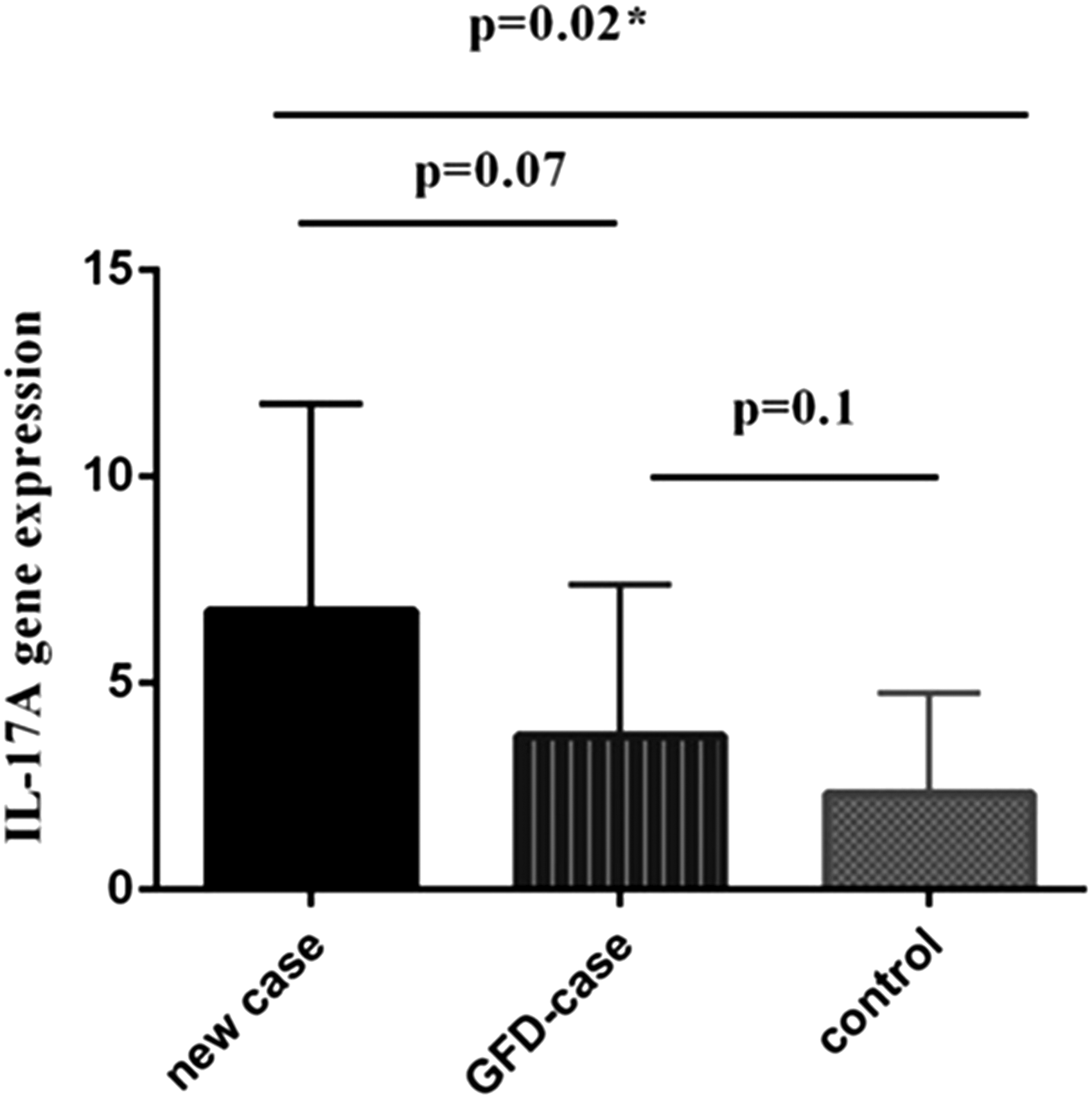
Expression level of IL17A gene in the studied groups.
Expression of IL21
IL21 gene expression in the three considered groups showed that although IL21 gene expression in the treated and untreated groups was higher than in the control group, these differences were not statistically significant (p = 0.3 for both comparisons). Moreover, also the IL21 expression was not significantly higher in untreated group compared with the treated group (p = 0.7) (Fig. 2). Bivariate Spearman's correlation showed significant correlation between osteoporosis and IL21 expression in untreated CD patients (r = −0.92, p = 0.008), and bloating (r = 0.28, p = 0.04) in treated CD-patients. This suggests that these patients may suffer from other intolerances (i.e., lactose) or that they are not in complete remission. No correlation with histopathological abnormality in CD patients was reported (Table 4).
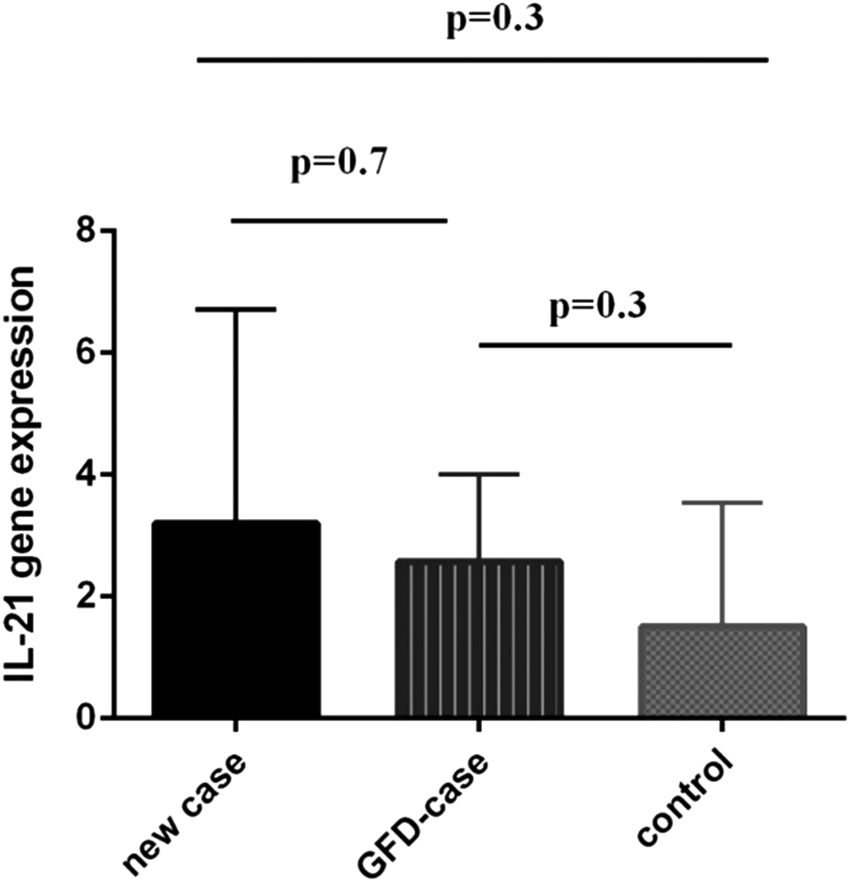
Expression level of IL21 gene in the studied groups.
Expression of Trx
Trx gene expression showed that the expression of Trx gene in the treated patients and in the untreated group is not statistically different from the control group (p = 0.4 and p = 0.3, respectively) (Fig. 3). However, bivariate Spearman's correlation showed a significant correlation between osteoporosis (r = −0.89, p = 0.01) and weight loss (r = −0.80, p = 0.05) with Trx expression in untreated CD patients (Table 5).
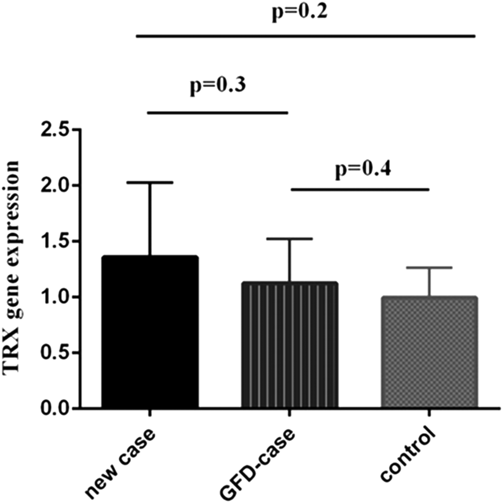
Expression level of Trx gene in the studied groups. Trx, thioredoxin.
Correlations between IL17A, IL21, and Trx
Correlations between messenger RNA (mRNA) expressions of IL17A, IL21, and Trx genes were examined by Spearson's correlation test. As illustrated in Figure 4, in treated-CD patients the expression of IL17A showed a significant correlation with IL21 (r = 0.40, p = 0.007) and in untreated-CD patients the expression of IL21 showed a significant correlation with Trx (r = 0.83, p = 0.009).
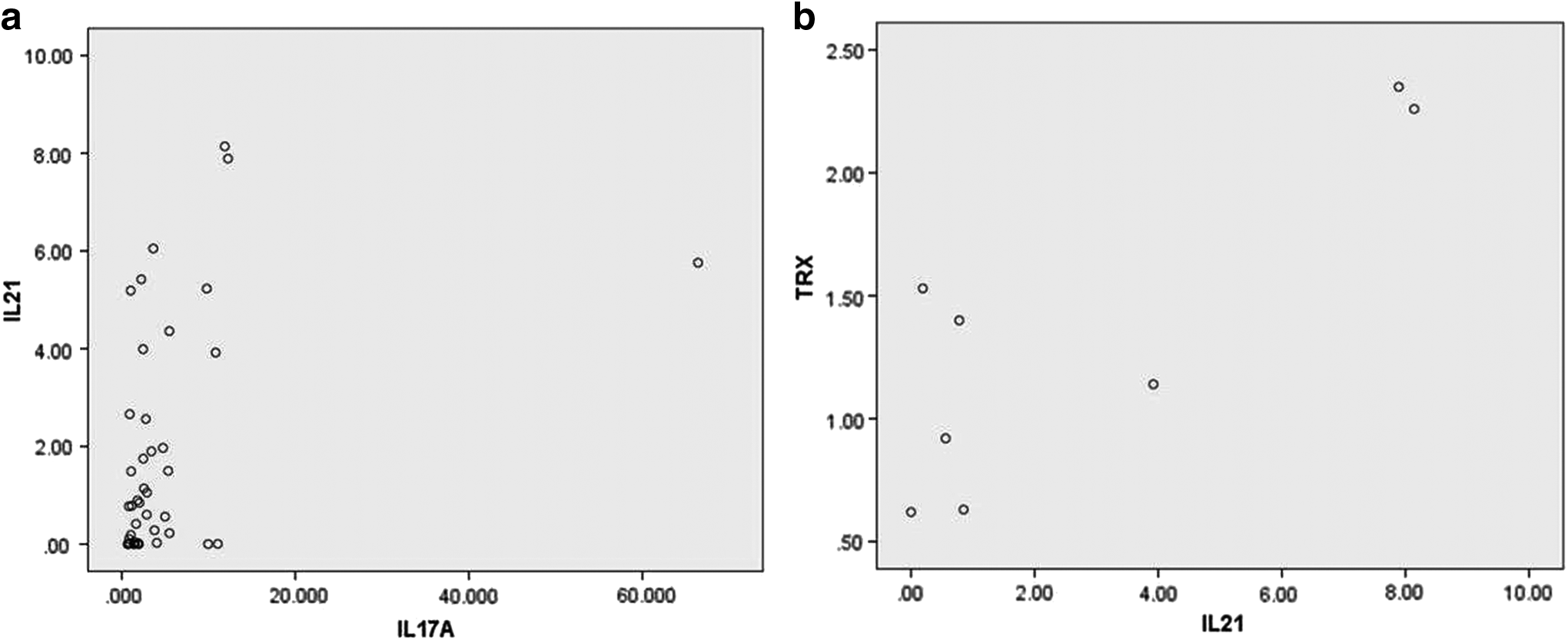
Correlation between mRNA expressions of IL17A, IL21, and Trx1 genes.
Serum Trx
As illustrated in Figure 5, in contrast to the expression of Trx gene in small intestine tissue, the serum concentration of Trx in treated CD patients was significantly higher than the healthy subjects (p = 0.001). However, bivariate Spearman's correlation showed no statistical correlation between serum Trx and symptoms and histopathological abnormality in CD patients (Table 6).
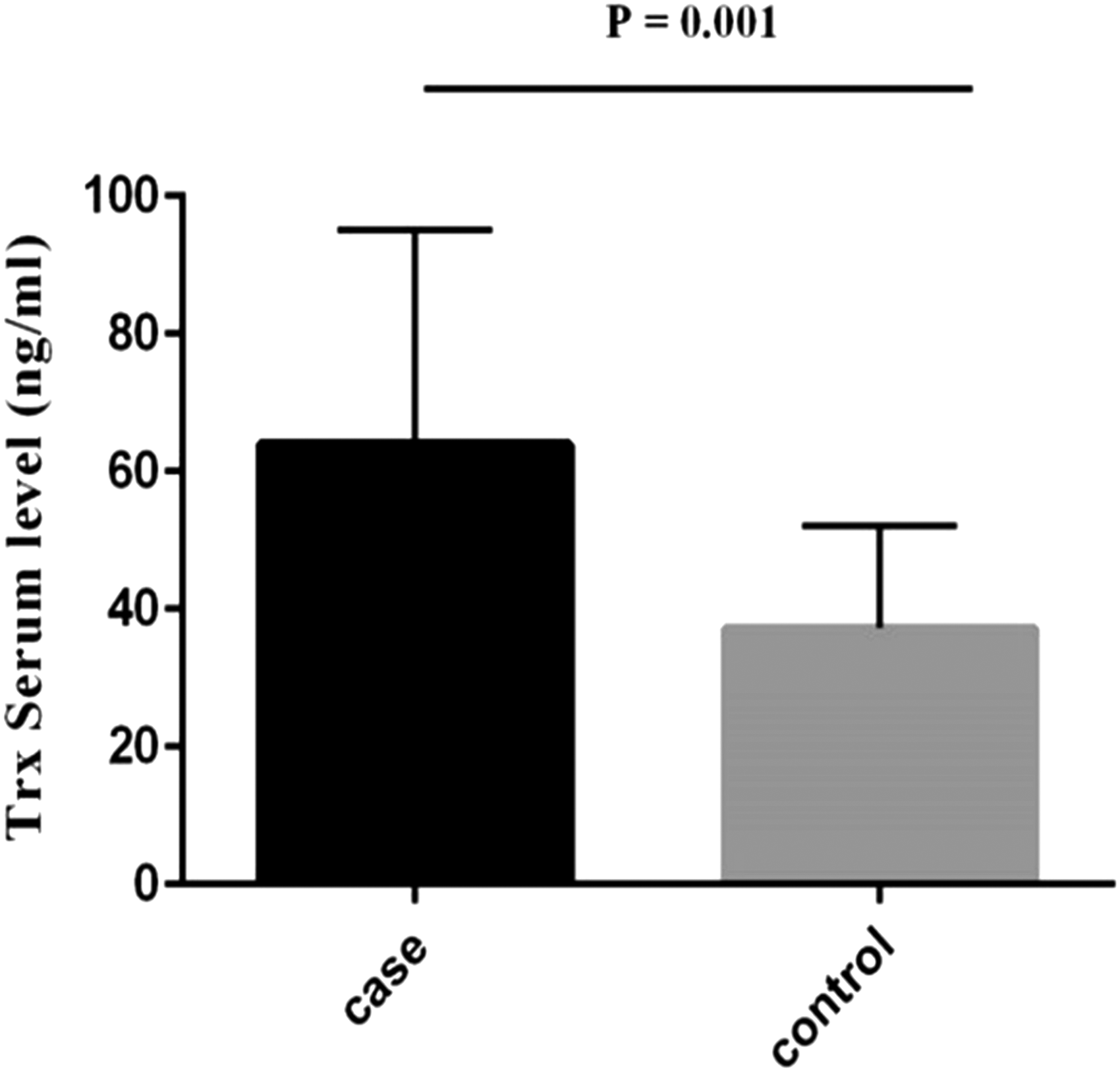
Comparison of Trx serum level in treated patients and healthy controls.
Correlation of mucosal damage with IL17A, IL21, and Trx expression
Out of 60 treated patients on a gluten-free diet (between 6 months and 2 years), the histological evaluation showed that 25 (41.7%) were Marsh 0, 14 (23.3%) Marsh 1, 3 (5%) Marsh II, and 18 (30%) were Marsh III. In eight untreated cases, three were Marsh II and five were Marsh III. All were seronegative. This heterogeneity can be explained taking into account the time frame of the treatment (from 6 to 24 months) that can delay a complete damage remission or to an involuntary/unconscious intake of gluten from food (i.e., restaurants or other stores), although all the treated patients were on a strict gluten-free diet (GFD). According to the Spearman's correlation test, treated CD patients display a significant moderate inverse correlation between IL-17A expression and mucosal severity damage. However, the correlations between IL-21 and thyroidoxin expression and mucosal damage were not significant. In untreated CD cases, none of the genes were correlated with the mucosal damage (Table 7).
Discussion
In this study we investigated the expression level of IL17A, IL21, and Trx genes and Trx serum level in three groups of patients including active-CD patients (i.e., patients with active CD), treated-CD patients (i.e., patients on gluten-free diet), and healthy control group. The results showed that IL21 and Trx gene expression in studied groups was not statistically different compared with controls, whereas IL17A was statistically different. Moreover, the serum level of Trx was significantly higher in treated-CD patients compared with control group (p = 0.001).
A study by Castellanos-Rubio et al. published in 2009 showed that the expression level of the cytokines associated with Th1 and Th17 (IL-17A, IL-17F, IL-6, IFNγ, IL-23A) was significantly increased in intestinal biopsy samples in patients with active CD compared with the control group (Castellanos-Rubio et al., 2009). In our study, the expression of IL-17A in the untreated and treated celiac groups was significantly increased compared with the control group (p = 0.02). In a study by Sapone et al. that appeared in 2010, 13 patients with active CD and 11 gluten-sensitive individuals were evaluated. The authors observed that mucosal expression of IL-17A and TCRγδ+ CD3+ cells expressing the CCR6 marker significantly increased in active celiac patients compared with gluten sensitive individuals and control group (Sapone et al., 2010) and this result is in agreement with our results.
In a study by Lahdenperä et al. in 2012, the level of IL-17A in four groups including seropositive children, untreated CD patients, treated CD, and children with T1D was investigated and the results showed that the mucosal expression of IL-17 was elevated in untreated CD compared with other study groups. In that study, the role of IL-17 in inducing apoptosis of the gut epithelial cells was investigated and the authors found that the incubation of the epithelial Caco-2 cells with IL-17 increased the gene expression of the antiapoptotic BCL2, therefore IL-17 might protect the mucosal layer from atrophy. The authors suggested that an increased gene expression of IL-17 in CD patients may be employed as an effective biomarker to detect the phase of the disease activity (Lahdenperä et al., 2012). In agreement with previous study, our results did not emphasize significant correlation between IL-17A and histopathological abnormality in both treated and untreated CD patients. Therefore, our results suggest that IL-17A should not have a role in the atrophy of villi.
In agreement with our study, Borrelli et al. (2016) also showed that the expression of IL-17A in active CD patients is higher than potential celiac patients and controls. In any case, further studies on the origin of IL-17A secretion and its exact role in the pathogenesis of CD are required. Moreover, many evidences have emphasized that Th17 cytokines are important for the induction of innate and adaptive host responses and contribute to host protection against a variety of pathogens at the intestinal mucosa (Guglani and Khader, 2010). Most notably, a reasonable balance between pathological and protective expression of Th17 responses at the intestinal mucosa that defines immunity or inflammation is becoming increasingly clear. However, additional investigations to study this fine balance in detail will be required to define the effect of the antigens on intestinal mucosa.
IL-21 plays an important role in the regulation of innate and acquired immune response and a key role in the pathogenesis of mucosal damage in CD (Borrelli et al., 2016). In a study by Fina et al. in 2008, an increased expression of IL-21 in 43 untreated celiac patients compared to controls was reported. Borreli et al. in 2016 compared the expression level of IL-21 in 76 patients with active CD, 90 potential CD, and 58 controls and they found that IL-21 expression, similarly to the control group, was decreased in potential CD compared with patients with active CD. Also, in lamina propria of potential celiac patients the density of IL-21-producing cells was reduced compared to those with active CD, although similar to control group. Our study, aimed at evaluating the expression of IL-21 in treated and untreated celiac patients, showed that although there is an increase of IL-21 expression in untreated celiac patients (compared to treated and control group) this difference was not statistically significant.
Trx gene expression is regulated by Th1 cell cytokines, including IFN-γ. This regulation interferes with PI3K/AKT, JAK, and ERK pathways, thus inducing the activation of transcription factors. Under stress conditions, Trx protects immune cells against apoptosis by regulating the production of IL-4 and IFN-γ, and helps to maintain hemostasis in immune cells (Kim et al., 2008). In 2011, Pennisi et al. measured the expression level of Trx1 proteins and TXNRD1 (thioredoxin reductase 1) in the blood and cerebrospinal fluid of 26 patients with multiple sclerosis and compared them to healthy subjects using western blot techniques. The results showed that the gene expression level of Trx1 expression in blood and cerebrospinal fluid in subjects with multiple sclerosis was significantly increased, but expression level of TXNRD1 protein was significantly decreased. Maurice et al. (1999) also evaluated the amount of Trx1 protein in plasma and synovial fluid (SF) in RA patients. Results showed that Trx1 level in SF, in the plasma and mRNA expression of Trx1 were significantly higher than Trx1 in healthy subjects, and concluded that Trx1 may be associated with the disease activity. Jin et al. (2011) demonstrated that human Trx can decrease with high specificity the disulfide bond of TG2 in vitro. Secreted IFNγ from specific gluten-T cells in the mucosa of CD patients can induce the secretion of Trx at levels capable of activating oxidized TG2. Regarding the role of Trx molecule in inflammatory and autoimmune diseases, our study was the first study that investigated the Trx gene expression in three groups of people including untreated celiac patients, treated CD, and healthy controls. The mRNA level of the Trx in untreated subjects compared with treated patients and healthy controls was not statistically significant (p = 0.3 and p = 0.4, respectively).
In our study, Trx serum levels were measured only in treated-CD patients and healthy control group, notwithstanding, results showed that Trx serum level was significantly higher than healthy controls (p = 0.001). This result opens new perspective in the use of this molecule as a potential biomarker of active CD and point to Trx as an interesting therapeutic target for potentially treating CD and related symptoms.
Conclusions
The results of this study showed that the expression level of IL17A gene in untreated and treated CD groups significantly increased compared with the control group. Moreover, serum level of Trx was significantly higher in treated CD patients than in control group. Statistical analysis indicated the absence of a significant correlation between the expression of IL17A, IL21, and Trx genes and severe histopathological abnormalities. Our finding suggest the use of IL17A and Trx as potential biomarkers for CD disease, even in the absence of other histopathological findings, opens new perspectives for developing new treatments for CD disease. However, further studies will be required to confirm our findings and clarify the molecular mechanisms underlying the expression changes (at mRNA and protein level) observed in the present study.
Footnotes
Acknowledgments
This research has been derived from the MSc thesis of M.F. and financially supported by Gastroenterology and Liver Diseases Research Center, Research Institute for Gastroenterology and Liver Diseases, Shahid Beheshti University of Medical Sciences (Tehran, Iran).
Author Disclosure Statement
The authors declare no conflicts of interest.