
Abstract
Background:
Osteoarthritis (OA) is a common chronic degenerative joint disease characterized by articular cartilage degeneration and synovitis. CircRNAs are increasingly being recognized as functional endogenous RNAs with a stable structure and high tissue specificity. Recent studies have shown that some circRNAs may be involved in the initiation and progression of OA and that there is differential expression of circRNAs in chondrocytes in vitro isolated from patients with OA.
Purpose:
In this study, we aimed to determine if circRNA levels in the peripheral blood of Chinese Han patients with OA would be diagnostic based on the previous in vitro studies.
Methods:
We collected peripheral blood samples from 25 patients suffering from OA and 25 healthy controls and measured hsa_circ_0032131_CBC1 RNA levels through quantitative RT-PCR (qRT-PCR). The statistical basis for evaluating the diagnostic value was to calculate the area under the receiver operator characteristic (ROC) curve.
Results:
The results of the qRT-PCR for hsa_circ_0032131_CBC1 were consistent with those of the microarray analysis. The ROC curve shows that hsa_circ_0032131 holds diagnostic value for OA (0.8455, p < 0.01).
Conclusions:
Our research indicates that differentially expressed circRNAs may be involved in the development of OA and could be used diagnostically.
Introduction
Osteoarthritis (OA) is a common and chronic debilitating disease of joints in elderly individuals with a higher incidence than other forms of arthritis (Mobasheri et al., 2017b). OA is characterized by gradual degeneration of the hyaline cartilage, accompanied by the degeneration of the surrounding synovial membrane and subchondral bone, which affects the integrity of the articular surface (Mobasheri et al., 2017a). Chronic inflammation is involved in the pathological process of OA (Robinson et al., 2016).
Articular cartilage is a type of avascular cartilage with nonmigrating and nonproliferating characteristics; its ability to endogenously regenerate is limited, and these factors likely underlie OA progression (Budd et al., 2017). Common causes of OA include joint damage, age, gender, genetic factors, and mechanical stress (Loeser et al., 2012). OA associated with an increasing medical burden during these years. A recent study showed that OA threatens the health of ∼3.1 million persons worldwide; and Australia is expected to spend >$2.9 billion on OA health care by 2030 (Ackerman et al., 2018).
The main clinical symptoms of OA are persistent pain and progressive disability of the affected joint (Glyn-Jones et al., 2015). Some clinical guidelines recommend early interventions for OA such as physical activity, oral and topical NSAIDs, and intra-articular injection (Hochberg et al., 2012). Despite the potential effects of symptom relief, eventually these measures fail and the pathological progression of OA continues with most patients requiring arthroplasty to maintain functionality (Hunt et al., 2014, 2017).
Recently, a novel RNA type circRNAs have garnered increased attention from scientific researchers. These circRNAs are without a 5′ cap and a 3′ poly-A tail, with a highly conserved closed continuous loop structure (Lasda and Parker, 2014). They are a kind of noncoding RNA that are highly prevalent in the cytoplasm of eukaryotic cells (Jeck and Sharpless, 2014). Owing to the closed ring structure feature, circRNAs have the ability to resist the digestion of RNAses (Suzuki and Tsukahara, 2014). CircRNAs were once regarded as aberrant splicing products without additional biological function (Cocquerelle et al., 1993). However, this view has changed since Hansen and his research team found the “miRNA sponge” function of the circRNA for miR-7 in the brains of mice (Hansen et al., 2011). They demonstrated that the sex-determining region Y (Sry) acts as a miR-138 sponge, and also showed an miRNA sponge absorption effect by circRNAs is a common phenomenon (Capel et al., 1993).
Since then, an increasing number of studies have confirmed the biological functions of numerous circRNAs. Since then, increasing studies have confirmed that circRNAs had important biological functions, including miRNA “sponge,” functional proteins induction, and translation function. Moreover, as well as dysfunction of circRNAs was related to the occurrence and progression of many different human diseases, for example, Alzheimer's disease in the nervous system (Lukiw, 2013), coronary artery disease (Zhao et al., 2017), rheumatoid arthritis (Zheng et al., 2017), and tumor (Liu et al., 2017).
The diagnosis of OA is still based on clinical manifestations and imaging of the joints. It is, however, still difficult to detect OA at early stage (Bijlsma et al., 2011). Clinical manifestations generally occur only after significant cartilage degeneration. Therefore, an effective early diagnostic is needed to be able to initiate treatment to slow the prognosis of OA. The key molecules and mechanisms leading to OA progression, however, remain largely unclear. Thus, it is essential to have a greater depth of understanding of OA pathogenesis. Recent studies have found that many circRNAs are associated with OA, indicating that they may play important roles in the pathology of OA (Zhou et al., 2018).
Our previous studies used high-throughput techniques to analyze circRNA expression profiles of knee chondrocytes grown in vitro from OA patients and controls. We found a significant difference in the expression of hsa_circRNA_0032131 between cases and controls. This RNA is encoded at chr14: 61995792-61997313, and its associated gene symbol is PRKCH.
Materials and Methods
Inclusion and exclusion of subjects
All specimens were selected randomly among the inpatients and outpatients in the First Affiliated Hospital of Xi'an Jiaotong University during the time period from May 2017 to October 2017, all subjects are Chinese Han nationality. In this study, the participants with OA were diagnosed according to the OA stiffness and pain index of Western Ontario and McMaster University (Ehrich et al., 2000), whereas patients with a history of other bone and joint diseases were excluded. Finally, 25 OA patients and 25 healthy individuals matched for age and gender were randomly divided into experimental and control groups. The study was approved by the Human Ethics Committee of Xi'an Jiaotong University, project approval number (2016-28), and all subjects signed informed consent.
Collection of peripheral blood samples
Three milliliters of peripheral blood was drawn from each donor before breakfast by peripheral venipuncture and gathered in ethylenediaminetetraacetic acid (EDTA) anticoagulant vacutainers, kept in the −80°C degree refrigerator until used for RNA isolation. For the next quantitative RT-PCR (qRT-PCR) assays presented in this study, RNA >1 μg in 100 μL of total blood is sufficient.
Isolation and extraction of RNA
Total RNA was extracted from the peripheral blood samples of 25 OA patients and 25 healthy controls using the TRIzol reagent (Invitrogen; Thermo Fisher Scientific, Inc., Carlsbad, CA) according to the product instructions. The purity and concentration of the RNA were detected by a NanoDrop ND-1000 instrument (Thermo Scientific, Waltham, MA). RNA amplification, labeling, and hybridization were performed using a Cy3-dCTP kit according to the manufacturer's product description. The quality and integrity of the extracted RNAs were evaluated using a high-resolution electrophoresis system (Agilent 2100 Bioanalyzer; Agilent Technologies, Palo Alto, CA).
qRT-PCR detection of hsa_circ_0032131_CBC1
To further validate the results of our previous research on microarray analysis, the significant differentially expressed gene hsa_circRNA_0032131 was selected as a target gene to undergo qRT-PCR. Total RNA from peripheral blood samples was reverse transcribed into cDNA using superscript II reverse transcriptase (Invitrogen; Thermo Fisher Scientific, Inc.) according to the product's technical guidelines. The selected circRNA was analyzed using the Applied Biosystems® Quant Studio™ 7 Flex Real-Time PCR System (Invitrogen™ Life Technologies™, Carlsbad, CA). The PCR conditions were as follows: predenaturation at 95°C for 30 s, 40 cycles at 95°C for 5 s, primer annealing at 60°C for 30 s, extension at 72°C for 30 s, and primer specificity was confirmed at the melting curve stage. According to our previous study of human chondrocyte, the primer sequence of hsa_circ_0032131_CBC1 (forward 5′-TACCTGGCTCCATGAAGATGC-3′, reverse 5′-CCTCCTTGCACATTCCGAAG-3′). The fold changes were examined using the 2−ΔΔct method. The PCR results were investigated using Quant Studio™ Real-Time PCR Software Version 1.1 (Life Technologies Holdings Pte. Ltd.). The Mann-Whitney-Wilcoxon test was used to determine the extent of significance of differences in gene expression screened between OA patients and healthy controls. Primers were synthesized by Shanghai Sangon Biotechnology Ltd.
Data analysis
Data were analyzed by relative quantification 2−ΔΔct method. Statistical analysis was performed by SPSS19.0 (SPSS, Chicago, IL) and p < 0.05 was considered statistically significant. The results were expressed as mean ± standard deviation.
Results
RT-PCR verification of differential expressed circRNA
Based on our previous chip analysis data (screening for differentially expressed circRNA in the cartilage of OA patients and its diagnostic value), we choose to evaluate the levels of five circRNAs, including hsa_circ_0032131_CBC1 RNA using qRT-PCR (Table 1).
Five circRNAs Were Selected for Validation According to Fold Change >2 and Original Signal Value >100
FC, fold change.
Receiver operator characteristic curve analysis of hsa_circ_0032131_CBC1 among OA and controls
We found elevated levels of hsa_circ_0032131_CBC1 in patients versus controls, and a receiver operator characteristic (ROC) curve analysis was performed to evaluate the potential diagnostic value of this marker, which showed it could distinguish patients with OA from healthy individuals with high sensitivity (area under the curve [AUC]: 0.8455, p < 0.01). The consistency of hsa_circ_0032131_CBC1 in chondrocyte and peripheral blood indicates that the differential expression of hsa_circ_0032131_CBC1 in patients with OA and healthy controls has some significance. Thus, it may be useful as a biomarker for OA (Fig. 1A). When hsa_circ_0032131_CBC1 = 9.365CT, the Yoden index reaches a maximum of 0.55, the sensitivity and specificity were 0.90 and 0.65, respectively (Fig. 1B).
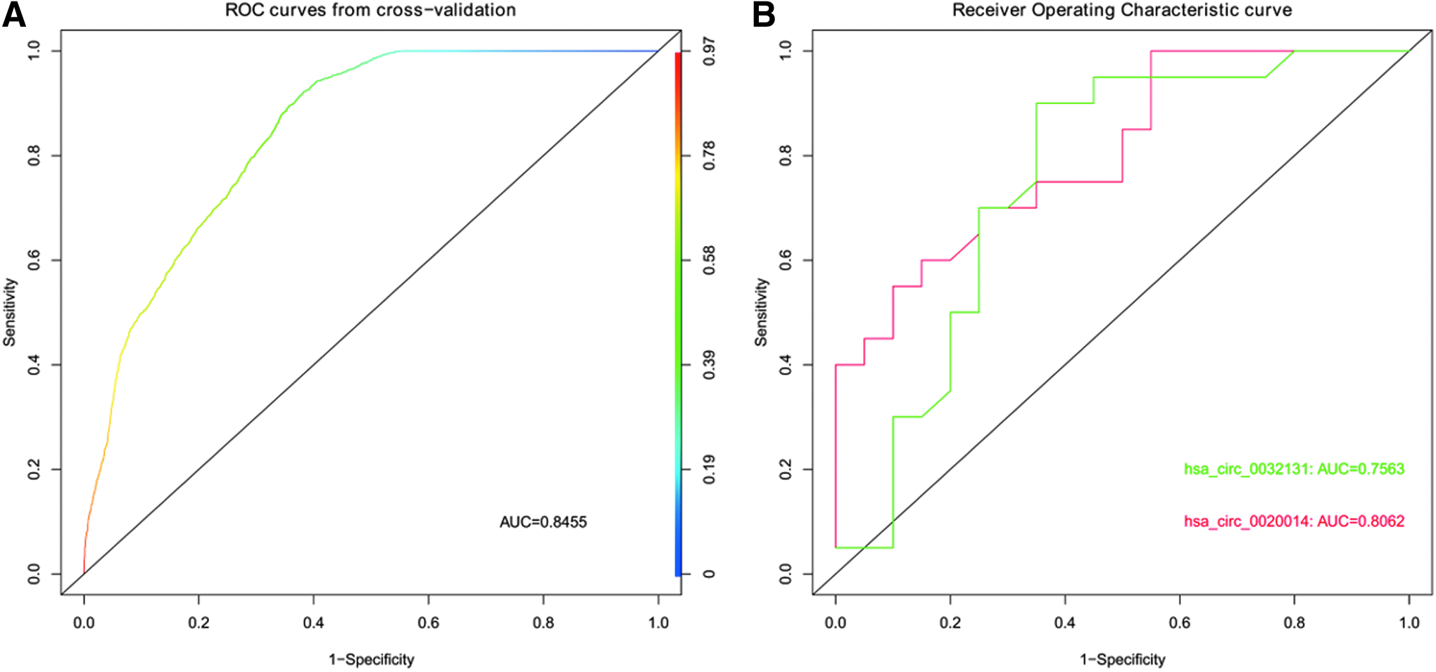
ROC curve of hsa_circ_0032131_CBC1.
Discussion
Since CircRNAs were first detected in RNA viruses in the 1970s (Wilusz and Sharp, 2013) thousands of circRNAs have been identified in transcriptomic libraries of various species through the use of RNA microarray technology. circRNAs because of their stability are candidates for diagnostic biomarkers (Li et al., 2015b; Xuan et al., 2016; Zhao et al., 2016). To date, diagnosing OA is still based on clinical manifestations and imaging features (Guo et al., 2014), clinical changes usually occur after cartilage degeneration, often with joint pain, stiffness, deformation, and other consequences; and are not sensitive enough to diagnosis cases at an early stage.
Our previous microarray analysis performed on cultured chondrocytes, which identified hsa_circ_0032131_CBC1 as a potential marker of OA were validated in this study. In this study, using a qRT-PCR we confirmed the upregulated expression of hsa_circ_0032131_CBC1 in peripheral blood of OA patients compared with healthy controls, which was consistent with the results of chondrocyte verification. Moreover, in our research, the level of hsa_circ_0032131_CBC1 in peripheral blood showed its high ROC AUC value (0.8455, p < 0.01) and potential diagnostic value.
We predicted the targeting miRNA of hsa_circ_0032131_CBC1 using miRanda software, we found that hsa_circ_0032131_CBC1 has 50 targeted miRNAs. Next, we sequenced and screened targeted miRNAs according to tot energy. Tot energy means the dimer formed by miRNAs and circRNAs, the smaller the tot energy, the closer the combination between them. According to tot energy, the predicted miRNA associated with hsa_circ_0032131_CBC1 in the top 10 are listed as follows according to the degree of close relationship: hsa-miR-7108-5p, hsa-miR-622, hsa-miR-4655-3p, hsa-miR-6755-5p, hsa-miR-4747-3p, hsa-miR-525-3p, hsa-miR-1182, hsa-miR-505-5p, hsa-miR-6811-5p. Mikkelsen et al. (2019) reported that hsa-miR-622 is upregulated in primary conjunctival melanoma and is associated with tumor metastatic spread. In the analysis of miRNA expression profiles in hypoxic lung cancer cells, Geng et al. (2016) pointed out that hsa-miR-622 was downregulated in hypoxic cells, and the target genes of miR-622 were predicted to be G3BP1 and CELF2. Shi et al. (2016) reported that hsa-miR-505-5p is upregulated in aortic tissue of patients with aortic stenosis. During the remaining 40 target miRNAs of hsa_circ_0032131_CBC1, Pereira et al. (2018) pointed out in the article that hsa-miR-223-3p was downregulated in cemento-ossifying fibroma compared with normal bone. In a miRNA expression profiling research on remission and relapse after treatment in RA patients, Fernandez-Ruiz JC et al. (2018) indicated that hsa-miR-432-5p was upregulated and the target gene of it is associated with RA recurrence related. In addition, some scholars reported that miR-16-5p was associated with OA. The expression of miR-16-5p in OA cartilage was significantly higher than that of healthy controls, it could induce the expression of matrix metalloproteinase, decrease the aggregation of type II collagen and the expression of proteoglycan, miR-16-5p inhibition could reverse the effects (Li et al., 2015a). Wang et al. (2018) indicated that miR-16-5p is upregulated in OA cartilage in an integrated bioinformatics analysis of miRNA expression characteristics of OA, it was also confirmed by Genomes and Panther signaling pathways and the enrichment of Kyoto Encyclopedia of Genes that miR-16-5p is associated with cell signaling and cell regulation.
In 2015, Wang et al. (2015) reported that the PRKCH gene is associated with RA, systemic lupus erythematosus, ankylosing spondylitis, and OA, and our study found that the target gene for hsa_circ_0032131_CBC1 is PRKCH. The miR-16-5p described earlier was confirmed to be associated with OA. Is there some kind of link between miR-16-5p and the target gene PRKCH of hsa_circ_0032131_CBC1? We have completed the study of differential expression profiles of circRNA in OA and healthy controls, and qRT-PCR verification of the selected hsa_circ_0032131_CBC1 in chondrocytes and peripheral blood of OA is performed, respectively. Our challenge is to further investigate the function of differentially expressed genes to reveal the mechanism of the circRNA in the initiation and progression of OA.
Conclusion
To sum up, it was the first exploration about the expression and diagnostic value of hsa_circ_0032131_CBC1 in OA for the first time. In our current research, total RNAs were extracted from peripheral blood of OA patients and healthy controls for further validation of hsa_circ_0032131_CBC1. We can draw the following conclusion that hsa_circ_0032131_CBC1 can be detected in peripheral blood at a relatively low cost, fast, and minimal invasive. Therefore, hsa_circ_0032131 has potential as a biomarker for the diagnosis of OA. However, the relationship between targeted miRNAs of hsa_circ_0032131_CBC1 and the target gene PRKCH needs to be further explored and the pathogenesis of OA is expected to be further elucidated.
Footnotes
Author Disclosure Statement
No competing financial interests exist.
Funding Information
Thanks to the National Natural Science Foundation of China (81602811) for funding this research, and Project Support of Shaanxi Natural Science Foundation (No. 2014 JQ2-8054).