
Abstract
Abstract
Introduction
Case Reports
Patients presented here did not abort, and therefore came to the hospital.
Case 1
A 25-year-old, second gravida who had had one previous full-term normal delivery admitted to the Post Graduate Institute of Medical Sciences (PGIMS), Rohtak at 20 weeks' gestation with missed abortion. She was referred after 48 hours by her private practitioner after failed induction with prostaglandins.
On admission, her vital signs were stable with a pulse rate of 80 beats per minute and blood pressure of 110/72 mm Hg. Her hemaglobin (Hb) was 7 g%. On examination her abdomen was tender, and uterine height corresponded to 16–18 weeks' gestation. Bimanual examination revealed that cervical os was tightly closed and fresh bleeding was present. Ultrasonography (USG) revealed a fetus of 16 weeks' maturity with absent cardiac activity. A small amount of free fluid was present in the abdomen.
On paracentesis, 5 cc of unclotted blood was aspirated. With a diagnosis of ruptured uterus, it was decided to perform a laparotomy. On opening, the hemoperitoneum was ∼500 ml. The pregnancy was in the left rudimentary horn, which was in process of rupture. The gravid horn was not communicating with the cervix and was attached to it with a thick fibrous band. The left tube, ovary, and round ligament were in relation to the lateral aspect of the gravid rudimentary horn. Excision of the left gravid rudimentary horn of the uterus along with left fallopian tube was performed. Postoperative period was uneventful.
Case 2
A 20-year-old primigravida was admitted with abdominal pain at 18 weeks' gestation. She had been married for 2 years and gave no history of dysmenorrhea. On examination, her general condition was good, with a pulse rate of 76 beats per minute and blood pressure of 110/70 mm Hg. Her Hb was 6 g%. There was tenderness in her lower abdomen. Bimanual examination revealed a tender cystic mass corresponding to a 16-week pregnant uterus on the left side, and the uterus was felt separately. USG showed a live fetus corresponding to 17 weeks' maturity in the rudimentary horn, and an empty uterus. A small amount of free fluid was present.
Emergency laparotomy was performed with the patient under general anesthesia. The uterus was unicornuate with the left horn rudimentary and gravid. It was attached to the main body of the uterus by a firomuscular band. Excision of the gravid horn with the fetus in situ along with the tube was performed preserving the ipsilateral ovary (Fig. 1). Intraoperatively, one unit of blood was transfused. The patient's postoperative period was uneventful and she was discharged on the 5th day.
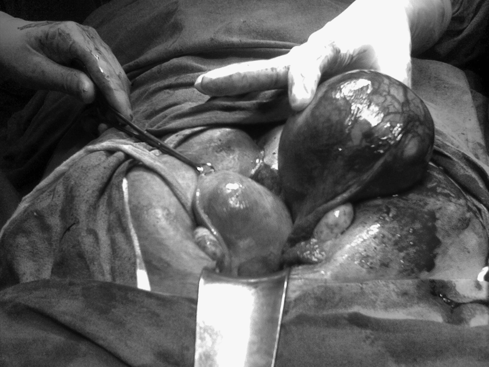
Operative photograph showing normal uterus and rudimentary horn.
Case 3
A 32-year-old woman, (gravida 4 para 3 abortus 0) was admitted in hypovolumic shock with a history of amenorrhea of 4 months' duration. She had had three full term normal deliveries. On examination she was extremely pale with a pulse rate of 100 beats per minute and systolic blood pressure of 70 mm Hg. Her Hb was 4 g%. There was abdominal distention with tenderness and guarding. Bimanual examination revealed fullness in all fornices. On paracentesis, unclotted blood was present. Along with resuscitative measures laparotomy was performed. There was hemoperitoneum of ∼1.5 L and the fetus was lying in the peritoneal cavity. The right noncommunicating horn ruptured at fundus with the placenta partially attached to it. Excision of the ruptured horn with right-sided salpingectomy was performed. Three units of fresh blood and one unit of fresh frozen plasma were transfused. The patient made an uneventful recovery and was discharged on the 8th day.
Case 4
A 25-year-old woman (gravida 2 para 1 abortus 0) reported with a history of amenorrhea of 4 months' duration and pain in abdomen of 3 days' duration. On examination her vital signs were stable. Her Hb was 9 g%. On abdominal examination there was cystic mass corresponding to 16 weeks' gestation more toward the right side. On bimanual examination, the same mass was felt on the right side and a bulky uterus was felt separately. USG confirmed that a live fetus corresponding to 16 ± 1 weeks' maturity was lying in the rudimentary horn and that the uterus was empty. On laparotomy, there was a unicornuate uterus with the right horn gravid and rudimentary, and attached to the main body of the uterus by a fibromuscular band. Excision of the rudimentary horn was performed with the fetus in situ along with right-sided salpingectomy. The patient's postoperative period was uneventful.
Discussion
A unicornuate uterus with rudimentary horn is a rare Mullerian abnormality. This uterine anomaly may cause many gynecologic and obstetric complications, including infertility, recurrent abortions, preterm deliveries, and rupture of the uterus. The incidence of uterine malformation has been quoted as 1:1500-2000. 3 Approximately 90% of rudimentary horns are noncomunicating and the pregnancy is presumed to occur by transperitoneal migration of sperm or fertilized ovum from the opposite side. 4 The attachment of the rudimentary horn to the main uterus varies from a fibromuscular band to an extensive fusion between two horns. 5 In the present series, pregnancy was in a noncommunicating horn and the rudimentary horn was attached to the uterus by a fibromuscular band in all the 4 cases. In a recent review, it was found that 85% of pregnancies occupied a noncommunicating horn, 30% of gestations progressed to term or beyond, 50% of pregnant uterine horns ruptured, with 80% of these events occurring before the third trimester. 6
The timing of rupture may vary from 5 to 35 weeks depending upon the thickness of the myometrium. In some cases, pregnancy may terminate in missed abortion or intrauterine fetal death caused by a decrease in blood supply and a defective endometrium as seen in Case. 1 of this series. Rupture is associated with catastrophe, and 80%–90% of the ruptures occur in second trimester. 5 There have been case reports of fetal survival in late rupture of rudimentary horn pregnancies. In one case, surviving twins were born 8 days apart. 2
Early diagnosis is of the utmost importance. Some cases were diagnosed only after an attempt to terminate the wrongly diagnosed intrauterine pregnancy, as happened in Case 1 of this series. A careful bimanual palpation of a mass extending outside the uterine angle (Baart de la Faille's sign) or displacement of the fundus to the contralateral side with rotation of the uterus and elevation of the affected horn (Ruge Simon Syndrome) and deviated uterus with palpable contralateral pelvic adnexa in the first trimester should arouse suspicion of uterine anomaly.7,8
Ultrasonographic criteria for diagnosis of RHP suggested by Tsafrir et al. includes gestational sac surrounded by myometrium by the side of a normal empty uterus, a pseudo-pattern of an asymmetrical bicornuate uterus, and noncommunication of the gestational sac with the endometrial cavity and the cervix. Additionally hypervascularization typical of placenta accreta on color flow Doppler may support the diagnosis of RHP. 9 Ectopic pregnancy in the tube and interstitial pregnancy can be mistaken for RHP. A tubal pregnancy will not show a ring of myometrium surrounding the gestational sac. An ectopic pregnancy in the rudimentary horn or an interstitial pregnancy may be confirmed by the eccentric location of the uterine sac and failure to demonstrate the sac in longitudinal section during USG. 10 In a review of 266 rudimentary horn presentations (210 gynecologic and 156 obstetric), the sensitivity of USG was 26% and decreased with advancing pregnancy age. 11 Magnetic resonance imaging (MRI) has proven to be a useful, noninvasive tool for the diagnosis of Müllerian abnormalities.12,13 If USG remains inconclusive, use of MRI has been suggested. Both clinically and radiologically, the diagnosis is more accurate in the first and early second trimesters.
In the past, the majority of cases were diagnosed after rupture of the rudimentary horn. However, with the advent of investigative methods such as ultrasound scan, MRI, and laparoscopy, the diagnosis is more often being made before rupture. In the present series, 2 cases were diagnosed preoperatively, whereas in the others diagnosis was confirmed at the time of laparotomy. Even previous normal deliveries do not exclude the possibility of an RHP. In the present series, out of 4 patients, 3 had had normal deliveries in the past.
The traditional treatment of RHP is laparotomy and excision of the rudimentary horn along with its tube, to prevent future tubal ectopic pregnancy. At the time of operation, the key to the diagnosis is the position of round ligaments on the lateral side of the gestation tumor. Several case reports describe a successful laparoscopic approach to RHPs diagnosed in the first trimester. Laparoscopy is the most accurate diagnostic tool that carries significant advantages in effective surgical management, thereby avoiding laparotomy. 14 At Sharma Post Graduate Institute of Medical Sciences, cases of RHP are being managed by laparotomy at the present time. However such cases can be considered for laparoscopy in future especially in thermodynamically stable patients. In all cases of RHP, evaluation of renal system is indicated because of the high incidence of associated urologic anomalies.
Conclusions
Every effort should be made to diagnose this condition before pregnancy occurs, or at least before rupture occurs, and the rudimentary horn should be excised. Patients with failure of induction should be investigated with a high index of suspicion.
Footnotes
Disclosure Statement
No competing financial conflicts exist.