
Abstract
Abstract
Background:
An unusual presentation of urethral diverticulum (UD) complicating pregnancy is presented. The diagnosis and management are challenging. UD during pregnancy should be managed conservatively.
Case Report:
A 28-year-old, gravida 2, during her 31st gestational week, presented with a painful mass in her vagina of 1 month's duration. She experienced a progressive worsening of pain and difficulty with urination. A digital cervical examination revealed the presence of an anterior vaginal cyst compatible with UD with an infectious complication during her pregnancy. This cyst was drained and the patient was prescribed antibiotic therapy with a second-generation cephalosporin for 7 days.
Results:
After drainage of the diverticulum, the patient had no more symptoms. She had good recovery of her urinary function immediately after the procedure. Her urine culture was negative for infection. Reevaluation after 1week showed that this patient remained asymptomatic and had no complaints. A genital examination did not reveal any abnormalities.
Conclusions:
Physical examination and early identification of UD during pregnancy can enable appropriate treatment.
Introduction
Urethral Diverticulum (UD) affects from 0.6 to 6.0% of women. A clinical examination is the first step in the diagnosis. UD frequently causes persistent urinary symptoms and might be associated with complications, such as lithiasis infection and malignant transformation.1–4 The diagnosis and management are challenging because of the rare nature of this condition, its varied presentations and differential diagnoses, and the possibility of misdiagnosis.
UD during pregnancy should be managed conservatively with expectant management, antibiotics, aspiration, and incision and drainage if necessary.
Case
A gravida 2 patient, in her 31st gestational week, presented with a painful mass in her vagina for 1 month's durations, with a progressive worsening of pain and with difficulty urinating. She did not have any fever. A clinical examination showed that her vital signs were normal: she was lucid and oriented; and had an arterial pressure of 110 × 70 mmHg, a respiratory rate of 24 breaths per minute, and a heart rate of 96 beats per minute. An examination of the abdomen revealed that the uterine fundus was palpable 8 cm from the umbilical mark and the fetal heart beats were 148 beats per minute, with good perception of fetal movement. Gynecologic examination revealed externalization of the anterior vaginal mucous membrane, below the middle urethra, in the form of a ball with a maximum diameter of 3.0–3.5 cm—in a 2-cm (+2) relation to the remaining hymenal meatus—which was painful upon palpation. (Image 1 from Fig. 1). A moderate amount of purulent material came out of that area. Her urine was clear. The clinical diagnosis was compatible with UD with an infectious complication during this pregnancy. The UD was drained (Image 2 from Fig. 1). She then received antibiotic therapy with a second-generation cephalosporin for 7 days.
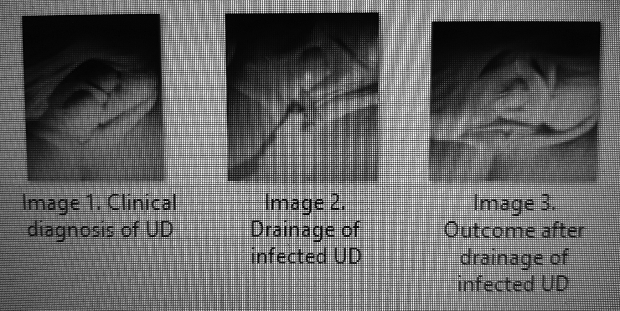
Image enabling clinical diagnosis, drainage and outcome after drainage of urethral diverticulum.
Results
This patient had no more symptoms after UD drainage (Image 3 from Fig. 1). She had good recovery of her urinary function immediately after the procedure. Her urine culture was negative for any infection. A 1-week follow up revealed that she remained asymptomatic and had no complaints. A genital examination did not show any abnormalities.
Discussion
Clinical relevance
“The diagnosis of the female Urethral Diverticulum is directly related to the surgeon's desire to discover it.”—Moore (1952) 5
The clinical relevance of this case refers to the importance of anamnesis and physical examination during the pregnancy–puerperal cycle, as a fundamental step for early diagnosis of morbidities, such as infected female UD. The technology for complementary tests helps the diagnosis; however, a clinical examination in obstetrics is a fundamental step in the context of both the health of the mother during childbirth and the health of the child.
In addition, the good technique of a simple gynecologic procedure, such as drainage of a urethral abscess during pregnancy, can prevent the progression of infection.
Etiology, diagnosis, treatment, and follow-up
The etiology of UD remains uncertain, although most are felt to be acquired rather than being congenital. The most-commonly accepted theory suggests that a UD originates from an obstructed periurethral gland that becomes infected, forming an abscess. The abscess eventually ruptures into the urethral lumen, forming an ostium. This process may be repetitive, with reinfection and reobstruction. 6 Other possible etiologies include obstetrical trauma and iatrogenic injury from urethral instrumentation, periurethral bulking agents, or midurethral slings. 7
For an asymptomatic women, the condition can be managed conservatively, whereas treatment for symptomatic women usually involves a diverticulectomy. Potential complications from diverticulectomy include diverticulum recurrence, stress incontinence, urethrovaginal fistulae, urethral stricture, and recurrent urinary-tract infections. 7 The diagnosis and management are challenging because of the rare nature of this condition, its varied presentations and differential diagnoses, and the possibility of misdiagnosis. 8
A clinical examination is the first step in making a diagnosis. Any patient presenting with genitourinary complaints or symptoms of a vaginal mass should undergo a thorough physical examination with careful inspection and palpation of the anterior vaginal wall. Visualization of the mid and distal anterior vaginal wall is facilitated by performing a split-speculum examination. Approximately half of patients with UD present with a palpable anterior-wall mass. 9
Early identification of UD during pregnancy may enable treatment and possibly labor with a vaginal delivery. Yet, there are situations in which a delayed diagnosis may affect the mode of delivery, resulting in a need for a cesarean section. 8
UD during pregnancy should be managed conservatively with expectant management, antibiotics, aspiration, and incision and drainage if necessary. 8 Most patients with UD present with vague urinary symptoms or pelvic pain that have often been refractory to previous treatments, at times leading to delays in diagnosis. 8
In a series of cases—published a few years ago—however, that were diagnosed and managed adequately, according to current recommendations, the clinical presentation of urethral diverticulum during pregnancy was a paraurethral mass, urinary incontinence, irritative symptoms, urinary-tract infection, urethral pain and discharge, and voiding difficulty. The diagnosis of UD during pregnancy was made by transvaginal ultrasonography, and cystoscopy; after pregnancy, the diagnosis was made using a voiding cystourethrogram. Management during pregnancy involved antibiotics, diverticulum aspiration, and incision and drainage. 10
Surgical excision of the UD should be avoided during pregnancy, as there is a greater risk of hemorrhage (resulting from venous congestion) and the subsequent trauma of a vaginal delivery confers risks of wound breakdown, infection, and possible development of a urethrovaginal fistula. 10
Postnatal follow-up is important. In women with persisting symptoms, elective transvaginal excision of the UD with layered closure should be performed. Small and asymptomatic UD can be managed conservatively with annual follow-up examinations. In older women, the association with urethral carcinoma is a good reason to perform a diverticulectomy, even if the symptomatology is minimal. 10
Differential diagnosis
Suburethral and periurethral masses may be of benign or malignant etiologies. Examples of benign masses that can seem similar to UD include vaginal-wall inclusion cysts, Gartner-duct cysts, Skene-gland cysts, urethral caruncles, urethral prolapse, anterior vaginal-wall prolapse (cystocele or urethrocele), ectopic ureteroceles, vaginal leiomyomas, granulomas, and hamartomas. There have also been case reports of endometriosis presenting as a periurethral mass. Malignant lesions, such as urethral carcinoma, vaginal carcinoma, and vaginal leiomyosarcoma, might also mimic UD. The correct diagnosis can be confirmed by vaginal examination, imaging, biopsy, or surgical excision. 7
Conclusions
This case report demonstrated that anamnesis, local examination and drainage of the purulent material may be the first approach in pregnant women with infected urethral cysts. It is important that the physician be invested in the physical examination and recognizes an infected urethral cyst. Drainage, associated with antibiotic therapy, may be a good option to prevent the progression of infection during pregnancy.
Footnotes
Acknowledgments
The authors would especially like to thank the pregnant woman who kindly agreed to share her case.
Author Disclosure Statement
No financial conflicts of interest exist.
Funding Information
No funding or cost-sharing of the project has come from any company or educational institution.