
Abstract
Abstract
Bozzini, Clarisa, Emilio O. Picasso, Graciela M. Champin, Rosa Maria Alippi, and Carlos E. Bozzini. Structural and material mechanical quality of femoral shafts in rats exposed to simulated high altitude from infancy to adulthood. High Alt Med Biol. 17:50–53, 2016.—The growth of the body and bone mass and the mechanical properties of appendicular bone are impaired in immature rats exposed to different simulated high altitudes (SHA) (1850–5450 m) between the 32nd and the 74th days of postnatal life. Now, we report the effects of exposure to 4100 m on the above cited variables in female rats from infancy (age: 1 month) to adulthood (age: 8 months) to define the occurrence of catch up and to establish whether the effects of altitude are transient or permanent. The ex vivo right femur was mechanically tested in three-point bending. Body weight and length, and structural (loads at yielding and fracture, and stiffness) and architectural (diaphyseal cross-sectional area, cortical area, and cross-sectional moment of inertia) properties were measured at 2, 4, 6, and 8 months of exposure to SHA. The negative influence of hypoxia on all variables was similar at different ages or, in other words, the difference among ages was maintained at any extent of hypoxia. Hypoxia did not affect the elastic modulus, thus suggesting that the mechanical properties of the bone tissue were maintained. Catch up did not occur. The resulting osteopenic bone remained appropriate to its mechanical function during the entire exposure to SHA.
Introduction
U
The effect of hypoxia on body mass growth rate has been associated with hypophagia because of reduced appetite (Alippi et al., 1983; Elia et al., 1985; Westerterp-Plantenga et al., 1999). It has been suggested (Bozzini et al., 2005) that body mass growth retardation during exposure to SHA could be due to the reset of a hypothetical body mass set point. A reduced body mass may be adaptive to high altitude by reducing oxygen needs (Mortola, 1993).
Undernourished rats induced by voluntary subnormal food intake during exposure to SHA (Bozzini et al., 2013) show important changes in the mechanical properties (biological responses to loads) of the appendicular skeleton. In studying the mechanical behavior of bone, it is important to distinguish between the mechanical properties of the bone as a structure (structural behavior of the whole bone) and of the bone tissue itself (material behavior). The material behavior of a specimen is independent of its geometry and reflects the intrinsic properties of the material. In contrast, the structural behavior of a specimen reflects both the morphology and the material properties of the specimen.
Mechanical tests performed on femur diaphyseal shafts obtained from rats in the growth phase exposed to SHA revealed the development of a lower than normal bone mass and changes in the architectural properties of the bone, without alterations in the material properties of the bone tissue (Bozzini et al., 2013). These alterations were directly related to the level of the exposed altitude when exposure was extended between the 32nd and the 74th day of postnatal life.
The purpose of the present investigation was thus directed to explore the behavior of both body mass and bone mechanical properties during a prolonged stay at SHA, from infancy (age: 1 months) to adulthood (age: 8 months). The study could thus define the presence of catch up in one or both parameters during the latter phase of the exposure period and define whether the effects of altitude on body mass and bone mechanical quality are transient or permanent.
Materials and Methods
Animals and experimental design
Female Sprague-Dawley rats aged 30 days and weighing 91.86 ± 9.86 g were used as experimental subjects. They were housed in stainless steel, wire-bottomed cages under a natural light–dark in a temperature-controlled (22–24°C) room. The rats were divided into two groups of 28 animals each, normoxic (NX) and hypoxic (HX). Hypobaric hypoxia was induced by placing HX animals into simulated altitude chambers. Exposure was chronic (minimum 22–23 h/day) with a daily interruption to replace food and water, clean animal cages, and perform experimental maneuvers when necessary. During weekends, exposure was continuous.
HX rats were exposed to an air pressure of 456 mmHg, which corresponds to 4100 m of simulated altitude. NX rats were maintained at 760 mmHg (0 m). Groups of seven HX and seven NX rats were euthanized at 2, 4, 6, and 8 months of the experimental period. Body mass was then recorded. The femurs were dissected and cleaned of adhering soft tissue, weighed in a Mettler scale, and stored at −20°C wrapped in gauze soaked with Ringer's solution in sealed plastic bags, in accordance with Turner and Burr (1993).
Biomechanical testing
On the day of testing, each bone was thawed at room temperature before analysis. To assess cortical bone mechanical properties, the right femur was tested in three-point bending (Turner and Burr, 1993; Hogan et al., 1999), which combines compression and tension.
Each bone was placed horizontally with the anterior side facing down on two transverse supports and central along its length. Load was applied perpendicularly to the long axis of the bone until fracture. The test machine (Instron Model 4442; Instron Corp., Canton, MA) was operated in stroke control at a constant rate of 5 mm/min to obtain the load (W)/deformation (d) curves, showing both the elastic (linear) and the plastic phases separated by the yielding point (departure from linearity). The test enables graphic determination of the main structural mechanical properties of bone shafts as beams (Turner and Burr, 1993) which essentially measures the resistance to both deformation (stiffness) and fracture (strength).
Micromorphometrical determination
Micromorphometrical determination of the horizontal and vertical, external (H, B) and internal (h, b) diameters of the elliptic-crown-shaped fracture sections enabled calculations of the bone diaphyseal geometrical properties. Measurements were taken with a digital caliper with the aid of a magnifier 40×.
The geometric properties were determined as follows: [a] Bone length and diameters: the femur length was measured directly using a stereomicroscope (Stemi DV4 Stereo Microscope; Carl Zeiss MicroImaging, Gottingen, Germany) with an accuracy of ±100 μm; [b] Mid-diaphyseal cross-sectional area (CSA): CSA was calculated by applying the equation “π (HB − hb)/4.” Second moment of inertia of cortical bone (with reference to the anterior–posterior bending axis, xCSMI) was estimated by the equation “π (B3H − b3h)/64.”
Bone material properties (elastic modulus) was calculated from structural and geometric properties and not directly determined by mechanical means: “WyL3/48dy.Ix” (dy = maximal elastic deflection, L = distance between supports, Ix = xCSMI, Wy = load at the yielding point).
Ashing of the specimens
The left femur of each animal was ashed at 600°C in a muffle furnace for 18 hours and the ash weight obtained. The tissue degree of mineralization (α), which expresses the percentage of mineral substance in the dried bone, was calculated as the ratio ash weight/dry bone weight.
Statistical analysis
Results are summarized as mean ± standard error of mean (SEM) and considered statistically significant at the level of p < 0.05. Comparisons between parameters were performed by one-way analysis of variance (ANOVA) and the Student–Newman–Keuls test by using GraphPad Software (GraphPad Software, Inc., San Diego, CA). Multivariate analysis of variance (MANOVA) was used to evaluate the consolidated effect of the material bone properties responding to age and altitude. Simple regression analysis tested the relationship between bone mass and body mass.
Results and Discussion
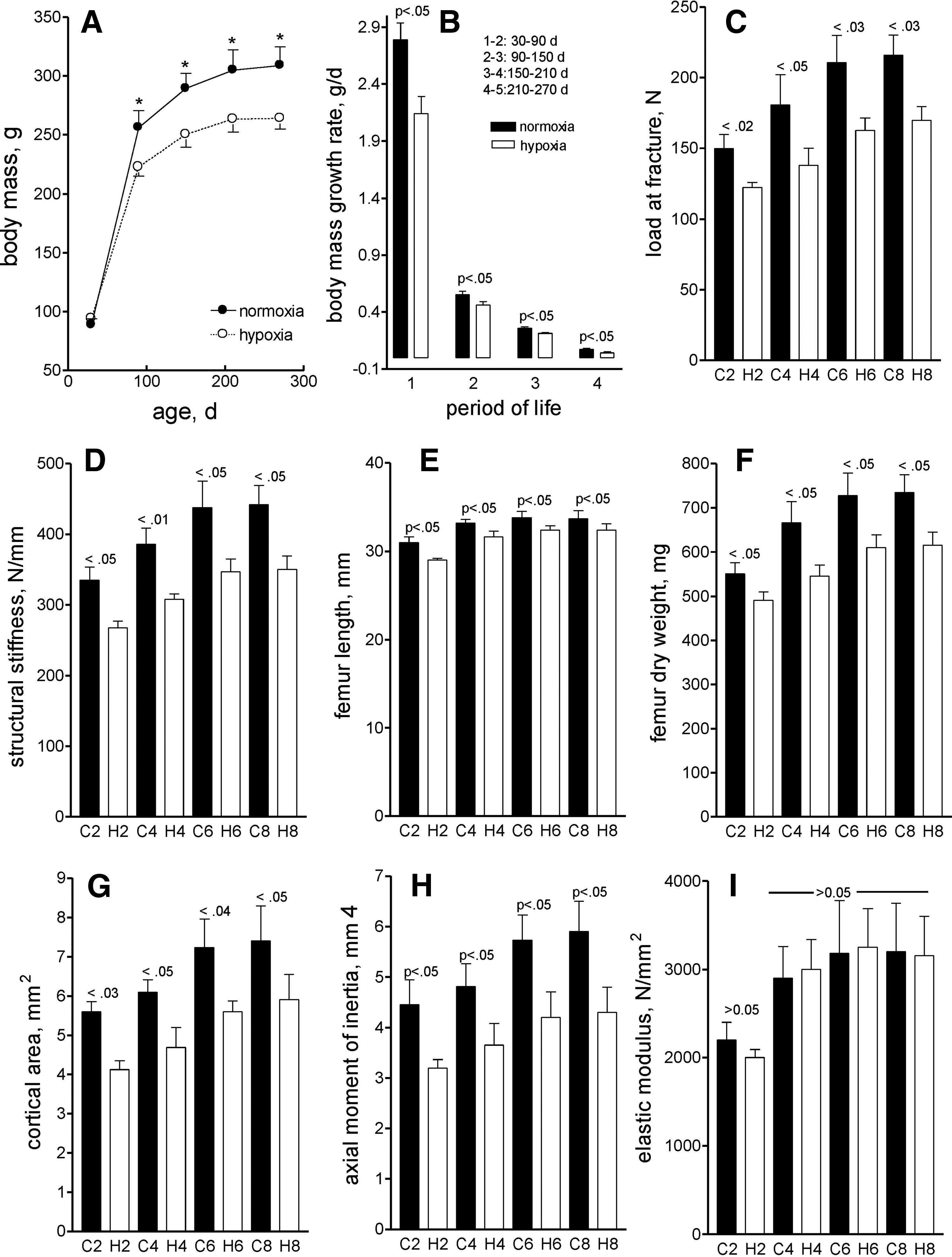
Pre-experimental body mass did not differ between groups. As expected, the body mass growth rate was negatively affected in rats exposed to SHA (Fig. 1A). While control rats grew at a rate of 2.79 ± 0.15 g/day between the 30th and 90th days of life (Fig. 1B), body mass weight gain was 2.14 ± 0.15 g/day in HX rats. Growth slopes decreased with time in both groups, being almost negligible after the age of 7 months. The negative influence of hypoxia on growth was similar at different ages or, in other words, the differences among ages were maintained at any extent of hypoxia. This behavior indicates that catch up growth did not occur in HX rats during the latter part of the experimental period. These data support and extend our previously reported study (Bozzini et al., 2013) performed in weaning rats that were exposed to different simulated altitudes (between 1850 and 5450 m) during 42 days.
In this study, the negative effect of hypoxia on body mass growth rate was directly related to the level of altitude. Therefore, body mass attained different values at the end of the exposure period. The results of the present experiment, in which rats were exposed to a simulated altitude of 4100 m, suggest that the subnormal body mass induced by hypoxia is sustained as long as the permanence of animals in the altitude is maintained, giving support to the concept of the existence of a “hypoxic body mass,” always lower that the normoxic one for gender and age.
The femoral functional integrity in both NX and HX rats was assessed in the present experiment by a structural strength test that measures how well the whole bone can bears loads. Both the structural strength, which represents the load required to fail the whole bone (Fig. 1C), and the structural stiffness, which is a measure of the resistance to deformation under the applied load (Fig. 1D), increased with time until an age of ∼6 months, remaining stable from this time on. Despite the growth and development in bone properties, the hypoxic rat bone properties were always compromised as shown in figures.
The changes of structural strength of the femur were paralleled to those described for the body mass. A high positive correlation (r = 0.98, p < 0.001) was found between the load at fracture and the body mass when all animals were tested together. The high coefficient of determination (r2 = 0.96) of this association suggests that body mass should be regarded as a first-order factor in the determination of bone structural quality. The negative influence of hypoxia on this variable was maintained at any extent of hypoxia.
The weakening of the femoral shaft induced by hypoxia was accompanied by the negative effects on femoral length and weight (Fig. 1E, F), the cortical area (Fig. 1G), and xCSMI (Fig. 1H), which indicate that the bone mass and the mid-diaphysis cross sections were significantly affected. The elastic modulus (Fig. 1I), a good indicator of the bone tissue stiffness, indicates that the material bone quality was not affected by hypoxia, probably because the degree of mineralization of the tissue remained unaltered.
Growing rats exposed to SHA probably reach an adequate degree of acclimation by reducing their body mass and, consequently, the oxygen needs. This type of acclimation does not apparently change with time of exposure because the body mass, as shown in this study, does not catch up and the animals reach adulthood with a lower than normal body mass (hypoxic body mass) for age and gender. The material quality of the bone tissue, as least in the appendicular bone, remains unchanged during the hypoxic exposure. The structural quality of the bone, however, is reduced when the hypoxic bone is compared to the normoxic one, thus making the bone less stiff and less resistant to fracture. Changes at the level of the geometric or architectural properties of the hypoxic bone, expressed by reduced cross-sectional area, cross-sectional cortical bone mass, and xCSMI, should be the main responsible factors for its less weak condition.
The high positive correlation found between bone strength and both body mass and cortical bone mass should indicate that the architectural changes of the bone might represent a physiological adaptation to the subnormal body mass. It is conceivable that the reduced loads applied to the weight-bearing bones because of the reduced body mass should negatively influence bone modeling, thus reducing bone mass and creating an osteopenic bone, although biomechanically adapted to function. In summary, it is suggested that long-term exposure to SHA negatively influences the structural quality of the femoral shaft without altering its material quality; however, the resulting bone appears to remain appropriate to its mechanical function.
Footnotes
Acknowledgments
This investigation was supported by Research Grants from the National Research Council (CONICET, PIP11220130100479CO) and the University of Buenos Aires (UBACYT, PID 200201130100126). C.E.B. and R.M.A. are Career Investigators from CONICET.
Author Disclosure Statement
The authors do not have any institutional or commercial affiliations that might pose a conflict of interest regarding the publication of this article.