
Abstract
Internalization of monoclonal antibodies (MAbs) binding to the targeted cells has drawn great attention to both scientists and anti-cancer drug developmental professionals. Internalization of conjugated MAb is thought to be one of the major mechanisms for tumor cell destruction, and can be studied using several biochemical and microscopic approaches. Here we report a new method based on papain digestion followed by flow cytometry (FCM). This method can identify whether the binding MAb has internalized into the cell, with an additional advantage of accurately quantifying the internalized MAb without altering cell morphology after papain digestion. With this method, we studied the internalization degrees of 3A4 (a mouse anti-human CD45RA MAb) at different time points: 5.3% (15 min), 7.3% (30 min), 36.9% (60 min), 69.2% (120 min), and 72.6% (180 min). This methodology can facilitate our understanding of the efficiency of MAb internalization and allows us to evaluate the targeted killing capacity of the MAb. Our technique can serve as a reference model for future targeted drug development using MAbs. In summary, we established a simple and useful evaluation tool for MAb drug development and research.
Introduction
Internalization can be studied using several biochemical and microscopic approaches.(7–9) Fluorescence microscopy(8) and confocal microscopy(10) are key methods, amongst them, providing distinct images of the internalized process. Investigators can justify whether the binding MAbs have internalized into cells by localizing the fluorescent signals.(9) However, they are qualitative analytical methods, and subsequently it is impossible to determine the degree of internalization. Radio-labeled binding is also used to observe the degree of internalization,(11) but it includes the disadvantage of adopting radioactivity exposure. Additionally, the use of radioisotopes is highly regulated and the waste generated can pose a disposal problem.(12) Pronase(13) or protease K(14) are able to digest the cell surface-bound protein and have been applied in internalization studies. However, both are non-specific hydrolytic enzymes and have the potential of resulting in cell damage or even cell death. It is therefore difficult to achieve an accurately quantitative determination of the internalized MAbs.
Papain is a cysteine protease hydrolase enzyme present in papaya. In immunology, papain is known to cleave the Fc portion of immunoglobulins (antibodies) from the Fab portion.(15) Fab fragments which are isolated from papain digests of monoclonal antibodies have a wide variety of uses in analytical and in in vivo and in vitro diagnostic applications.(16)
The antigen-binding site of an antibody is located in the Fab portion; however fluorochrome is conjugated through the Fc portion.(17) Fluorochrome will be cleaved with the Fc portion if the MAb is present on the cell surface following papain digestion. The fluorescence will remain and can be detected by FCM or fluorescence microscopy if the MAb has been internalized into the cells (Fig. 1).
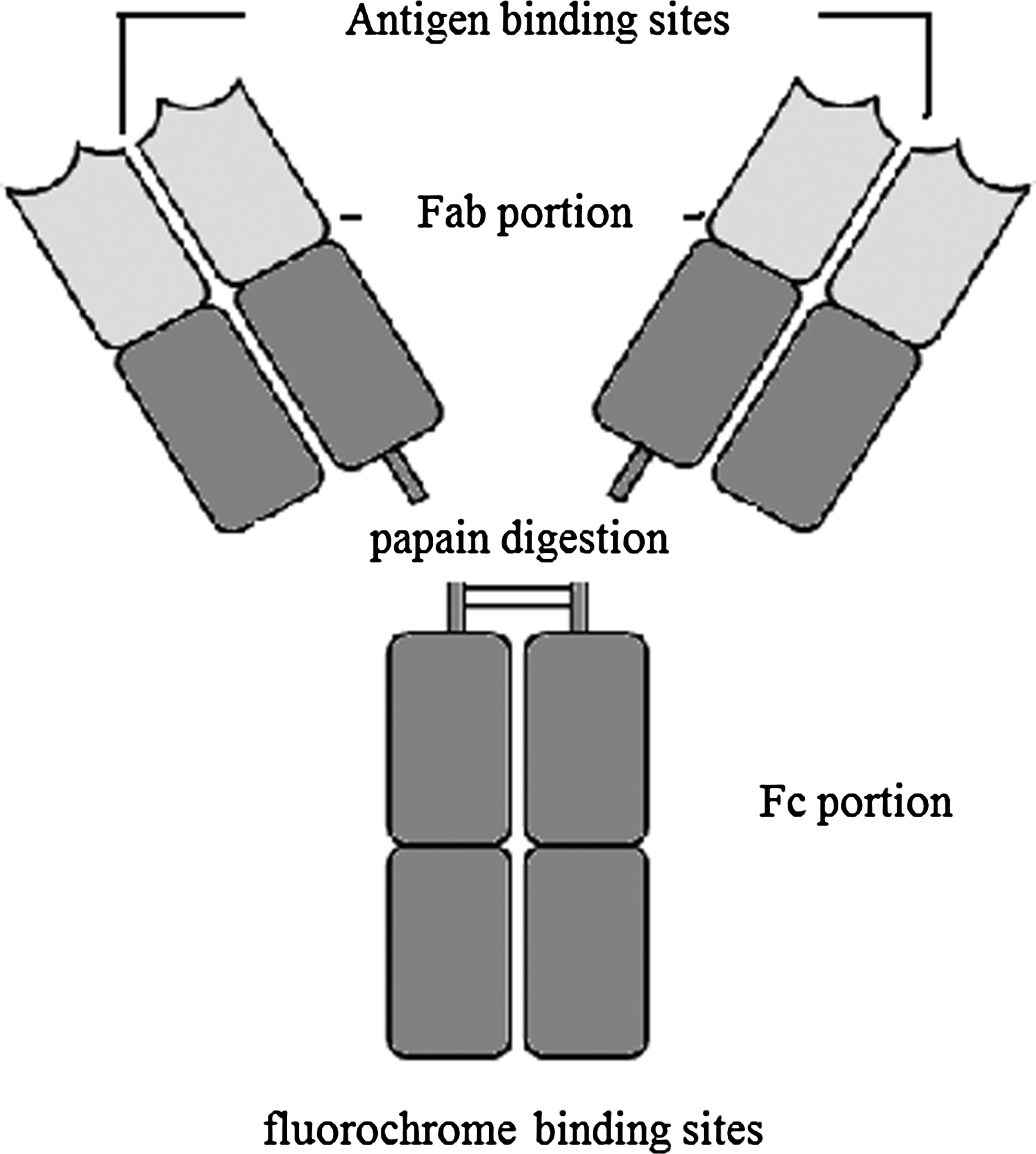
Model of papain-digested antibody.
In this paper, we have established a new method to quantitatively measure the internalization degree of the binding MAb on the cell surface based on papain digestion followed by FCM analysis.
Materials and Methods
Cell culture
KG1a (human bone marrow acute myelogenous leukemia cell line; ATCC, Manassas, MD) cell line was cultured in Iscoves modified Dulbecco medium (IMDM, Invitrogen, Carlsbad, CA) supplemented with 10% fetal bovine serum (FBS, Sijiqing, Hangzhou, China) at 37°C, 5% CO2, in a humidified incubator.
Preparation of FITC conjugated 3A4 (3A4-FITC)
ZCH (Zhejiang Children's Hospital)-6-3A4 (3A4) is a new monoclonal antibody against the human CD45RA antigen generated in our laboratory. It was classified into the CD45RA category at the Seventh International Workshop and Conference on Human Leukocyte Differentiation Antigens (HLDA7) in 2000.(18) 3A4 hybridoma cells were injected into the peritoneal cavity of BALB/C mice (Laboratory Animal Center, Zhejiang University, HanghZhou, China) primed with sterile paraffin fluid for the preparation of 3A4 ascites. 3A4 MAb was purified from ascites by high performance protein G sepharose affinity column (Invitrogen). 3A4-FITC was prepared by the modified Marshall method.(17)
Establishment of method for determining antibody internalization using papain digestion and FCM analysis
Regular method
KG1a cell line was used to evaluate the internalization capabilities of 3A4. The target cells were harvested and adjusted into a concentration of 107/mL. In this process, cells were set with 100 μL of cell suspension for each tube (106/tube). KG1a and 3A4-FITC (1μg/mL) were incubated at either 37°C or 4°C for 30 min. Next, cells were gently washed twice with PBS to remove any excess antibody. Tubes at each temperature were treated with either papain (Roche, Basle, Switzerland) at a concentration of 2 mg/mL for 15 min at 37°C (papain group) or no agent (control group), respectively. The cells in the control group were fixed with 1% paraformaldehyde (PFA, Invitrogen) in PBS and analyzed by FCM (Becton Dickinson, Franklin, NJ) directly. The cells in the papain group continued to be incubated with papain for another 15 min at 37°C; then the cells were washed twice with PBS and fixed with 1% PFA before FCM analysis.
Method for identifying MAb internalization degrees at different time points
KG1a (106/tube) and 3A4-FITC (1 μg/mL) were incubated at either 37°C or 4°C for durations of 15, 30, 60, 120, and 180 min, respectively. Tubes at each temperature were treated with either papain for 15 min at 37°C (papain group) or no agent (control group), respectively. Next, the cells were gently washed twice with PBS to remove any excess antibody. The cells in control group were fixed with 1% PFA and analyzed by FCM directly. The cells in the papain group continued to be incubated with papain for another 15 min at 37°C; then the cells were washed twice with PBS and fixed with 1% PFA before FCM analysis. The outcomes of FCM analysis were validated by confocal microscopic scanning simultaneously.
Papain digestion and confocal microscopic scanning
In this process, eight tubes were set. The 106 KG1a cells were incubated with 1 μg of 3A4-FITC at either 37°C or 4°C with a 15 min and 60 min duration, respectively. After incubation, the cells were washed twice with PBS. Tubes at each temperature group were treated with either papain (papain group) or PBS (control group) for 15 min at 37°C, respectively. Cells of the two groups were fixed with 1% PFA for 15 min, followed by incubation with 100 μL permeabilizing solution (Becton Dickinson) for 10 min and addition of 5 μL of propidium iodide (PI, 50 μg/mL; Sigma, Saint Louis, MO) for another 5 min to stain the nucleic acid. Then the cells were analyzed by confocal microscopic scanning (LSM 510 Meta, Zeiss, Oberkochen, Germany).
Pronase digestion and FCM analysis
In order to compare the hydrolyzed function of papain and pronase, papain was replaced by pronase (Roche) at a concentration of 0.5 mg/mL in KG1a and 3A4-FITC. The steps described in the above procedure were then followed.
Data analysis
All results are expressed as the mean ± SE (standard error).
Result
Papain digestion and FCM analysis
Internalization at 37°C (condition permissive for internalization)
KG1a cells and 3A4-FITC were incubated at 37°C for 15, 30, 60, 120, and 180 min, respectively. The positive cell percentages and the mean fluorescence intensity (MFI) among control tubes were similar, with a mean level of (99.79 ± 0.09)% and 119.08 ± 6.39. In the papain group, the positive cell percentages and MFI increased as the incubation time was prolonged. The degrees of internalization at different time points were 5.3% (15 min), 7.3% (30 min), 36.9% (60 min), 69.2% (120 min), and 72.6% (180 min), respectively (Fig. 2).
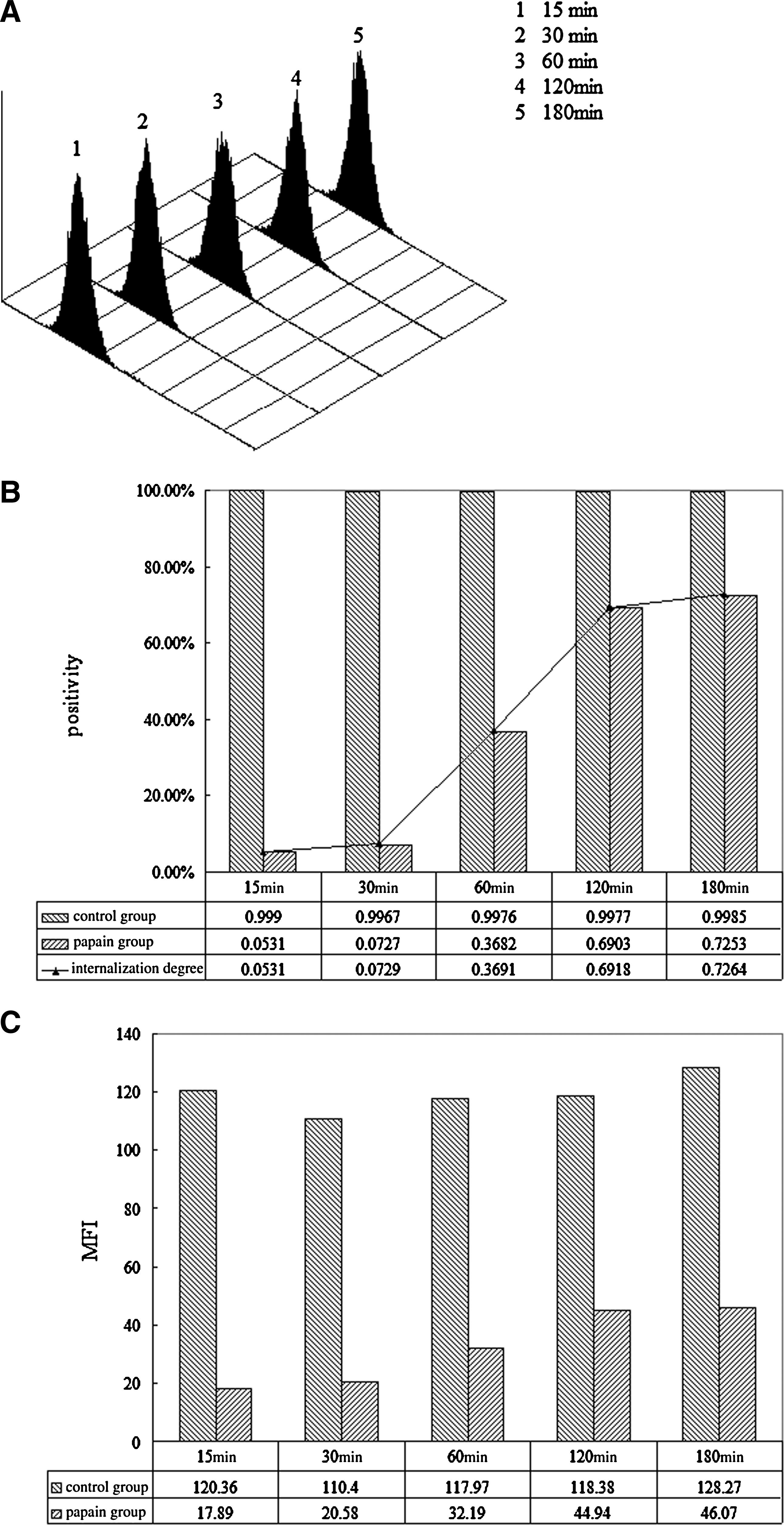
Comparison of positive cell percentages and MFI between control group and papain group incubated at 37°C at different time points. (
Cells incubated at 4°C (condition that prevents internalization) for different time points
106 KG1a cells and 1 μg of 3A4-FITC were incubated at 4°C for 15, 30, 60, 120, and 180 min, respectively. The positive cell percentage (1.42 ± 0.11%) and the MFI (11.05 ± 0.42) among the tubes in the papain group were significantly reduced compared with those in the control group (99.82 ± 0.08%) and (104.37 ± 2.52), respectively (Fig. 3).
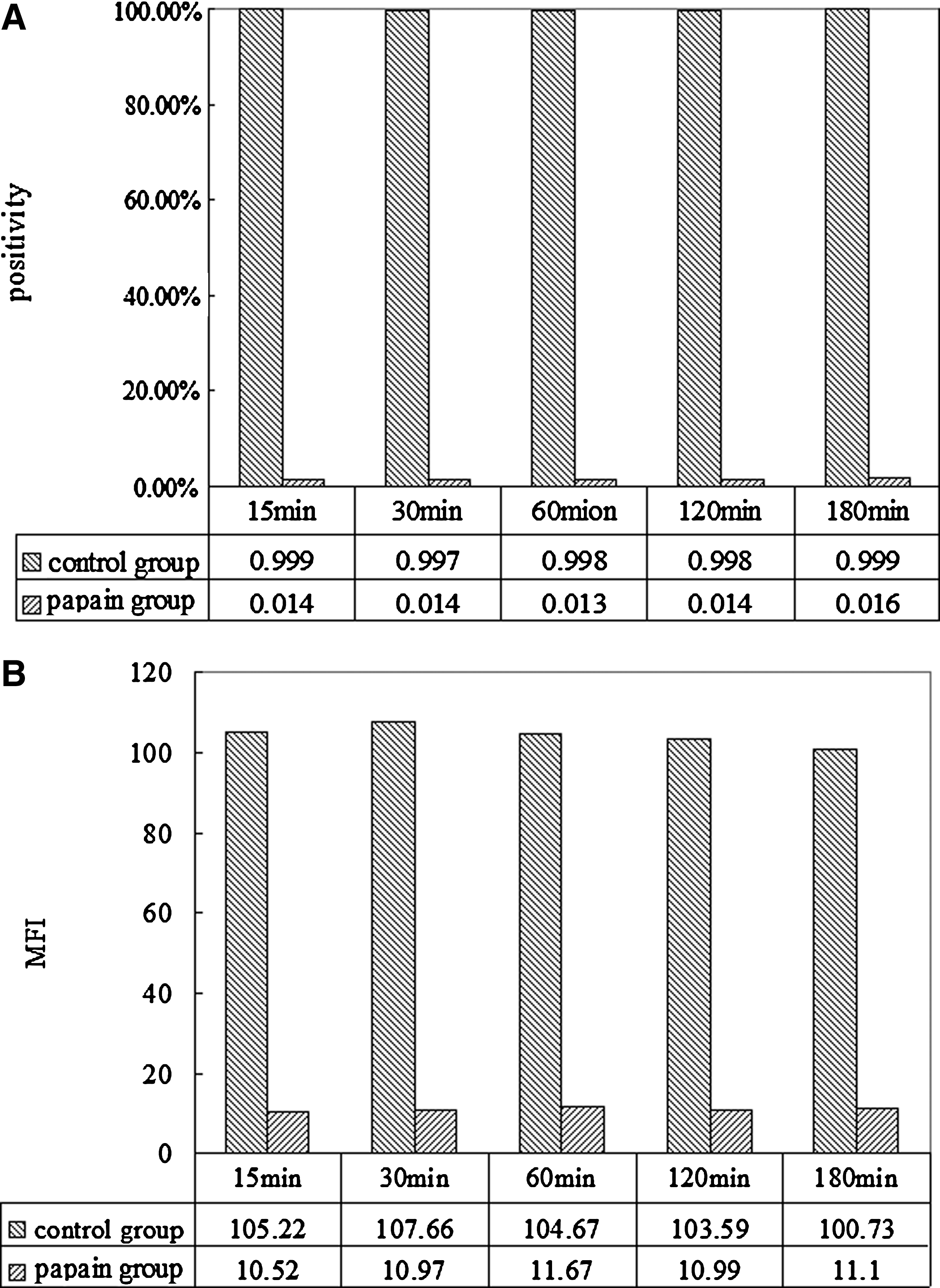
Comparison of positive cell percentages and MFI between control and papain groups incubated at 4°C for different time points. (
Pronase digestion and FCM analysis
FSC (forward scattered) and SSC (side scattered) are important indices for evaluating the cell morphology. From analyzing the two parameters (FSC/SSC) using dot plots, the morphology of cells (Fig. 4A) changed significantly after pronase digestion (Fig. 4B). Conversely, the papain group was unchanged between the testing (Fig. 4C) and the control (Fig. 4D) groups. Analysis of histogram drew the same conclusion (Fig. 5).

Plots of cell morphology with and without treatment of pronase (
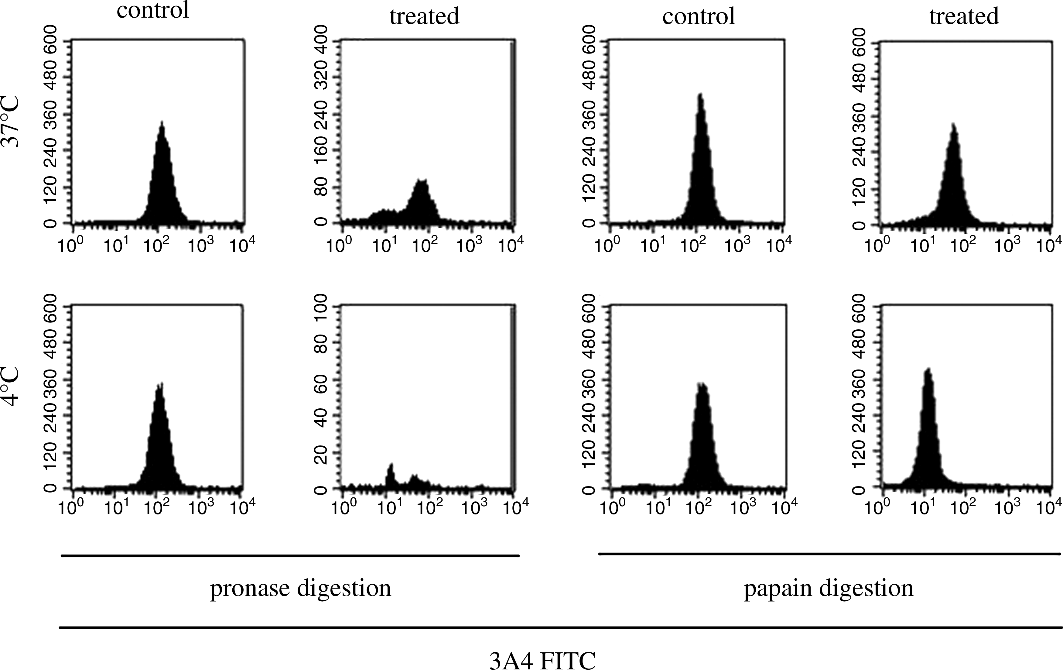
FCM analysis of cells incubated with 3A4 FITC at either 4°C or 37°C for 2 h. This indicated that both pronase and papain could cleave the fluorochrome-conjugated antibody on the cell surface but the former could severely damage the cell morphology.
Papain digestion and confocal microscopic scanning
A series of pictures were gained through confocal microscopic scanning, with the outcomes that were consistent to those demonstrated by FCM (Fig. 6). When incubation was conducted at 37°C, fluorescent signals decreased partially after papain digestion. However when incubation occurred at 4°C, fluorescent signals disappeared completely following papain digestion. Although confocal microscopic scanning could provide excellent images to determine the localization of the MAbs, it was unable to measure the fluorescent intensity and the percentage of the MAbs internalized.
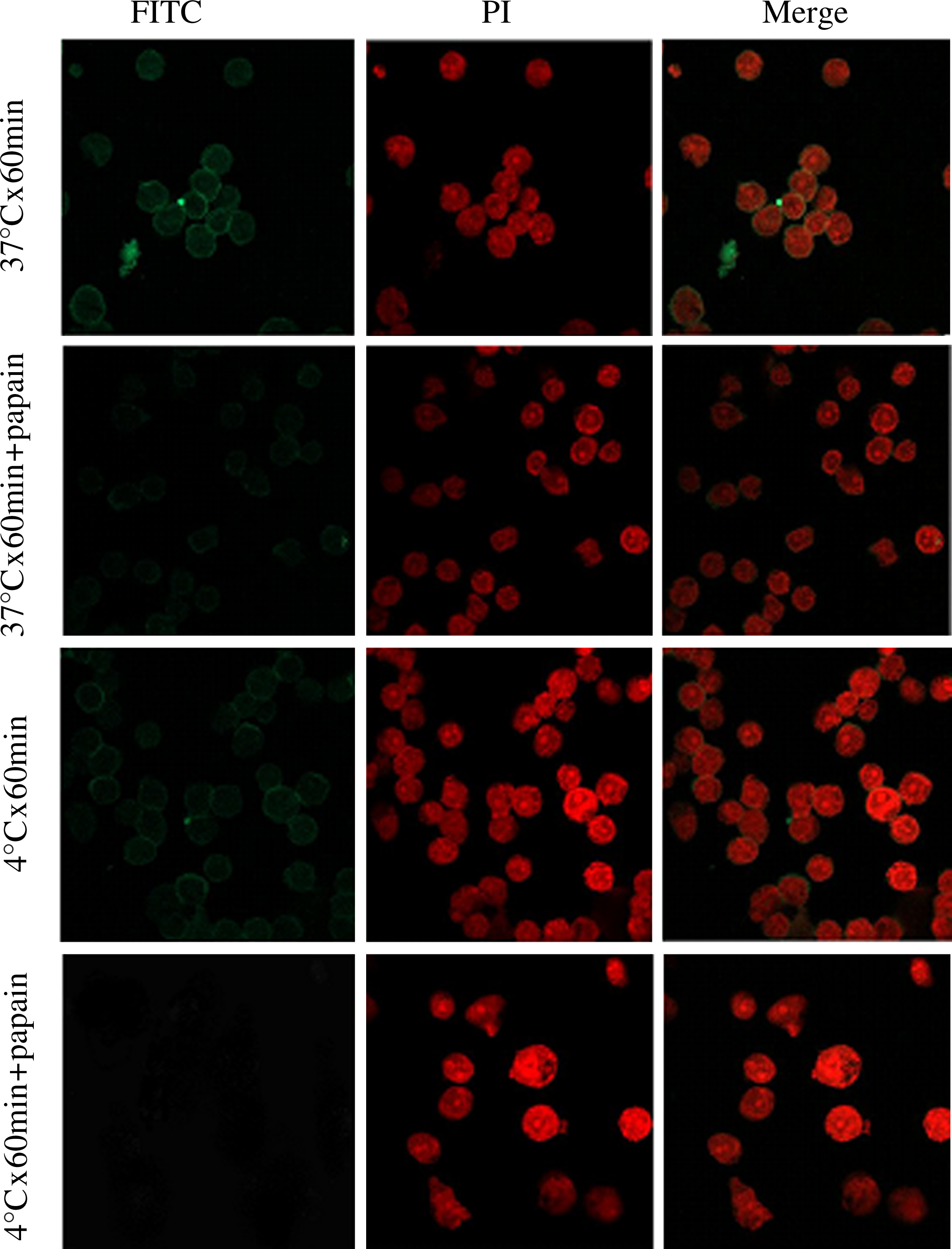
Analysis of 3A4 internalization by confocal microscopy. Internalization studies of 3A4 in KG1a cells were evaluated at either 37°C or 4°C for 60 min. When incubated at 37°C, the fluorescent intensity of the cells weakened partially in the papain group compared with that in the control group, indicating that the bound antibody partially internalized into the cells. While incubated at 4°C, fluorescent signals were absent in the papain group, indicating that the bound antibody did not internalize into the cells.
Discussion
Antigenic modulation includes shedding of antigen from the cell surface in response to interaction with antibody and internalization of Ag–Ab complexes. Each of these mechanisms will result in an apparent decreased antigen expression on the surface of the target cells, which might protect cells from destruction by antibody-dependent cell cytotoxicity (ADCC). However, internalization may provide an advantage for the therapeutic use of cytotoxic immunoconjugates.(19) Moreover, determining the internalization and the degree of MAb is required in the study of targeting drugs.
Pronase is a non-specific protease. The proteolytic activity it possesses extends to both denatured and native proteins, which are broken down into individual amino acids.(20) It has been applied in the fields of histochemistry and cell culture (detaching cells from culture dishes), glycobiology (analysis of glycoproteins),(21) and molecular genetics (extraction of phage DNA, isolation of plasmid DNA).(22) Pronase was used to remove the cell surface-bound MAb in the studies of internalization,(13) but has strong digestive actions that are likely to result in severe cell damage or even cell death.
Papain has a site-specific hydrolysation action on the Fab-Fc binding site of the antibody that yields three fragments after hydrolysis: two 50 kDa Fab fragments and one 50kDa Fc fragment. Papain has been widely used to digest Fab fragments from intact monoclonal antibodies. The purified Fab fragments offer a wide array of applications in both in vitro and in vivo diagnostic procedures. The mechanism by which it breaks peptide bonds involves deprotonation of Cys-25 by His-159. Asn-158 helps to orient the imidazole ring of His-159, allowing this deprotonation to take place. Cys-25 then performs a nucleophilic attack on the carbonyl carbon of a peptide backbone. This frees the amino terminal of the peptide, and forms a covalent acyl-enzyme intermediate. The enzyme is then deacylated by a water molecule, and the carboxy terminal portion of the peptide is released.(23)
Previous studies on internalization revealed that 37°C was the condition permissive for internalization and 4°C for the prevention of internalization. They also showed that the internalization was a time-dependent process,(5,6) which was consistent with our results. At 37°C, more MAbs were internalized into the cells with a longer incubation time in the first 2 h. In addition, our studies showed that the internalization was not significantly increased after 2 h of incubation, which were 72.6, 71.2, 52.9, and 46.1%, respectively, after 3, 4, 6, and 24 h of incubations. The positive cell percentages and MFI decreased slightly at 6 and 24 h, possibly due to the fluorescence quenching. At 4°C, all MAbs were bound to the cell surface; even when we prolonged the incubation time to 24 h no MAbs were further taken up into cells. We also used Raji (Human Burkitt's lymphoma cell line, ADCC) as the target cell line to study the internalization degree of 3A4, the outcomes of which were similar to those of KG1a cell line (data not shown).
Flow cytometry has been applied in a number of fields, including molecular biology, pathology, immunology, plant biology, and marine biology.(24) In contrast to the microscopic analysis, FCM can achieve quantitative analysis. Furthermore, FCM has the advantages of a rapid analysis of large cell numbers, a decrease in the analysis time, and cost reduction for assays. Where the disadvantage of this method may exist is that if the fluorochrome conjugation site is located at the Fab portion of the antibody, papain will not work and the method will not be applicable.
In conclusion, this study has shown that the application of papain digestion followed by FCM analysis was a novel method to evaluate the degree of internalization of the cell surface-bound MAb. This is a feasible and practical approach for quantifying internalization of the MAb bound to the cells targeted.
Footnotes
Acknowledgments
This study was partially supported by grants from the National Natural Scientific Fund of China (no. 30971283), the Zhejiang Provincial Fund of Natural Science (no. Z205166), and the Zhejiang Provincial Fund of Science and Technology Bureau (no. 2007C23007). The authors would like to thank Hongqiang Shen, Baiqin Qian, and Ning Zhao for their help with the FCM technology. The authors would also like to thank Dr. Godfrey Chan at Queen Mary Hospital (Department of Pediatrics, University of Hong Kong) for his critical comments and helpful suggestions on the manuscript.