
Abstract
Nup96 is a component of the Nup107–160 complex, the largest subunit of the nuclear pore complex. Nup96 is generated as a precursor protein with Nup98. However, the mechanism by which Nup96 contributes to cell function is not clear. We report here on the preparation of a monoclonal antibody (MAb) directed against mouse Nup96. The antibody was produced by the hybridization of mouse myeloma cells with lymph node cells from an immunized rat. The antibody, MAb 4H5, specifically recognized Nup96, as evidenced by immunoblotting using the whole cell lysates. In immunostaining using MAb 4H5, a nuclear rim staining pattern was observed. This antibody will be useful in immunoblotting and immunolocalization experiments, as well as further analyses of the biological function and cellular dynamics of this protein.
Introduction
Nup96 is one of the components of the Nup107–160 complex. It is synthesized as a 186 kDa precursor, and proteolytic cleavage of this precursor produced two nucleoporins, Nup98 and Nup96.(10)
Here we report on the production and characterization of a monoclonal antibody that specifically recognizes Nup96. This antibody, 4H5, would be useful for immunoblotting and immunolocalization studies, and should aid in the elucidation of the physiological function of this nucleoporin.
Materials and Methods
NIH3T3 cell cultivation
Mouse NIH3T3 cells were cultured in Dulbecco's modified Eagle's medium (DMEM) containing 10% fetal bovine serum (FBS) at 37°C under 5% CO2 atmosphere.
Plasmid construction and purification of recombinant protein
To generate a GST-tagged Nup96 fragment (amino acids [aa] 881–953 of mouse Nup98–Nup96 precursor) expression vector in Escherichia coli, a DNA fragment was amplified by PCR and subcloned into the BamHI and SalI site of pGEX6p. To express GST-Nup96 fragment, E. coli strain BL21, which had been transformed with pGEX6p-Nup96, was grown in Luria-Bertani medium containing 100 μg/mL ampicillin at 37°C. Expression was induced by the addition of 1 mM isopropyl-β-d-thiogalactopyranoside and incubated for 14 h at 20°C. Cells were harvested by centrifugation and resuspend in lysis buffer (50 mM Tris-HCl [pH 8.3], 500 mM NaCl, 1 mM EDTA, 2 mM DTT, 0.2 mM PMSF). They were then sonicated and the extract was clarified by centrifugation. Then, the supernatant was incubated with glutathione-sepharose (GE Healthcare, Buckinghamshire, United Kingdom) at 4°C. The recombinant protein that bonded with sepharose was eluted with elution buffer (100 mM Tris-HCl [pH 8.3], 100 mM NaCl, 1 mM EDTA, 2 mM DTT, 20 mM glutathione, protease inhibitors).
Immunization of rat and production of monoclonal antibody
The anti-Nup96 rat monoclonal antibodies were acquired based on the rat lymph node method established by Sado and colleagues with minor modifications.(11–13) A 10-week-old female WKY/IZM rat was injected via the hind footpads with 200 μL of an emulsion containing 400 μg of recombinant GST-Nup96 fragment and Freund's complete adjuvant. After 3 weeks, cells from the lymph nodes of a rat immunized with the antigen were fused with mouse myeloma Sp2/0-Ag14 cells at a ratio of 10:1 in a 50% polyethylene glycol (PEG4000, Merck, Darmstadt, Germany) solution. The resulting hybridoma cells were plated on 96-well plates and cultured in HAT selection medium (a GIT medium [Wako Pure Chemical Industries, Osaka, Japan] that included 10% FBS, 10% BM condimed H1 [Roche, Indianapolis, IN], 10 mM hypoxathine, 0.4 mM aminopterin, 1.6 mM thymidine). After 7 days post-fusion, the hybridoma supernatants were screened by means of an enzyme-linked immunoadsorbent assay (ELISA) against GST-Nup96 fragment. Positive clones were subcloned and rescreened by ELISA and immunostaining on NIH3T3 cells.
ELISA
Recombinant GST-fused Nup96 fragment (10 μg/mL) in T-TBS (20 mM Tris-HCl [pH 7.5], 150 mM NaCl, 0.05% Tween-20) was adsorbed to the surface of 96-well flexible microplates (Nunc, Roskilde, Denmark) for 1 h at 37°C. To avoid non-specific binding, the plates were blocked with 1% bovine serum albumin (BSA) in T-TBS. The hybridoma supernatants were incubated for 1 h at room temperature, then washed twice with T-TBS. The plates were incubated for 30 min at room temperature with alkaline phosphatase-conjugated anti-rat IgG antibody. After washing with T-TBS three times, immunoreactivity was visualized by means of a pNPP phosphatase substrate system (KPL, Gaithersburg, MD).
Immunoblotting
The whole cell lysates of NIH3T3 were separated by 7% SDS-PAGE and then electrophoretically transferred to nitrocellulose transfer membrane (GE Healthcare). The membrane was blocked for 1 h at room temperature with blocking solutions containing 3% skim milk in TBS (20 mM Tris-HCl [pH 7.5], 150 mM NaCl), and then incubated overnight with hybridoma supernatants of the anti-Nup96 rat antibody. After washing with T-TBS, the membrane was incubated for 30 min with HRP-conjugated anti-rat IgG (Jackson ImmunoResearch Laboratories, West Grove, PA). After washing with T-TBS, the membrane was developed by treatment with ECL Western Blotting Detection Reagents (GE Healthcare).
Immunofluorescence
The NIH3T3 cells were fixed with 3.7% formaldehyde in PBS for 15 min and permeabilized with 0.5% Triton X-100 in PBS for 5 min at room temperature. After treatment with blocking solutions (PBS containing 1% BSA, 50 mM glycine, and 2% normal horse serum), the samples were incubated overnight with the primary antibody (anti-Nup96 antibodies) diluted in blocking solution. After washing with PBS, the cells were incubated with Alexa 488-conjugated donkey anti-rat IgG (1:100; Invitrogen, Eugene, OR) diluted blocking solution as a secondary antibody for 30 min and washed with PBS. DAPI diluted in PBS was applied and incubated for 10 min to visualize cell nuclei.
SiRNA experiments
The siRNA duplex used for silencing Nup96 (UCAUCGCCUUGCCCUUCUUTT) and control siRNA (UCUAAUUCAACAAGAAUUGTT) were purchased from Nippon EGT (Arakawa, Japan). NIH3T3 cells were transfected using Lipofectamine RNAiMAX (Invitrogen).
Results and Discussion
The GST-fused mouse Nup96 fragment was initially expressed and purified from a bacterial source. This fragment contains N-terminal 73 aa of Nup96 in 186 kDa precursor of Nup98 and Nup96 (Fig. 1). A 10-week-old female WKY/IZM rat was immunized via the hind footpads with only a single injection. In this method, a single injection of antigen is sufficient for immunization. Hybridomas, obtained after fusing lymphocytes from a rat medial iliac lymph node with mouse myeloma cells, were tested for the production of monoclonal antibodies (MAbs) that react with the recombinant Nup96 fragment in an ELISA. Fifty-five supernatants, positive clones on ELISA, were examined by Western blotting for specificity using the lysate of 293f cells overexpressing of GST-Nup96.
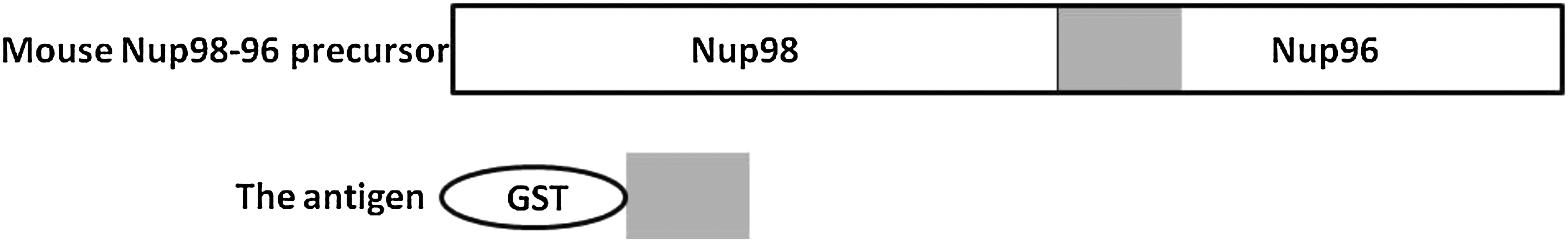
Schematic of mouse Nup96. Top, Nup98–96 precursor. Bottom, GST-fused Nup96 fragment is expressed in E. coli as recombinant glutathione S-transferase (GST) fusion proteins.
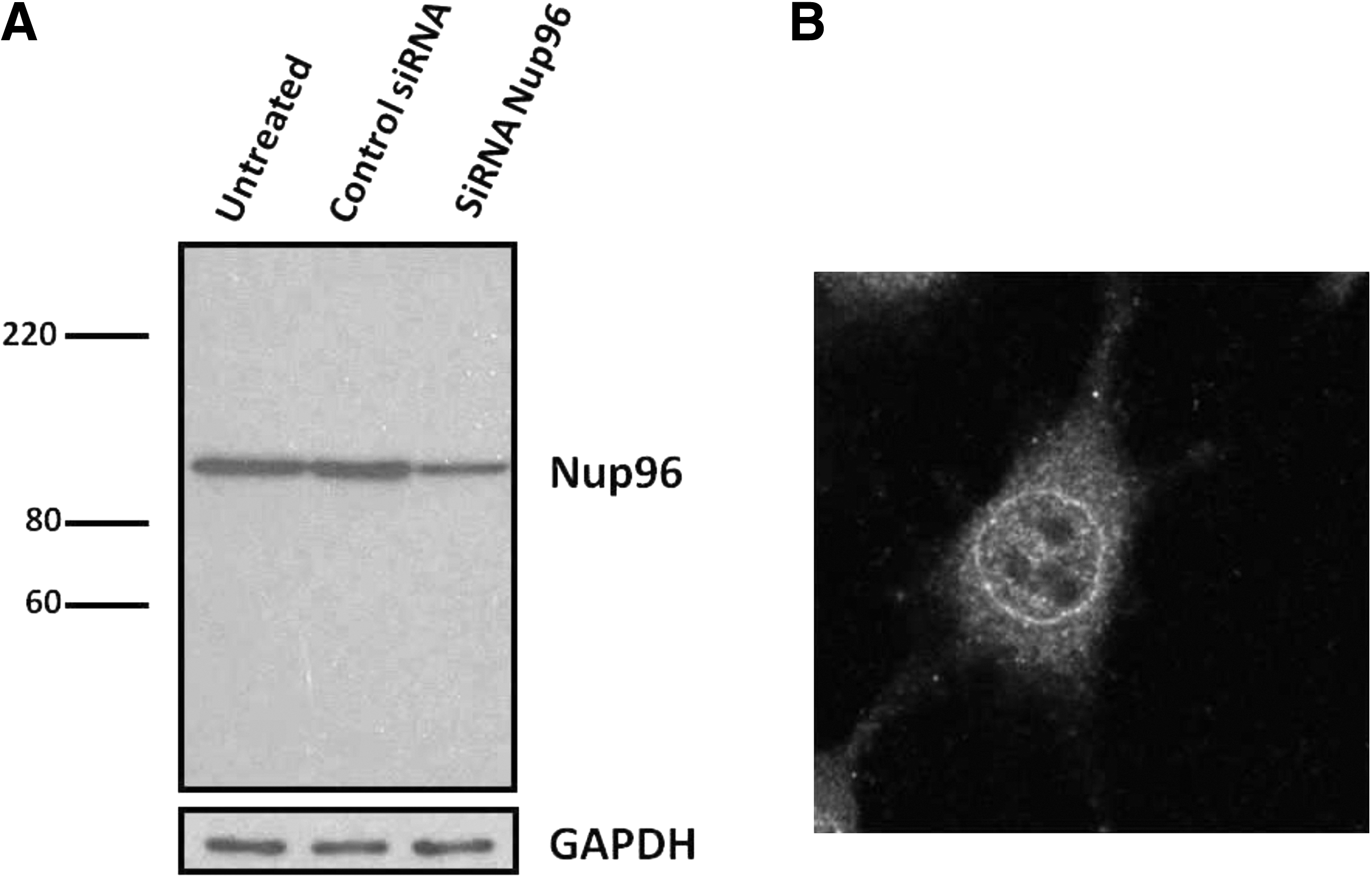
One of these antibodies, designated MAb 4H5, was selected for further study. MAb 4H5 detected endogenous Nup96 in NIH3T3 cell lysates as a single band, which was specifically downregulated by Nup96 siRNA (Fig. 2A). This antibody was next characterized by immunostaining with the mammalian cells. An indirect immunofluorescence study using MAb 4H5 revealed a predominant punctuate nuclear rim staining pattern that would be expected from a protein associated with the NPC in NIH3T3 cells (Fig. 2B). Moreover, immunostaining of HeLa cells with MAb 4H5 also resulted in a nuclear rim staining pattern similar to that for NIH3T3 cells (data not shown).
(
Although it has been reported that Nup96 is a component of Nup107–160 subcomplex, the function of each component is not still clear. The monoclonal anti-Nup96-specific antibody (MAb 4H5) developed in this study would serve as an aid in elucidating the functional roles of this nucleoporin.
Footnotes
Author Disclosure Statement
The authors have no financial conflicts to declare.