
Abstract
Abstract
The available toxicity data of benzalkonium chloride (BKC) clearly shows that it is toxic; however, the weight of evidence favors the view that at doses encountered in nasally and orally inhaled pharmaceutical preparations it is well tolerated. The adverse toxicological data predominantly come from in vitro and animal studies in which doses and exposure periods employed were excessive in relation to the clinical doses and their posology and, therefore, not directly applicable to the clinic. The conflict between the in vitro and animal data and the clinical experience can be reconciled by understanding some of the physicochemical properties of BKC, the nasal and respiratory tract microenvironments, the doses used, and the posology.
Introduction
M
The lack of consensus arises from a variety of factors, including that BKC is a complex and variable mixture of different carbon chain length compounds that affect the quality of the compound used in the various investigations, its anti-microbial activity, its protein binding, and its ability to form micelles; species differences in sensitivity to BKC; the duration of exposure in experimental studies often exceeds that experienced by humans; the use of different endpoints to monitor toxicity; and the physiological differences between in vivo and in vitro environments.
This review is intended to integrate the in vitro and in vivo animal data with the human data and also the physicochemical properties of BKC. The interplay between the physicochemical properties of BKC and the micro-environment of the nose and respiratory tract is key to understanding the toxicity assessment of BKC as a preservative in orally and nasally administered pharmaceuticals.
Toxicologically Relevant Physicochemical Properties of BKC
BKC is a quaternary ammonium antiseptic and cationic surfactant comprising a mixture of alkyldimethylammonium chlorides. The structure of BKC is shown next. The CAS Registry Number is 8001-54-5, and the average molecular weight is 360.
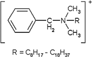
The USP30-NF25 describes BKC as a mixture of alkyldimethylammonium chlorides of the general formula: [C6H5CH2N(CH3)2R]Cl, in which R represents a mixture of alkyls, including all or some of the group beginning with n-C8H17 and extending through higher homologs including n-C16H33.. On an anhydrous basis, the content of the n-C12H25 homolog is not less than 40.0%, the content of the n-C14H29 homolog is not less than 20%, and the total content with each of these homologs together comprises not less than 70.0% of the total content. The European Pharmacopoeia requires that BKC should contain not less than 95% and not more than 104% alkyldimethylammonium chloride as C22H40NCl.
There can be considerable variation in the percentage composition of the different chain length homologues; the percentages of C12 and C14 in three marketed ophthalmic preparations in Europe were 59%/49%, 64%/34%, and 58%/42%, respectively,(2) and the percentages of C12/C14 in four aerosol preparations were 72%/28%, 55%/29%, 65%/35%, and 77%/23%, respectively.(3)
BKC analogues with different alkyl chain lengths possess different degrees of toxicity, protein binding, inhibition of histamine release, and antimicrobial activity.(4–8) Solutions containing C12, C14, C16 alkyl chain lengths, or a mixture (C12 60%–70%, C14 30%–40%, C16 <5%) induced severe damage to the mucosal epithelium of slugs, and the irritation increased with decreasing alkyl chain length. In addition, they showed a similar ranking in the bovine corneal opacity and permeability assay.(5)
A similar series of BKC compounds was used to determine their cytotoxicity in cultured normal rabbit corneal epithelial cells and in an in vivo corneal transepithelial electrical resistance (TER) assay. All of the BKC homologs displayed a concentration (0.0025%, 0.005%, and 0.01%)-dependent cytotoxicity after a 60-second exposure. Cytotoxicity of the C12 and C16 homologs was milder at all concentrations, and the cytotoxicities were found to be between those of C12 and C14 homologs. In the in vivo TER assay, a concentration-dependent corneal barrier dysfunction was noted for all BKC homologs; among these, C12 exhibited the lowest corneal impairment, whereas C14 induced the most severe corneal dysfunction.(6) This type of parabolic activity was also seen in an earlier study(4) where C14 was more effective in ablating neurones of the myenteric plexus than either C12 or C16 BKC homologs.
The antimicrobial activity of BKC is primarily dependent on the alkyl composition.(9,10) The antimicrobial activity of BKC has been shown to be more marked against Gram-positive than Gram-negative bacteria. However, the presence of BKC in multi-dose bottles of albuterol sulfate did not prevent hospital outbreaks of lower respiratory tract colonization and infection with Buckholderia cepacia.(11) The antimicrobial activity of BKC can be enhanced by the addition of other preservatives such as phenylethyl alcohol.
A preservative system comprising BKC and phenylethyl alcohol was found to be far superior to combinations of BKC plus disodium edentate or potassium sorbate plus disodium edentate.(12) The antimicrobial activity of BKC is diminished by protein binding, and the bactericidal activity of BKC in a solution of bovine serum albumin depended on the amount of unbound BKC.(10) The binding to bovine serum albumin increased with an increase in the carbon chain length: C14-BKC bound to bovine serum albumin 2.5–3.7 times more than C12-BKC. In a 100 μg/mL solution of C12-BKC containing 10 mg/mL bovine serum albumin, the amount of unbound BKC was 38.4 μg/mL: an approximate 60% decrease. BKC binding to bovine serum albumin is parabolically dependent on the alkyl chain length, with an optimal chain length of around C16.(8) In 5 mg or10 mg/mL BKC solutions in milk, 25% of the BKC was bound to the milk protein.(13)
In addition to modulating cytotoxicity, antimicrobial activity and protein binding differences in the alkyl chain length of BKC can selectively inhibit histamine release from mast cells induced by polyamines.(7) C13 and C14 side chains were more effective than C16 in inhibiting polyamine-induced histamine release from rat peritoneal mast cells.(14)
BKC is an amphiphatic molecule and tends to form micelles at concentrations exceeding a substance-specific critical micellular concentration (CMC). The CMC of BKC depends on: the chain length ranging from 881 mg/L for C12 and 23 mg/L for C16(15); the tonicity of the medium, increasing the concentration of sodium chloride and decreasing the CMC; and complexation with other molecules such as cyclodextrins that can shift the CMC higher.(16) These CMCs bridge the concentration range in pharmaceutical preparations and would likely reduce the presence of free BKC molecules at higher concentrations; evidence of this effect has been observed in cell culture systems.(17)
It is apparent that, depending on the source of origin, batches of BKC can contain differing amounts of the various homologues.(9) Some of the discordant results described in the following sections could, in part, arise from the use of BKC solutions with differing alkyl compositions, resulting in different protein binding properties, cytotoxicity and functional aspects, and the BKC concentration inducing a parabolic effect on protein binding and micellular formation.
In Vitro Toxicity and Mutagenicity
The in vitro cytotoxicity and mutagenicity of BKC has been evaluated in a wide variety of cells and approaches by using a wide range of doses, including those considered excessively high and artificial. The cytotoxicity studies detailed next have been conducted in plant, fish, and mammalian cells, including normal and transformed human cells. The mutagenicity assays include the Ames assay, chromosomal aberration, and DNA damage assays such as the comet assay.
The cytotoxicity of various concentrations and durations of exposure to BKC has been investigated in single-layer cultured human fibroblasts by using the MTT assay. Cells were exposed by removing the medium and replacing it with a solution of BKC in phosphate buffer; after the appropriate contact time, the solution was removed, rinsed in phosphate buffer, and replaced with MTT solution. The range of tested concentration (0.0001%–0.5%) after a 2-minute contact time resulted in a dose-dependent cytotoxicity, and increasing the contact time for a 0.001% BKC solution resulted in a time-dependent increase in cytotoxicity. A 2-minute contact time decreased cytotoxicity significantly below 0.001%; whereas during a contact time from 1 to 20 minutes, a continuous increase in toxicity occurred from 14.4% to 33.0%. A concentration of 0.01% BKC resulted in ∼50% viability after a 2-minute exposure.(18)
The MTT assay has also been used to determine the cytotoxic nature of BKC toward human keratinocytes in monolayer culture. The BKC was applied for 24 hours in serum-free medium. The calculated 50% loss of viability was 0.004 mM (equivalent to a concentration of 0.00014%).(19) In a modified MTT assay, the cytotoxic and membrane-toxic potential of BKC in U937 cells has been described, the BKC was diluted in RPMI supplemented with heat-inactivated fetal calf serum, and a 50% reduction in viability occurred at 6 and 0.6 μg/mL concentrations for 1- and 24-hour exposure periods, respectively. The membrane integrity was determined by using an arachidonic acid release test and resulted in 10% arachidonic acid release values of 19.3 and 1.62 μg/mL, respectively, for incubation periods of 1 and 24 hours.(20) In both studies, marked cytotoxicity was noted after lengthy incubations in BKC solutions; however, in the latter study,(20) concentrations up to 1.1 μg/mL even when exposed up to an hour exhibited no significant toxicity.
The cytotoxic effects of BKC on normal human epidermal keratinocytes of neonatal foreskin (NHEK) and a normal human skin fibroblast cell line NB1RGB have been reported.(21) Confluent cultures were incubated for 1 hour in a mixture of BKC in phosphate buffer and medium (1:4). BKC showed different results in NHEK and NB1RGB cell cultures, and the ED50 were 3.9 and 62 μM, respectively. The difference in activity was explained on the basis of the serum protein concentration in the culture medium. The medium for the NB1RGB cells contained 10% (v/v) of fetal calf serum (7.1 mg protein/mL), which was much higher than for the NHEK cells (26 μg protein/mL).
Smith and Alexander(22) determined the cytotoxicity of BKC in Balb/c 3T3 embryonic mouse cells. Confluent cultures were exposed for 1 hour to varying concentrations of BKC in medium containing 2.5% fetal calf serum and 2.5% newborn calf serum and assayed for cytotoxicity by using the neutral red assay. The EC50 (50% viability) was 18.4 μg/mL (0.0003681% BKC concentration).
Cell viability was determined by the MTT assay, and cell morphology was determined by electron microscopy in explant cultures of human nasal epithelium exposed to varying concentrations of BKC in phosphate-buffered saline (PBS) for 15 minutes.(23) The clinically used concentration (0.01% BKC) significantly reduced cell viability, with only 14%–19% of nasal epithelial cells surviving the treatment. Electron microscopy revealed that nasal epithelial cells showed the loss of microvilli, destruction of cell membranes, and poor cytoskeletal alignment.
Deutschle et al.(17) exposed suspensions of BEAS-2B cells to varying concentrations of BKC in PBS for 2 hours and determined viability by using trypan blue dye exclusion and DNA damage by the comet assay. There was a rapid dose-dependent reduction in cell viability from 92% at a concentration of 0.002% to 0% viability at 0.007%; cytotoxic effects were not evident at concentrations higher than 0.02%. This “U”-shaped response curve was believed to be the result of the formation of micelles of BKC at concentrations higher than 0.02%. In the comet assay, BKC in concentrations between 0.002% and 0.02% resulted in a stepwise increase in tail movements. This effect was not present at a concentration of 0.05%. The DNA damage not only occurred at concentrations typically found in commercially available nasal preparations but was also associated with significant cytotoxicity.
In other assays, cytotoxicity was observed in a neutral red uptake assay where 50% viability was obtained between 5.8 and 15.0 μg/mL for a 24-hour exposure and between 1.5 and 4.0 μg/mL for a 72-hour exposure; whereas in an MTT assay, 50% viability was observed at 4.7 μg/mL for a 12-hour incubation period. BKC was negative in the Ames assay at 1000 μg/plate (tester strains TA 1535, TA 100, TA 1537, TA 98, and G46-uvrB) and in the CHO chromosomal aberration assay.(24,25) BKC did not induce chromosomal aberrations in Syrian hamster embryo cells and was reported negative in two mutagenicity assays (rec-assay and reversion assay).(26)
Eight commercial ophthalmic preparations containing 0.004% to 0.02% BKC each tested negative in the Ames assay (tester strains TA 98, TA 1538, TA 1537, and TA 100) with and without metabolic activation. However, in the Rosenkranz Escherichia coli DNA polymerase A- assay, one compound containing 0.02% BKC demonstrated a genotoxic potential; this also showed that when BKC is diluted in distilled water reparable DNA damage occurs at concentrations of 0.02% and higher.(27)
In a further study, BKC had been tested in four genotoxicity assays: Ames assay, comet assay with primary rat hepatocytes, and in micronuclei assays with human peripheral blood lymphocytes and root tip cells of Vicia faba. The Ames assay was negative in indicator strains TA 98, TA 100, and TA 102 with and without metabolic activation up to cytotoxic doses. In the comet assay, primary rat hepatocytes were cultured in various concentrations of BKC diluted in minimal essential medium for 60 minutes. A slight but significant difference in tail movement indicating DNA damage was only seen at the highest dose tested (1.0 mg/L: 0.0001%) and that resulted in a slight decrease in cell viability (9%–14%). In the human peripheral lymphocyte assay, only 1 and 3 mg/L concentrations of BKC induced significant increases in the number of micronucleated cells. BKC also induced a significant increase in the number of micronucleated cells in V. faba at a concentration of 10 μg/mL (0.001% concentration), and there was a decrease in the rate of cell division at concentrations of 1 μg/mL and greater.(28)
The in vitro studies clearly show that BKC is cytotoxic at concentrations typically found in commercial ophthalmic and nasal preparations. The degree of cytotoxicity was generally similar across the various studies, with typical 50% viability values/concentrations of 1–20 μg/mL (0.001%–0.02%). The degree of cytotoxicity was also time dependent, with higher values associated with longer incubation periods. In addition, where it was possible to compare culture conditions, the presence of protein was associated with greater cell viability, which was probably linked to the reduction in the concentration of free BKC due to protein binding. The majority of the in vitro cell culture studies employed monolayer cultures that may greatly overestimate the cytotoxicity of BKC.
Yanochko et al.(29) showed that cell viability after 30 minutes of exposure is approximately 1000 times less for a monolayer culture of conjunctival cells than for a stratified cell culture with an air interface. The in vitro studies also show that BKC is not mutagenic; however, it is able to induce DNA damage after prolonged exposures. The relevance of these in vitro findings to assessing human safety is uncertain.
In the in vitro studies described earlier, the BKC was administered in a variety of ways; it was dissolved in PBS, PBS with serum, serum-free culture medium, or culture medium containing serum. These differing in vitro conditions with the presence or the absence of protein may contribute to the degree of cytotoxicity observed.
General, Dermal, and Ocular Toxicology Data
The general toxicology of BKC, including oral, dermal, and ocular safety, has been reviewed because oral safety issues may arise from the fraction of the nasally applied or orally inhaled dose that is swallowed; whereas dermal and ocular safety issues may arise from the inadvertent exposure of the face from incorrect use of the delivery device.
The acute toxicity of BKC is moderate with oral LD50 in mice of 150–175 mg/kg and LD50 in rats of 14.5, 13.9, 240–300 mg/kg, and 1.42 g/kg for IP, IV, oral, and dermal routes of administration, respectively.(20,24,31,32) The mortality data in rats, given that BKC in single doses ranged from 79.4 to 316.2 mg/kg, indicated the LD50 to be 234.3 ± 26.5 mg/kg and the only pathological changes observed in this series were those of a pure gastrointestinal irritant.(30)
In a 12-week study in Sprague-Dawley (SD) rats and a 52-week study in beagle dogs,(13) animals were orally/gastrically administered solutions of BKC (50 and 100 mg/kg in rats and 12.5, 25, and 50 mg/kg in dogs) in distilled water and in milk. The rats dosed with 50 and 100 mg/kg in water or in milk were normal in appearance; however, the growth rate of the rats in the 100 mg/kg group was depressed throughout the experiment, and their final body weight averaged 29% less than that of the water control group. There were no significant microscopic lesions attributable to BKC in any of the treatment groups. In the dog study, one of the three dogs that received 50 mg/kg in water died and all three of the dogs receiving 25 mg/kg in water died. Slight-to-moderate hyperemia of the small intestine and pyloric portion of the stomach was observed in dogs receiving 50 mg/kg doses in milk.
No significant macroscopic lesions were observed in dogs receiving 12.5 or 25 mg/kg doses in milk. Moderate-to-severe irritation of the small intestine was observed in all dogs receiving 50 and 25 mg/kg doses in water and in two of the three dogs receiving 12.5 mg/kg doses in water. The dogs receiving 50 and 25 mg/kg doses in water also had moderate-to-severe irritation and congestion of the stomach and intestines. No microscopic lesions were noted in animals dosed with BKC in milk.
The effects of a diet containing 0.0%, 0.015%, 0.031%, 0.062%, 0.125%, 0.25%, or 0.5% BKC on groups of albino rats (24 per group) over a 2-year period have been investigated.(30) Diet at levels tested below 0.5% produced no demonstrable effects on growth, food consumption, measured blood parameters, incidence of pathological lesions, or longevity compared with the control animals. A study also reported on mongrel dogs fed diets containing BKC: Two dogs were fed 1.0% and single dogs were fed 0.031%, 0.062%, 0.125%, 0.25%, or 0.5% BKC for 15 weeks. Animals at the 1.0% and 0.5% concentrations did not survive the experiment and showed signs of gastroenteritis.
The dermal toxicity has been extensively investigated since it is a common preservative in many cosmetics and healthcare products. BKC at concentrations up to 0.1% free compound is considered safe as a cosmetic ingredient.(32) BKC has been listed by the FDA as an inactive ingredient in drug products,(33) including topical shampoos (maximum potency 0.2%) and topical lotions (maximum potency 0.1%). Adverse reactions such as erythema typically occur at much higher doses and after longer periods of exposure than those likely to occur from inadvertent use of either nasal or oral inhalation devices. The irritation potential has been tested(34) on human forearm skin from healthy volunteers by increasing concentrations of aqueous formulations of BKC (0%–16%).
The various concentrations were applied for 24 hours, and a non-uniform spotty erythema became visible after patch application of a 2% solution. The maximal response was observed on day 2 after an application of 16% BKC. In mice, the application of a 1 or 2 mg BKC prepared in ethanol over the ventral and dorsal surfaces of the ears induced a long-lasting inflammatory response and a subsequent increase in the thickness of the ear that was maximal 6 hours after treatment.(35) Also in mice treated by direct epicutaneous application of 12.5 μL of 2% BKC solution to both dorsal and ventral sides of the ear once a day for 3 days, BKC was determined to be a dermal irritant but not a contact allergen based on the number of cells and their flow cytometric profile obtained from draining lymph nodes.(36)
The effect of lifetime dermal application of BKC in mice and rabbits was investigated.(37) In mice, 0.02 mL of either 8.5% or 17% BKC was dropped on the dorsal skin between the shoulder blades twice a week on a 1-inch square area. In rabbits, the same volume and concentrations were applied to the anterior left ear. In mice, BKC application caused ulceration, inflammation, and scarring in many animals but no tumors. In rabbits, fibrosis, inflammation, and ulceration but no skin tumors were observed.
The potential effect of BKC (0.025%, 0.1%, and 1.0%) solutions on the permeability of canine oral mucosal to organic compounds has been investigated by Siegel and Gordon(38); the 0.025% BKC solution produced a mixed response, having no effect on the permeability of propanol, 1,7-heptanediol, ethylene glycol, mannitol, inulin, and three molecular-weight dextrans (20, 75, and 250 kDa) and only a modest effect on the permeability of acetamide, urea, glycerol, and sucrose. The higher doses (0.1% and 1.0%) had a more marked effect on the permeability of the organic compounds.
BKC is widely used as a preservative in topical ophthalmic preparations at concentrations varying from 0.004% to 0.02%, with 0.01% being the most common clinically used concentration. The FDA considers BKC an inactive ingredient in many approved drug products, including ophthalmic ointments and suspensions (maximum potency 0.02%). Instillation of a drop of a 0.1% or 0.01% aqueous solution of BKC into both eyes of albino rabbits twice daily for 7 days resulted in no gross damage; however, at higher concentrations (2.0%, 1.0%, and 0.5%), there was a dose-dependent increase in corneal damage, including conjunctival necrosis, ulceration, and haziness of the cornea. By light microscopy and scanning electron microscopy, minor cytotoxicity occurred in corneas treated with five daily administrations of 0.1% BKC.(39) On instillation of 0.003, 0.01, 0.03, or 0.1 mL of a 0.1% aqueous solution of BKC directly to the central corneal surface of rabbits and the subsequent effects evaluated over 21 days by using the Draize scale, only negligible irritation was observed.(40)
The corneal toxicity of 0.01% BKC and 0.55% hydroxyethyl cellulose was evaluated in rabbits and dogs after multiple instillations. Slit-lamp and light microscopic examination revealed corneal epithelial changes in rabbits but not in dogs after hydoxyethyl cellulose,(41) with sloughing of the superficial epithelial cells and slight loss of polarity of the basal cells. Formulations containing 0.01% BKC and hydroxyethyl cellulose in concentrations between 0.3% and 0.8% produced similar results but not with the individual compounds. The corneal epithelial damage in the rabbit was believed to be the result of the increased viscosity of the hydroxyethyl cellulose formulation, prolonging the contact time of the BKC with the cornea.
Changes in permeability of the corneal epithelium have also been reported; after administration of ophthalmic formulations containing BKC, these changes appear to be pH- and dose dependent.(42) Corneal permeability was assessed with radiolabeled inulin that was instilled 15 minutes after a pH 4 or neutral pH buffer eye drop containing either 0.01% or 0.02% BKC. In neutral pH solution, 0.01% BKC did not significantly alter corneal permeability, but the 0.02% solution increased corneal inulin concentration by a factor of seven compared with controls. In pH 4 buffer, however, both the 0.01% and 0.02% BKC solutions increased corneal permeability by 10- and 18-fold, respectively. Structural changes in the epithelium due to BKC have been reported(43); 5 and 15 minutes after instillation of 0.01% BKC in vivo, cell damage and separation were observed in the superficial layers of the corneal epithelium.
A high proportion of the ocular toxicity studies have been conducted using rabbits, a species shown to be more sensitive than other species, including humans.(44,45) The latter administered two drops (50 μL each) of a BKC solution (0.0025%, 0.005%, 0.01%, 0.02%, and 0.05%) to one eye of rabbits and human volunteers, whereas the contralateral eye received saline drops and acted as the control. Corneal permeability was measured by determining the degree of anterior chamber fluorescence after fluorescein drops were applied to the corneal surface. In rabbits, corneal permeability increased with a rising BKC concentration; at a concentration of 0.01%, the anterior chamber fluorescence level increased 1.8 times over the control. In human subjects, 0.01% BKC produced no significant permeability changes; however, 0.02% BKC caused a 1.23-fold permeability increase.
The deposition of a 0.01% BKC solution used as the preservative in a multi-dose container for the treatment of chronic obstructive pulmonary disease (COPD) has investigated the deposition on the face of a mannequin from the misuse of the Respimat© oral inhalation device.(46) A low deposition on the face (mean 3.8% of emitted aerosol, range 1.9%–6.8%) and eyes (mean 0.1%, range 0%–0.4%) from various misuse scenarios is estimated. This study suggests that if the Respimat device is misused, there is likely to be little potential for unwarranted side effects resulting from ocular or dermal deposition because of the small volume of BKC solution involved, the intermittent nature of such misuse events, and the low velocity of the generated aerosol.
The ocular deposition observed in this in vitro study could be greater than that likely to occur in a clinical situation because the patient may blink or turn their head away when the spray is fired accidentally. In the case of nasal sprays that typically have a spray volume of 50–100 μL per actuation and a high spray velocity, it may be prudent to minimize the potential for inadvertent exposure to the eyes, particularly in patients with a compromised corneal surface, although the dose of BKC is expected to be low and in the range commonly used in ophthalmic preparations. The potential for dermal side effects from the misuse of nasal sprays is small because of the low concentration of BKC (in the context of skin irritation) and the intermittent nature of such events.
In vitro Respiratory Tract Toxicology
The ciliostatic effect of BKC nasal formulations has been investigated in numerous in vitro studies by using changes in ciliary beat frequency as the endpoint in animal and human tissue explants, cell cultures, and suspensions. Some of the relevant studies are detailed in Table 1. However, in other studies, it is frequently difficult to clearly separate the effect of BKC since the commercial nasal spray formulations used contained other preservatives and excipients that may have influenced the outcome of the study. In addition, some of the studies lack the appropriate BKC-free controls. In general, where the studies are adequately controlled BKC appears to decrease ciliary beat frequency in vitro and conflicts with the findings of in vivo studies where a direct comparison can be made.
Includes comparison with human mucociliary clearance.
Includes comparisons with rabbit mucociliary clearance.
The effects of varying concentrations of pharmaceutical-grade BKC alone or with EDTA on the ciliary beat frequency of chicken embryo tracheas have been determined.(47) Exposure to BKC produced a concentration- and time-dependent decrease in the ciliary beat frequency. After 20 minutes of incubation, 0.01% BKC had no effect on ciliary beat frequency whereas exposure to 0.025% and 0.05% BKC reduced the ciliary beat frequency by 10% and 75%, respectively. The addition of 0.01% EDTA to 0.01% BKC solution reduced the ciliary beat frequency after 20 minutes of incubation to ∼75%.
In a later study,(48) the effects of 0.006% BKC and 0.1% EDTA on the ciliary beat frequency in chicken embryo tracheas or mucociliary clearance in healthy volunteers were investigated. The ciliary beat frequency in the chicken embryo tracheas was reduced by 50% with 20 minutes of incubation, whereas there was no significant reduction in the mucociliary clearance measured in healthy volunteers. Although this study does not delineate the effect of BKC alone since there was no BKC-alone treatment group, it does show that there is an inconsistency between in vitro and in vivo findings. In addition, the earlier study(47) showed that 20 minutes of in vitro exposure to concentrations of BKC typically found in marketed nasal and oral inhalation formulations did not alter the ciliary beat frequency in chicken embryo tracheas.
The effects of 0.01% BKC on the ciliary beat frequency of embryo chicken tracheas and the effect on the rheological properties of purified pig gastric mucus glycoprotein have been reported.(49) The effect of BKC was variable; in some experiments, it was well tolerated whereas in others it appeared ciliotoxic and it did not alter the rheological properties of the purified gastric mucus glycoprotein.
Tracheal rings from chicken embryos have also been used to investigate the effects of 0.01% and 0.02% USP quality BKC in Locke-Ringer solution and the effect of Lock-Ringer solution and saline on the ciliary beat frequency.(50) The ciliary beat frequency in the Locke-Ringer solution remained around 100% of the initial frequency during the 60 minutes of the experiment. Physiological saline decreased ciliary beat frequency with time, leading to 40% of the initial value after 60 minutes of incubation. A 0.01% solution gradually decreased ciliary beat frequency to almost zero in 60 minutes, whereas a concentration of 0.02% stopped ciliary beat frequency within 40 minutes. This study clearly shows that under experimental conditions even saline can reduce ciliary beat frequency over time, especially those durations that greatly exceed that experienced in the clinic.
The effects of varying concentrations of BKC (0.00125%, 0.0025%, and 0.005%) in Locke-Ringer solution on rat and guinea pig tracheal rings and human mucosal explants of maxillary and ethmoid sinuses have been reported.(51) The rat was the most sensitive species where BKC caused a decrease in ciliary beat frequency in a concentration- and time-dependent manner. The highest concentration of 0.005% completely stopped the ciliary activity in <5 minutes; whereas in the guinea pig, irreversible inhibition was seen at 0.005% in 40 minutes. In comparison, the impairment of ciliary beat frequency in human mucosa was not dramatic as a 0.005% concentration decreased ciliary beat frequency by ∼60% after 60 minutes of exposure. In this study, the ciliostatic effects of BKC were much less pronounced in human respiratory mucosa than in rats or guinea pigs. Species differences were also noted by van de Donk et al.,(52) who described a significant difference in BKC sensitivity between chicken embryo tracheas and human adenoid tissue slices.
Morimoto et al.(53) studied the effects of varying concentrations of BKC in Locke-Ringer solution on the ciliary beat frequency in cultured rabbit tracheal mucosa and tracheal mucociliary transport determined by an in vivo endoscopic method in rabbits exposed by nebulization to an aerosol containing BKC. The ciliary beat frequency was slightly elevated (not significantly) during a 60-minute culture period with 0.01% BKC and was unchanged by a 0.02% solution. The ciliary beat frequency rapidly decreased in the presence of 0.03% solution and was completely stopped by a 0.05% concentration. The mean mucociliary transport rates were slightly increased by the administration of a 0.01% solution, not changed by a 0.03% concentration, but significantly decreased with treatments of 0.05% BKC.
Boek et al.(54) also examined the effect of 0.01% BKC in Locke-Ringer solution on the ciliary beat frequency in tracheas from chicken embryos and in cryopreserved human sphenoidal sinus mucosa and reported a more rapid and pronounced effect on ciliary beat frequency in the human mucosal tracheas than that in the chicken embryo tracheas. Ciliostasis was reached within ∼20 minutes in the human mucosal tracheas and by 50 minutes in the chicken embryo tracheas. In this study, the effect of physiological saline on ciliary beat frequency was also determined and was found to be mildly ciliostatic.
Alberty and Stoll(55) examined the effect of 0.022% BKC in a placebo solution (composition not described) on cell suspensions of nasal epithelial cells obtained from healthy volunteers and reported no significant decrease in ciliary beat frequency from that of the placebo solution.
The effects of a range of common excipients/preservatives in nasal formulations, including BKC alone and in combination with EDTA, on the ciliary beat frequency in chicken embryo tracheas have been described.(56) The tracheas were exposed to NaCl, 0.01% or 0.02% BKC, or a 0.01% solution with 0.1% EDTA for a 15-minute period and thereafter allowed to recover until 60 minutes. Saline caused a 26% decrease in ciliary beat frequency that returned to normal values by 60 minutes. In contrast, 0.01% and 0.02% BKC induced ∼50% decrease in ciliary beat frequency after 15 minutes of incubation and returned to 20% and 43% of their original values by 60 minutes, respectively.
Changes in ciliary beat frequency after single and repeated BKC exposures of confluent human nasal epithelial cell cultures have been reported. Cultures treated with 0.015% or 0.001% BKC for a 15-minute period were not significantly decreased. After 360 minutes of recovery in culture medium, these cultures had a significantly decreased ciliary beat frequency (38% and 24%, respectively). Long-term exposure (360 minutes) to a 0.01% solution of BKC resulted in ciliostasis after 180 minutes, whereas 360 minutes of incubation with a 0.001% solution resulted in ∼70% decrease in ciliary beat frequency.
To determine the effect of repetitive administrations of BKC, cultures were exposed for 15 minutes on 5 consecutive days to concentrations of 0.001%, 0.002%, 0.005%, 0.01%, or 0.02%. The highest concentration tested (0.02%) showed immediate and non-reversible ciliostasis after the first exposure period. Lower concentrations (0.01%, 0.05%, 0.0025%, and 0.001%) showed ciliostasis at later time points. The lowest concentration (0.001%) resulted in ciliostasis after five single exposures of 15 minutes on 5 consecutive days.(57)
Two studies(58,59) compare the in vitro ciliary beat frequency with mucociliary clearance in humans. Stanley et al.58 compared the ciliary beat frequency in epithelial strips from the inferior nasal turbinates in healthy volunteers exposed to betamethasone nasal drops containing 200 μg/mL BKC (0.02%) and 100 μg/mL EDTA with the individual components in fresh culture medium. The EDTA had no significant effect on cilia up to 1 mg/mL. BKC at concentrations of 200 and 100 μg/mL caused ciliostasis within 5 minutes; at 20 μg/mL (0.002%), ciliostasis occurred after 60 minutes; and at 8 μg/mL, ciliary beat frequency was 57% of control values after 360 minutes. The results of cultures exposed to the formulated nasal drops paralleled those of the BKC concentration. In the in vivo studies, 15 minutes after administration of three drops of betamethasone, nasal mucociliary clearance and ciliary beat frequency did not change significantly (see Clinical Studies section).
This study shows that although ciliotoxicity can be demonstrated in vitro, the results do not correspond to those in the clinic. The second study reported the effects of a defined BKC solution on the ciliary beat frequency, mucociliary clearance, and induction of pro-inflammatory effects.(59) The quality of the BKC was described as C12/C14/C16 = 40/50/10. The ciliotoxicity was assessed in isolated nasal epithelia from 15 healthy volunteers. A pronounced ciliotoxic effect of 0.01% and 0.025% BKC was observed; after 120 minutes, the ciliary beat frequency was approximately 30% and 5% of the control (Ringer solution), respectively. In contrast, there was no difference in mucociliary clearance in healthy volunteers administered 0.05% BKC as a 200 μL nasal spray(59) (see Clinical Studies section); again, this shows a disconnect between in vitro studies and clinically based studies.
In other studies, the presence or lack of beating cilia has been used as an endpoint to investigate the effects of BKC on mucosal tissues. Steinsvag et al.(60) report a method to culture explants of human adenoid tissue that has been used subsequently in a number of studies with nasal formulations containing BKC.(61,62) In these studies, 1 mm2 adenoid tissue fragments were cultured for 10 days before treatment. Another explant study measured the effect of BKC on the mucociliary clearance in a preparation from the frog palate.(63)
Oximetazoline nasal spray (0.5 mg/mL) containing 0.15 mg/mL BKC has been administered to human adenoid explants either undiluted for periods of up to 30 minutes or in various dilutions for 10 minutes.(61) The test item (0.015 mL) was placed directly on the surface of the epithelial culture after the culture medium was removed. At the end of exposure, the culture dishes were refilled by adding 1 mL of medium. Saline was used as the control; no BKC-free oximetazoline was employed as an additional control. This lack of the additional control group may be considered a weakness since oxymetazoline has been shown to inhibit or reduce ciliary beat frequency(64,65) and appears to slow mucociliary clearance in humans.(66)
The endpoints investigated were morphological, and the presence or absence of beating cilia was determined by inverted phase microscopy. Placing undiluted nasal spray on the fragments resulted in a severe reduction in the number of fragments with beating cilia, with longer exposure periods being more toxic than the shorter periods. The addition of 0.015 mL of nasal spray containing 0.15 mg/mL BKC would give a dose of 2.25 μg/mm2 (225 μg/cm2). This dose is 75–2500 times the calculated nominal human dose based on delivered volume. The 10-minute incubation period was also used to determine the effect of various dilutions of the oxymetazoline formulation; the results show that over the 14-day culture period, there were no differences between the saline control and the 3% dilution and that up to 12 days of culture there were no differences between the 10% dilution and the saline control. Assuming the dose multiples estimated earlier, these results would tend to support the safety of BKC (rather than detract from it) at doses commonly found in prescription nasal spray products.
In a similar approach, the effects of four topical nasal steroids with and without BKC on cultured adenoid fragments and human neutrophils in vitro have been reported, where only 0.05 mL of test substance was placed directly on the epithelial surface and incubated for 10 minutes each day for up to 10 days.(62) The steroid formulations contained 310, 220, or 200 μg/mL BKC; the fourth steroid formulation contained no BKC. In addition, human leucocytes were incubated in varying concentrations of test substance and the effects on actin polymerization, degranulation, and oxidative burst were determined. The explants exposed to the steroids alone and the steroid formulation without BKC retained their morphological features, whereas those exposed to the formulations containing BKC possessed marked degenerative changes that closely paralleled those seen in the BKC containing saline solution.
The BKC-containing formulations inhibited neutrophil function in a time- and concentration-dependent manner; however, a decrease in neutrophil function was also seen with the steroid formulation containing no BKC. The concentration and dosage volumes in this study are 1.5–6 times those in an earlier study,(61) which indicates that this in vitro study was conducted by using extremely high doses well outside the range experienced in the clinical setting.
Taken together, these studies(61,62,67,68) show that BKC-containing nasal sprays can induce marked effects in cultured adenoid tissue fragments. However, these marked changes occur at extremely high doses that are not likely to be experienced in the clinic. Evaluation of these studies may be impaired by the lack of sufficient details: One(67) did not report the BKC concentration in the oxymetazoline formulations used, another(62) did not report the concentration of BKC in the saline control solution, and a further one(61) did not include a BKC control.
In an amphibian explant study, excised pieces (5 mm2) of frog palates were exposed in vitro to 30 μL of 0.01%, 0.05%, and 0.1% BKC solutions and mucoliary clearance was measured after a 10-minute incubation period. There were no significant changes in mucociliary clearance after incubation with the 0.01% solution, and higher concentrations resulted in significant decreases in mucociliary clearance. The BKC concentrations of 0.01%, 0.05%, and 0.1% represent doses of 12.0, 60, and 120 μg/cm2, respectively. The authors considered that the frog palate preparation more closely resembled the nasal mucosa of patients in vivo as it retains an intact mucus layer, unlike the studies with cultured adenoid tissues.(63)
In Vivo Non-Clinical Respiratory Tract Toxicology
The nasal and respiratory tract responses to BKC-containing solution have been investigated in a number of species and by various modes of administration, including by nasal instillation, nasal sprays, and inhalation. For the purposes of estimating dose to the nasal cavity, the surface area of the rat has been taken as 13.4 cm2,(69) rabbit as 113 cm2,(70) and monkey as 30.2 cm2.(71) For rat inhalation studies, the deposition efficiency in the nose is taken as 0.42; for the tracheo-bronchial region, 0.03; and for the lung, 0.06 of the inhaled dose; these values are the average of the ranges described for a deposition model.(72) The surface area of the rat ciliated airways is taken as 27.2 cm2.(73)
Commercial preparations of corticosteroids containing 0.02% BKC were administered to cynomolgus monkeys, and a preparation containing 0.01% BKC was administered to rats.(74) Two male and two female monkeys each received 2-m doses of 0.1 mL of drug product into the right nostril four times daily for 28 days. Two male and two female monkeys served as controls and were treated with 5% glucose spray. No abnormalities were noted in the nasal epithelium of the monkeys after light and electron microscopical examination.
The calculated nasal dose in this study would be 5.3 μg/cm2, representing a no-observed effect level (NOEL). In the rat study, six male and six female SD rats were exposed nose-only for 1 hour daily for 28 days, resulting in a steroid dose of 0.6 mg/kg/day and a BKC dose of 0.03 mg/kg/day. A further six males and six females were exposed to air and served as controls. No abnormalities were noted in the nasal epithelium rats. The inhaled dose of 0.06 μg/kg/day represented a NOEL after a 1 hour exposure to 0.01% BKC and a nasal dose of 0.13 μg/cm2.
Marttin et al.(75) treated a group of five male Wistar rats with a single intranasal instillation of a 0.01% BKC solution. Under anesthesia, 20 μL was administered unilaterally to the right nasal nostril from a cannula that was introduced into the posterior part of the nasal cavity via an incision in the esophagus. The left nostril acted as the control. After a 15-minute incubation period, the animals were killed and the nasal tissue was prepared for light and electron microscopy. In one of the five treated animals, no changes were observed; in the remaining animals, the anterior portion of the treated nasal cavity showed minor to major changes in ciliary and apical cell border appearance and moderate mucus extrusion. The calculated dose of BKC (0.15 μg/cm2) was associated with toxicity.
The effect of Beclomethasone or flunisolide preparations containing 220 and 310 μg/mL BKC, respectively,(68) was determined in lightly anesthetized rats that were administered 10 μL of the aqueous steroid sprayed into the right nostril and 10 μL saline into the left nostril twice daily for 21 days. An additional group of animals were treated with a budesonide nasal spray containing no BKC. The epithelium of the right nasal cavity showed marked mucosal differences to that of the left nasal cavity. There were areas with obvious morphological features characteristic of squamous metaplasia, and the lesions were larger and the most extensive in the anterior part of the nose, especially on the septum. No nasal changes were observed in the steroid preparation containing no BKC. The calculated doses of BKC administered with beclomethasone and flunisolide were 0.32 and 0.46 μg/cm2, respectively.
Kuboyama et al.(76) exposed groups of five male Wistar rats to saline (control), 0.01%, 0.05%, and 0.1% BKC solution. A 20 μL volume was administered by a micropipette to the left nostril every hour eight times a day. Sixteen to 18 hours after the final administration, the animals were killed and the nasal tissue was prepared for light microscopy. After exposure to the 0.01% solution, the nasal epithelium was unchanged from that seen in the controls. In the 0.05% and 0.1% groups, lesions were confined to the anterior nasal passages consisting of epithelial desquamation, degeneration, edema, or neutrophilic cellular infiltrate in the nasal mucosa. The changes were more marked in the 0.1% group. The calculated doses in the 0.01%, 0.05%, and 0.1% groups are 1.19, 5.97, and 11.9 μg/cm2, respectively, indicating that a cellular dose of 1.19 μg/cm2 represents the NOEL.
Lebe et al.(77) instilled 7 μL of a 0.01% BKC solution into both nostrils of a group of 11 Wistar rats twice daily for either 1 or 4 weeks by means of a micropipette. A group of 12 rats were similarly treated with saline and served as controls. At the end of the treatment period, nasal tissue was prepared for light and electron microscopy. The first findings observed in the BKC-treated animals were edema and mild-to-severe inflammatory cell infiltration in the nasal mucosa; these findings were more marked after a 4-week administration. Light microscopy also revealed an increase in vascular structures of the nasal mucosa, intraepithelial gland development, and squamous metaplasia. The calculated dose of BKC in this study is 0.1 μg/cm2 and it appeared toxic at this cellular dose.
Cho et al.(78) administered 7 μL of 0.01% and 0.1% BKC/saline or a saline solution per nostril twice daily for 1, 2, and 4 weeks in 4–5 week-old SD rats. The two BKC concentrations induced a time-dependent increase in the thickness of the nasal respiratory epithelium. There was no dose dependency, and the effects were primarily in the anterior nasal cavity where edematous changes with prominent inflammatory cell infiltration and proliferation of intraepithelial glands were evident. However, the group sizes (three animals) were small and the standard deviations were too large to draw statistically valid conclusions for a dose dependency. The saline controls appeared normal. The daily doses in this study were 0.1 and 1.0 μg/cm2 for the 0.01% and 0.1% BKC solution, respectively, with both epithelial doses being toxic.
Cureoglu et al.(79) evaluated the effect of a 0.001% BKC solution on the ultrastructure of the nasal epithelium in rabbits. Rabbits were administered 50 μL of 0.001% BKC twice daily to the left nostril for either 14 or 28 days; the right nostril was administered saline and acted as the control. Degenerative changes, including squamous cell metaplasia, nuclei deformation, and increased heterochromatin, were observed. The estimated daily dose (0.008 μg/cm2) was associated with toxicity.
Mathias et al.(80) used an intranasal dosing volume of 25 μL/nostril of either saline, 0.02% or 0.2% BKC to evaluate the expression of secreted total protein, lactate dehydrogenase, and interleukin 1α release in lavage fluid after a 30-minute exposure period in male SD rats. The 0.02% BKC concentration resulted in no significant difference from the saline control, whereas the 0.2% concentration produced marked and significant changes in biomarkers. The daily doses in this study were 0.75 and 7.5 μg/cm2 for the 0.02% and 0.2% BKC solution, respectively, with the low dose representing a NOEL.
Twelve female albino rats of the CD strain (mean weight 216 g) and 12 Syrian Golden hamsters were exposed to an aerosolized hair spray (9.9 mg/m3) containing 0.1% BKC(32) for 5 days a week (4 h/day) for 14 consecutive weeks. The estimated achieved dose in the rat study is 1.95 μg/kg/day. There were no significant differences in weight gain, hematologic values, and serum chemistry data between experimental and control groups. There were no exposure-related deaths, and neither gross nor microscopic changes attributed to BKC inhalation. Insufficient details are provided to calculate a dose to the nose and respiratory tract; however, the achieved dose of 1.95 μg/kg/day represents the NOEL.
Swiercz et al.(81) exposed female Wistar rats to aerosols of BKC to determine the acute or short-term effect on the respiratory tract. Three groups of five animals were exposed by nose-only inhalation to 52.8, 37.6 mg/m3 BKC for 4 hours and to 30.5 mg/m3 for 6 hours over 3 consecutive days. The controls were unexposed rats. The 4-hour exposure to the highest dose resulted in the death of two of the five animals; no deaths occurred in the 30.5 mg/m3 group, suggesting a steep exposure-response relationship. Exposures to BKC resulted in a significant increase in relative lung weight and concentrations of total protein, LDH, and IgE in bronchoalveolar lavage fluid, indicating severe damage to the lung. These findings resulted from the very high estimated daily doses of approximately 10.6 mg/kg/day for the 4-hour exposure to an aerosol concentration of 52.8 mg/m3 and for the 6-hour exposure to the 37.6 mg/m3 concentration, an estimated daily dose of 11.3 mg/kg/day.(82)
The acute effects of inhaled BKC were evaluated in mice.(83) Short exposure (30 minutes) to concentrations up to 19 mg/m3 resulted in a concentration-dependent decrease in tidal volume, with a concomitant increase in respiratory rate and neutrophilic lung inflammation indicating pulmonary irritation. The lowest dose (0.049 mg/m3) resulted in a significant decrease in tidal volume, whereas the highest dose represented the lowest-observed-effect-level (LOEL) for lung inflammation.
In the non-clinical toxicology program conducted to support the market authorization of tiotropium bromide monohydrate,(84) repeat-dose studies in mice, rats, and dogs and reproduction toxicity studies in rats and rabbits were conducted with control groups exposed to an aqueous solution of 0.01% BKC with either 0.01% or 0.05% EDTA. In the repeat-dose studies (Table 2), the incidence of respiratory tract lesions was low in the control animals, and the few that did occur were primarily found in the nasal cavity. In one mouse study, laryngeal squamous cell metaplasia occurred in 3–10 males but not in any of the 10 female mice.
Data obtained from reference 84; all studies involved exposures to 0.01% BKC.
Since no EDTA or BKC-free controls were used, it is difficult to assign these limited changes to the presence of BKC. No respiratory tract lesions were identified in dogs exposed for 52 weeks. In a 2-year carcinogenicity study in rats and mice that used control groups with and without BKC and air, it was possible to separate out the potential effects of prolonged BKC inhalation exposures. Rats and mice were exposed for 1 hour daily for up to 104 weeks. BKC did not induce tumors in the respiratory tract, and there were no differences between any of the three control groups with the exception of the larynx in the rat study. In the BKC-containing vehicle group, there was a significant increase in the incidence of very mild cranial squamous hyperplasia that was similar to that seen in the animals exposed to the test article.
Laryngeal changes in the rat are generally considered an adaptive and reversible response having little toxicological relevance.(85) The achieved inhaled BKC dose in the rat study is calculated as 0.818 μg/kg/day (based on a body weight of 377.5 g [average of male and female body weights at week 53 of study], a reported dose of 21.7 μg/kg/day tiotropium, and a BKC solution concentration of 0.01%). This inhaled dose would give a tracheobronchial dose of 0.0246 μg assuming a tracheobronchial deposition fraction of 0.03 (average of range reported by Hsieh et al.(72) and Cuddihy et al.(86)) and an epithelial dose of 0.74 ng/cm2. An inhaled dose of 0.818 μg would represent a nasal dose of 0.34 μg assuming a deposition fraction of 0.415 (average of range reported(89)) and an epithelial dose of 25.37 ng/cm2. The achieved inhaled BKC dose of 0.818 μg/kg/day represents the no-observed adverse effect level (NOAEL).
Clinical Studies
The effect of the presence of BKC in various nasal drug formulations has been investigated in healthy volunteers and patients with allergic rhinitis, non-allergic rhinitis, and rhinitis medicamentosa by using a variety of approaches, including mucociliary clearance, symptom scores, mucosal swelling, and nasal reactivity to histamine. Where there are data available on the concentration of BKC, the spray volume and the total number of daily actuations from which an estimated topical epithelial dose can be calculated (Table 3). The nasal dose calculation assumes that 100% of the spray volume is distributed over the entire nasal epithelium (160 cm2).(87) For calculation of the tracheo-bronchial dose, the surface area of the human ciliated respiratory tract is taken as 2690 cm2(88) and a deposition fraction is assumed to be 0.11.(89)
NC, not calculated.
Calculation based on epithelial surface area of 160 cm2.(87)
Total number of applications to both nostrils.
Dose estimate based on 100% deposition in nasal cavity. Information from Dudkiewicz-Wilczynska et al.(3)
Information from Romeijn et al.(50)
Single nostril application.
Information from U.S. Patent No. 4983595.
Information from Graf et al.(99)
Dose estimate based on 11% deposition in central lung region.(119)
Information from Pavia and Dewberry.(115)
van de Donk et al.(48) investigated the effects of a solution containing 0.006% BKC (commercial grade) and 0.1% EDTA. Measurements of mucociliary clearance were made in healthy volunteers before treatment and after a single dose by using either the saccharin (taste) test in 11 subjects or the indigo carmine (color) test in 12 subjects; in addition, measurements were also made in 10 subjects with no treatment. Results were similar with both saccharin and indigo carmine tests and there were no significant differences between the groups; the mean transport times before and after treatment varied from 8.7 to 13.3 minutes. A dose to the nasal epithelium could not be calculated from the information provided. This report also contained data from an in vitro study that showed that the BKC solution caused a decrease in the ciliary beat frequency in chicken embryo tracheas.
Holmberg and Pipkorn(90) studied the effect of BKC on mucociliary clearance in 14 healthy volunteers in a triple-blind, randomized, placebo-controlled, cross-over study. Measurements of mucociliary clearance were made before treatment, after a single dose, and after 1 week of continuous treatment with either a placebo containing BKC (0.022%) or a corticosteroid formulation also containing BKC, thus utilizing the saccharine dye test. The dose was two actuations into each nostril twice daily. The baseline nasal mucociliary clearance for the subjects varied from 3.62 to 11.9 mm/min (mean 6.99 mm/min), and repeat measurements in the same nostril before treatment revealed a variation of 16%. The mean value for mucociliary clearance before and after treatment was not statistically significant, indicating that BKC in this study was without effect. A dose to the nasal epithelium could not be calculated from the information provided.
Batts et al.(91) determined the effect of 0.01% BKC solution on human nasal clearance in healthy volunteers by using a radiolabeled saccharin solution. The BKC solution or control saline was administered as drops (total volume 0.3 mL) to one nostril to groups of six subjects. One hour after administration, mucociliary clearance was measured by gamma scintigraphy. The rate of clearance of the nasal spray was found to be biphasic. It was found that 25%–30% of the radiolabeled saccharin reached the nasopharynx immediately after dosing and that ∼50% of the dose was cleared within 20 minutes. The first phase was faster than the second and rarely lasted longer than 20 minutes. The clearance rate of the labeled saccharin solution was not significantly different from that of the BKC solution. The calculated dose based on 0.3 mL of 0.01% BKC to one nostril was 0.375 μg/cm2.
McMahon et al.(92) investigated the effects of a single dose or multiple doses of BKC in healthy volunteers. In the single-dose study, 34 subjects were administered either saline or saline and 0.02% BKC; the tests were conducted in random order one week apart. Mucociliary clearance was determined 10 minutes after administration by using the saccharin taste test. In the chronic study, 65 subjects were divided into three groups that were treated with saline, a marketed corticosteroid nasal spray containing BKC, or the placebo containing BKC plus other excipients present (not listed) in the marketed nasal spray. The subjects were treated daily for 2 weeks with two puffs into each nostril twice a day.
Before the study and at the end of treatment, mucociliary clearance was measured by using the saccharin test taste; ciliary beat frequency was measured on nasal smears taken from the inferior turbinate; and nasal airway cross-sectional areas by acoustic rhinometry and symptom scores were taken. In the single-dose study, the mean clearance time after saline was 620.7 ± 437.9 seconds and after BKC was 762.7 ± 459 seconds; the slight prolongation of the saccharin clearance time was only detectable by using a non-parametric statistical method.
The results show the typical wide range of clearance times seen in normal individuals and also that approximately a third of the subjects had shorter clearance times after BKC treatment. Subjects administered the BKC-containing placebo had no significant differences in any of the parameters tested compared with patients treated similarly with saline nasal spray and steroid nasal spray with BKC. Patients were treated with two 100 μL puffs into each nostril twice daily, representinged an estimated BKC dose of 1.0 μg/cm2.
Rizzo et al.(93) compared the effect of a 3-week use of a saline nasal spray containing 0.01% BKC with preservative-free saline on nasal mucociliary clearance rate in a double-blind, placebo-controlled, randomized, crossover, single-center trial in healthy volunteers. The sprays were administered three times per day to healthy volunteers; evaluations of mucociliary clearance were conducted at the beginning and the end of each 3-week period by gamma scintigraphy. Nasal mucociliary rate was impaired in both treatment groups, compared with baseline measurements. The authors could not conclude whether this difference was clinically meaningful and a dose could not be calculated from the data provided since the spray volume was not reported.
Riechelmann et al.(59) investigated the effects of BKC (described as C12/C14/C16 = 40/50/10) on the ciliary beat frequency, mucociliary clearance, and induction of pro-inflammatory effects. There was no difference in mucociliary clearance in healthy volunteers administered either 0.05% BKC or saline as a 200 μL nasal spray in each nostril four times daily for 8 days, nor were there any changes in nasal cytology (myeloperoxidase, tryptase, CD45+, and CD68+) or nasal secretions (cytokines IL-6, IL-8, epithelial neutrophil antigen-78, and neuropeptide substance P).
However, directly after the spray application, nasal irritation, hypersecretion, and a burning sensation compared with placebo were reported. Moreover, symptoms of persistent nasal irritation were higher with BKC than with placebo. The 0.05% BKC solution in this study would equate to a dose of 5 μg/cm2 nasal epithelium (200 μL per nostril four times daily and total nasal cavity surface area of 160 cm2). The concentration of 0.05% BKC is 2.5–5 times that typically used in nasal formulations and at this higher concentration, no significant effects were noted in vivo in healthy volunteers.
Storaas et al.(94) report two studies in healthy volunteers in which the effects of acute and sustained BKC exposures were examined. Nasal symptoms were also recorded. One nostril of each volunteer was exposed to 0.01% BKC by using a pool technique where 15 mL of solution was kept in the nostril for a period of 10 minutes, after which nasal lavages were carried out. The lavage fluid levels of α2-macroglobulin and fucose were measured as indices of mucosal exudation of plasma and glandular secretion, respectively. Acute exposures significantly increased the nasal output of fucose; α2-macroglobulin was unaffected.
Repeat exposures during 10 days did not alter the exudative responsiveness of the airway mucosa. Acute exposure was associated with an immediate smart or pain sensation; after 10 days of exposure, tachyphylaxis occurred, with no smarting or pain being reported. BKC was demonstrated to possess irritant-like properties by increasing the nasal secretion of fucose. This response may have been due, in part, to a 15 mL volume, which is approximately 30–75 times the normal volume administered intranasally, and the high dose, which is calculated as 18.75 μg/cm2 (15 mL 0.01% BKC over 80 cm2).
Graf et al.(95) investigated the effect of BKC in a decongestant nasal spray in a randomized double-blind parallel study with 20 healthy volunteers. The subjects were divided into two groups that were treated with decongestant with or without 0.01% BKC administered three times daily (0.1 mL per nostril; total 0.6 mL daily) equivalent to 0.375 μg/cm2. In this study, there was no placebo group. Both groups of patients had significant increases in mucosal swelling and nasal stuffiness after 30 days of treatment, which were indicative of rhinitis medicamentosa.
However, the changes in the group administered decongestant with BKC were more marked, indicating that it aggravated the rhinitis medicamentosa. In a subsequent study by Hallen and Graf,(96) patients were treated for 10 days with decongestant nasal spray with or without BKC after a 30-day rest period from an initial 30-day treatment with the same combinations of drugs. The mucosal swelling found with the decongestant containing BKC was significantly more than that found in the decongestant-alone group, and the group was also associated with a significant increase in nasal stuffiness.
In a later parallel, randomized, double-blind clinical study by Graf and Halllen,(97) 30 healthy volunteers were randomized to receive three daily administrations of 0.1 mL in each nostril of a decongestant with 0.01% BKC, decongestant alone, or placebo (saline solution with sodium phosphate and EDTA) for 1 month. The three variables studied were nasal mucosal swelling, symptom scores, and nasal reactivity, as estimated by histamine challenge. In the placebo and decongestant with BKC groups, there was no increase in nasal stuffiness or reactivity to a histamine challenge. There was a significantly greater mucosal swelling after treatment with the decongestant containing BKC compared with the other treatment groups. The calculated nasal epithelial BKC dose is 0.375 μg/cm2.
Braat et al.(98) administered patients with perennial allergic rhinitis, a steroid containing 0.02% BKC, 0.02% BKC plus placebo, or placebo alone for 6 weeks. Patients received two actuations intranasally, twice daily into each nostril. Before treatment and 6 weeks after treatment, two nasal biopsies were taken from each patient from the anterior tip of the inferior turbinate. Before treatment, at two weekly intervals during treatment, and 2 weeks after treatment, mucociliary clearance was measured. No statistically significant difference in mucociliary clearance was found between the different treatment groups at any of the measurement points, and there was no statistical relationship between the number of ciliated cells present and the treatment that the patients received. Scanning and transmission electron microscopy examination showed no adverse effects of BKC. The calculated nasal epithelial dose is 1.05 μg/cm2 assuming a spray volume of 95 μL.
Graf et al.(99) studied the effect of a decongestant with and without BKC (0.01%) in patients with vasomotor rhinitis. Both groups of patients sprayed 0.1 mL of the substances in each nostril three times daily for 10 days. This study showed that mucosal swelling did not occur after 10 days of administration of a decongestant with or without BKC; this is in contrast to previous studies in rhinitis medicamentosa patients and healthy subjects where these authors(94) report that BKC-containing decongestant was associated with significant mucosal swelling. The calculated nasal epithelial BKC dose is 0.375 μg/cm2.
Naclerio et al.(100) compared the nasal clearance after administration of two steroid nasal spray formulations, one with BKC and one without, in patients with allergic rhinitis. Mucociliary clearance and quality of life were determined before treatment and 2 weeks after treatment. At the end of treatment, there was no difference in the rate of clearance between the two formulations, although there was a significant difference in the amount of change from before to after treatment in favor of the formulation without BKC.
However, this difference was <1 minute and was probably without biological significance due to the variation seen in the individual clearance rates and the limited number of patients involved. In addition, after treatment with the formulation containing BKC, a significant improvement in the overall quality of life was seen but not after the BKC-free solution. For comparative purposes, a dose for this study could not be calculated from the information provided; however, the authors use their study to clinically support the adverse in vitro effects of BKC on mucociliary clearance.
Discussion
A review of the available toxicological data of BKC clearly shows that it is toxic and raises possible concerns about its safe clinical use(1,101–103); however, the weight of evidence favors the view that BKC at doses encountered in pharmaceutical preparations is well tolerated. The adverse toxicological data predominantly come from in vitro and animal studies in which doses and exposure periods employed were excessive in relation to the clinical doses and their posology and, therefore, may not be applicable to the clinic. The conflict between the in vitro and animal data and the clinical experience can be reconciled by understanding some of the physicochemical properties of BKC, the nasal and respiratory tract microenvironments, the doses used, and the posology.
The current clinical data show that typical nasal products using BKC at concentrations up to 0.02% are well tolerated by patients, with results comparable to saline control, and that BKC at concentrations <0.1% inhaled orally are also well tolerated. However, the European Medicines Agency(104) is considering revising the guidelines for BKC as an excipient in nebulized and orally inhaled respiratory medicines. The original limit was a delivered dose of 10 μg; the revision suggests a zero limit.
For nasally administered solutions, the posology of BKC is important in assessing its potential clinical toxicity. In patients, it is typically administered in one or two actuations per nostril, one to four times daily. Each actuation provides an initial concentration that rapidly declines through its high solubility, its dilution in airway fluid, its binding to proteins in airway secretions, the continual replenishment of airway fluid and secretions, and mucociliary clearance.(103) Batts et al.(91) report that 25%–30% of a nasally administered dose immediately reached the nasopharynx after dosing and that ∼50% of the dose was cleared within 20 minutes. The amount and the concentration of free BKC available in vivo are dependent on the quantity of protein present in and the volume of lining fluids.
The surface area of nasal cavity (both nostrils) is ∼160 cm2,(87) although delivery of nasal formulations is typically more toward the anterior portion of the nasal cavity.(89,105–107) The volume of nasal secretions present in the anterior nasal cavity has been estimated as 136 ± 17 μL for each nostril, whereas the total volume of nasal secretions in the entire nasal cavity is 800 ± 111 μL (including some undetermined portion of the upper pharynx) in healthy volunteers.(105)
Stanley et al.(58) calculated a nasal fluid volume of 400 μL based on a depth of the mucus and periciliary fluid layer of 25 μm and a total surface area of 160 cm2. The volume of nasal secretions can increase dramatically as a result of allergic rhinitis, experimentally from a baseline volume of 136 μL in the anterior nasal cavity of healthy volunteers; it was calculated to increase to 827 and 882 μL in each nostril after metacholine and histamine challenge, respectively.(105)
These results show that any unbound BKC could be diluted by a factor of 2–8 depending on the initial spray volume in each nostril. The amount of free BKC will depend on the amount of protein present in the nasal and lung fluid; in normal humans, the total protein in nasal fluids appears to be between 400 and 600 μg/mL.(108,109)
The amount of protein in the nasal secretion may vary during the day, for instance, a diurnal variation in IgG, IgA, and albumin described by Mygind and Thomsen.(110) The average concentrations during the day time (12 am and 4 pm) were 35 mg/100 mL IgG, 30 mg/100 mL IgA, and 170 mg/100 mL albumin; the corresponding values for the night (midnight and 04 am) were 140, 80, and 590. This level of protein suggests that protein binding in the nasal cavity may be in the region of 25%–65%.(10,13) Taking into account the immediate loss of 20%–30% of the initially administered dose, the 25%–65% protein binding, and the two- to eightfold dilution in periciliary fluid, potentially only 3%–30% of the original BKC content may be unbound and free to exert its effect.
The reduction in the dose of free BKC due to protein binding, dilution in lining fluid, and the brief intermittent nature of the exposure most likely accounts for the lack of clinically relevant effects in studies with healthy volunteers (Table 3). The concentration of BKC was between 0.01% and 0.05% with estimated epithelial doses of 0.375–5 μg/cm2. These values cover the range of doses and concentrations reported for marketed nasal formulations (Table 1).
In addition, extrapolation from healthy volunteers to patients with nasal symptoms requiring medication may be conservatively confounded by several factors, including differences in the volume and nature of the nasal secretions and differences in pH. The increase in the volume of nasal secretions in patients with rhinorrhea or rhino/sinusitis will reduce the concentration of free BKC and, hence, diminish the potential for toxic effects. The pH in the normal nasal cavity is reported to be in the range of 5.5–6.5 and can increase to 7.2–8.3 in sinusitis.(108) van de Donk et al.(48) have shown that reducing the pH of a BKC solution from pH of 7.4–6 may significantly decrease ciliary movement in excised guinea pig tracheal tissue, indicating that higher pHs associated with nasal disease may be less likely to impair mucociliary clearance.
The in vivo clinical exposure scenario is in marked contrast to in vitro conditions where the cells are bathed in a BKC solution for 5–60 minutes during which the concentration is unaffected by dilution or protein binding. In these cellular studies, the concentration of the BKC is the key parameter and actual cellular dose cannot generally be estimated. In cases where it has been possible to estimate dose (explants of defined area and administered volume), the doses have been 75–2500 times the nominal clinical dose (based on nominal delivered volume). It is, therefore, not surprising that such studies frequently show toxicity and are not directly relevant for assessing human safety.
However, in one study(61) where the explants were treated under conditions mimicking the clinical setting (10 minute exposure and 0.01% BKC), no differences from controls were observed. This in vitro study supports the clinical safety of currently approved nasal products, where the daily calculated nasal epithelial dose of BKC is up to 1 μg/cm2 delivered from up to eight intermittent administrations.
Although there are less safety data with inhaled BKC than with nasal administration, what data are available show that at clinically appropriate doses adverse events were not apparent. The concern for orally inhaled solutions containing BKC in current markets is low because of the minute volumes administered and deposition over a large epithelial surface area with estimated epithelial doses in the sub-nanogram range. In the ciliated airways (generations 1–16) of humans, the amount of airway fluid is estimated as 3.6 mL.(112) In airway disease, as many as half of the smaller airways may be plugged with mucous secretion, suggesting that the volume of airway lining fluid may approach 20 mL for the trachea and bronchi and 45 mL for the conducting bronchioles.(112)
This indicates that the amount of free BKC may be very limited due to substantial dilution and binding to proteins. However, the inclusion of BKC in nebulization solutions is associated with an increased incidence of bronchoconstriction in sensitive populations of severe asthmatics.(113) The periods of bronchoconstriction occurred after prolonged (typically 15–30 minutes) and repeated exposures to high doses of about 300 μg.(114) BKC-containing nebulizer solutions have now been replaced by BKC-free unit dose vial medications. In contrast, inhalation solutions containing 0.01% BKC used in the Respimat® inhaler are associated with low incidences of bronchoconstriction that are comparable to those experienced in patients using metered dose inhalers (MDIs).(115,116)
The Respimat Soft Mist™ inhaler delivers per actuation 12 μL of inhalation solution containing 1.1 μg BKC, and typically one to four actuations per day are necessary for effective medication and are well tolerated in asthmatic and COPD patients.(117,118) The amount of BKC delivered by Respimat is ∼200 times lower than that delivered by wet nebulizer formulations(118) and has been shown in clinical studies not to induce bronchoconstriction. The dose of BKC delivered in the clinical studies with tiotropium is 0.088 μg/kg/day assuming a 50 kg individual, which is 10-fold less than the estimated dose in the carcinogenicity studies in rats.(84) In the other rat BKC inhalation study(32) that was negative, the dose was 20-fold more than with the clinical studies. These two studies provide a safety margin of between 10- and 20-fold that may be conservative due to the one to four hour exposures compared with the brief intermittent daily clinical exposures. In the inhalation study where marked adverse respiratory findings were noted, the estimated doses were in excess of 10,000-fold of the clinical doses.
The weight of evidence supports the continued use of BKC as a preservative in currently approved multi-dose nasal and oral inhaled drug products, and further regulatory actions to limit or remove BKC from such drug products seem unwarranted and will negate the convenience of multi-dose products for patients.
Footnotes
Author Disclosure Statement:
No competing financial interests exist.