
Abstract
The most well-known forms of inflammatory bowel disease (IBD) that affect the entire gastrointestinal tract are ulcerative colitis (UC) and Crohn's disease (CD). The serum profile of inflammatory biomarkers and noncoding RNA and their role in the propagation of the inflammatory process remains controversial. Thus, this study was designed to examine the relationship between hematological profile, C-reactive protein (CRP), tumor necrosis factor-alpha (TNF-α), interferon-gamma (INF-γ), and the expression of LINC00641 and miR-378a in individuals with IBDs. In addition, we elucidated the correlation between the expression of LINC00641 and miR-378a and the biochemical variables analyzed. This retrospective study analyzed 94 unrelated participants. Group I included healthy controls, Group II consisted of participants diagnosed with UC, and Group III consisted of participants diagnosed with CD. Patients with IBDs experienced significant elevations in CRP, total leukocyte count, platelets, erythrocyte sedimentation rate, TNF-α, and INF-γ. However, participants with IBD had lower hemoglobin and albumin levels than healthy control participants. Moreover, the expression levels of LINC00641 and miR-378a were elevated in participants with IBD, with a significant difference between participants with IBD and healthy controls. The most striking observation was a clear association between serum LINC00641 and miR-378a levels and the biochemical variables assessed. This study demonstrated a positive correlation between the expression of LINC00641/miR-378a and TNF-α in patients with UC and CD patients. This study suggests that LINC00641 and miR-378a are prospective biomarkers and noninvasive screening tools for IBDs, which may help predict the progression of complications.
Introduction
Inflammatory bowel disease (IBD), a lifelong disease, has been a global health issue with a steadily increasing incidence in almost 7 million people worldwide (Kaplan and Windsor, 2021). The most well-known forms of IBD are ulcerative colitis (UC) and Crohn's disease (CD), which affect the entire gastrointestinal tract and frequently cause irreversible gut damage requiring hospitalization or medical procedures (Kaplan and Windsor, 2021; Lamb et al., 2019).
UC is an idiopathic and emerging risk factor for inflammation that affects the colon. The hallmark of UC is chronic progressive intestinal inflammation (Khalil et al., 2023; Lamb et al., 2019). CD frequently occurs anywhere in the digestive tract from the mouth to the anus (Baumgart and Carding, 2007; Baumgart and Sandborn, 2012). Prompt diagnosis of IBDs is crucial for its management and alleviation by improving prognosis and therapy (Matsuoka et al., 2018). This opens new avenues for exploring and scrutinizing novel candidate biomarkers for IBD diagnosis.
Tumor necrosis factor-alpha (TNF-α) and interferon-gamma (INF-γ), two essential proinflammatory cytokines in the pathogenesis of IBD, are associated with disease characteristics (Hiroshi et al., 2022). Proinflammatory cytokines are produced in excessive amounts in inflammatory situations, which not only enhance the host organism's immune response but also have negative impacts that can affect metabolic control and hemodynamic regulation (Jarczak and Nierhaus, 2022). Thus, inhibiting TNF-α and INF-γ is effective in causing and maintaining clinical reactions and IBD remission.
Long noncoding RNAs (lncRNAs) are noncoding RNA that encompass ∼200–600 nucleotides. However, they also regulate gene expression through many meaningful regulatory procedures (Ayeldeen et al., 2023; Statello et al., 2021). LINC00641 acts as an anticancer RNA, inhibiting bladder cancer progression by regulating miR-197-3p14 (Han and Zhang, 2022).
MicroRNAs (miRNAs) represent a noncoding form of RNA associated with biologic process varieties and modulate the expression of target genes (O'Brien et al., 2018). Furthermore, miRNAs are linked to several metabolic pathways and are used to diagnose diseases such as colorectal cancer and breast cancer (Chen et al., 2019; Khaliefa et al., 2023; Si et al., 2019). miR-378a contributes to various metabolic pathways by inhibiting the Wnt/β-catenin signaling pathway, which in turn inhibits cancer cells (Deldar et al., 2021; Yu et al., 2021).
To our knowledge, the differential expression of LINC00641 and miR-378a in individuals with IBDs and their correlation with C-reactive protein (CRP), TNF-α/INF-γ, and hematological profiles have not been explored. Thus, this study was designed to scrutinize the relationship between hematological profile, CRP, TNF-α/INF-γ, and the expression of LINC00641 and miR-378a in individuals with IBDs and explore their correlations and diagnostic potential for IBDs.
Materials and Methods
In this retrospective case–control study, 94 unrelated participants were evaluated during the period from March 2022 to January 2023, and written informed consent was provided for participation before enrollment in the study. Group I included healthy controls, Group II consisted of participants diagnosed with UC, and Group III consisted of participants diagnosed with CD. Endoscopy and clinical criteria were used in the diagnosis and differentiation of UC and CD. This retrospective study followed the ethical principles of the Declaration of Helsinki and the agreement of the Research and Ethics Committee (FMBSUREC/02102022/Abd-elhameed). Patients with acute or chronic renal failure, cardiac disease, cirrhosis, malignancy, or acute or chronic infections were excluded from the study.
All participants had fasting venous blood samples (10 mL) taken for routine analysis, including the hematology profile, CRP, and liver profile, and were stored at −80°C until LINC00641 and miR-378a expression analysis.
TNF-α and INF-γ assay
Serum TNF-α measurement was carried out using TNF-α ELISA kits provided by Avi-Bion (Orgenium Laboratories, Finland), according to Corti et al. (1992). Serum INF-γ were determined by using a commercially available enzyme-linked immunosorbent assay (ELISA) kit (R&D Systems, Inc., Minneapolis, MN) according to the manufacturer's instructions.
LINC00641 and miR-378a expression analysis
TRIzol reagent (Invitrogen) was used to isolate total RNA encoding miRNAs and lncRNAs from serum samples. A NanoDrop® (ND)-1000 spectrophotometer (NanoDrop Technologies, Inc.) was used to assess and evaluate isolated RNA. Using the miScript II RT kit (Qiagen, Valencia, CA), whole RNA was reverse transcribed in a final volume of 20 μL RT reactions. Primers specific for LINC00641 and miR-378a were used to amplify the cDNA using miScript SYBR Green Master Mix.
The amplification data were assessed using SNORD 68 and glyceraldehyde 3-phosphate dehydrogenase (GAPDH) as the housekeeping genes. Differential expression was assessed using the 2−ΔΔCt method (Livak and Schmittgen, 2001).
Statistical analysis
SPSS statistical software, version 22, was used to evaluate the data. Statistical data were expressed as mean ± standard error of the mean and percentages. One-way analysis of variance (ANOVA) was used to investigate differences between the groups under consideration. Post hoc multiple comparisons tests (Duncan) at P < 0.05 were used for pairwise comparisons. In addition, the correlations among the analyzed variables were assessed using Pearson's correlation coefficient, and diagnostic performance tests were determined using receiver operating characteristic (ROC) curve analysis.
Results
In this study, we included 30 participants with UC, 34 with CD, and 30 healthy individuals; the demographic and clinical data of the study participants are given in Table 1.
Demographic Data and Clinical-Pathological Features of the Studied Groups
CD, Crohn's disease; CDAI, Crohn's disease activity index; UC, ulcerative colitis.
Regarding the hematological and biochemical profiles, all participants with UC and CD had significantly higher rates of total leukocyte count (TLC), PLTs, and erythrocyte sedimentation rate (ESR) than healthy individuals (P < 0.05). However, the hemoglobin (Hb) and albumin levels in participants with UC and CD were considerably lower than those in healthy subjects. Participants with UC and CD showed elevated CRP levels compared with healthy participants (Table 2).
Hematological and Biochemical Characteristics of Studied Participants
Data are expressed as mean ± SEM; According to the Duncan multiple range test, different letters indicate statistical significance different means.
CRP, C-reactive protein; ESR, erythrocyte sedimentation rate; Hb, hemoglobin; SEM, standard error of the mean; TLC; total leukocyte count.
To determine whether LINC00641 and miR-378a were differentially expressed in the serum of UC, CD, and control individuals, their expression was measured using quantitative real-time polymerase chain reaction. In individuals with UC, the expression level of miR-378a and LINC00641 were elevated compared with that of healthy controls. Similarly, CD patients exhibited even higher levels of expression of these markers than UC patients. Furthermore, it is worth mentioning that serum TNF-α and INF-γ expression levels were significantly (P < 0.001) elevated in both UC and CD patients compared with healthy participants. TNF-α and INF-γ expression levels demonstrated a noticeable elevation in patients with CD versus those with UC (Table 3).
Expression Levels of LNIC00641, miR-378a, TNF-α, and INF-γ Regarding UC and CD Patients
Data are expressed as mean ± SE. According to the Duncan multiple range test, different letters indicate statistical significance different means.
TNF-α, tumor necrosis factor-alpha.
There were no significant differences (P > 0.05) in the expression levels of LINC00641 and miR-378a among UC patients with pancolitis, left-sided, and proctitis involvement (Table 4). In patients with CD, there were significant differences in the expression levels of LINCOO641 and miR-378a in patients with CD depending on the extent of the disease by colonoscopy (Table 5).
Differential Expression Levels of LNIC00641, and miR-378a Among UC Patient in Relation to the Extent of Disease by Colonoscopy
Data are expressed as mean ± SE. According to the Duncan multiple range test, different letters indicate statistical significance different means.
Differential Expression Levels of LNIC00641, and miR-378a Among CD Patients in Relation to the Extent of Disease by Colonoscopy
Data are expressed as mean ± SE. According to the Duncan multiple range test, different letters indicate statistical significance different means.
Pearson correlation analysis was performed to determine the relationship between LINC00641/miR-378 expression and the biochemical parameters of UC patients (Table 6). According to correlation analysis, miR-378a had substantial positive relationships with LINC00641, TNF-α, INF-γ, CRP, and TLC levels. Moreover, no statistical significance was observed between LINC00641 and TNF-α, INF-γ, CRP, or TLC levels (P > 0.05). In addition, miR-378/LINC00641 had a nonsignificant negative relationship with Hb levels in patients with UC (Table 6).
Pearson Correlation Coefficient Between Biochemical Variables in Study Participants
Significant linear correlation at P < 0.01 (2-tailed).
Significant linear correlation at P < 0.05 (2-tailed).
There was correlation between LINC00641/miR-378 expression and biochemical parameters of CD patients. According to correlation analysis, LINC00641 and miR-378 expression had substantial positive relationships with TNF-α, INF-γ, CRP, and TLC levels. In addition, miR-378/LINC00641 had a significant negative relationship with Hb levels in patients with UC (Table 6).
Diagnostic performance of serum LINC00641, and miRNA-378a
With an area under the ROC curve (AUC) of 0.843, serum LINC00641 was able to distinguish healthy controls from UC in ROC curve analysis, with confidence interval (CI) = 0.737–0.949, P < 0.001, sensitivity = 63.33%, specificity = 100% (Fig. 1a). Serum LINC00641 discriminated healthy controls from CD patients with AUC = 0.92, 95% CI = 0.836–1.004, with sensitivity of 88.24%, and 100% specificity (Fig. 1b). Also, serum LINC00641 discriminated CD from UC with AUC = 0.839, 95% CI = 0.734–0.943, P < 0.001, with sensitivity = 79.41%, specificity = 83.33% (Fig. 1c).
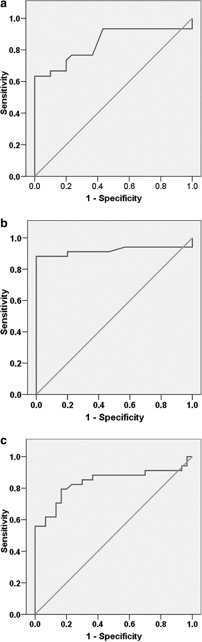
ROC analysis regarding LINC00641 between
On the contrary, with an AUC of 0.968, miR-378a discriminated healthy controls from UC with 95% CI = 0.906–1.030, P < 0.001, 96.67% sensitivity, and 100% specificity (Fig. 2a). Moreover, with an AUC of 0.974, serum miR-378a distinguished controls from patients with CD with 95% CI = 0.914–1.027, P < 0.001, 97.06% sensitivity, and 100% specificity (Fig. 2a). In addition, it distinguished from UC with AUC = 0.922, 95% CI = 0.836–1.007, P < 0.001, with sensitivity = 91.18% and specificity = 100% (Fig. 2c).
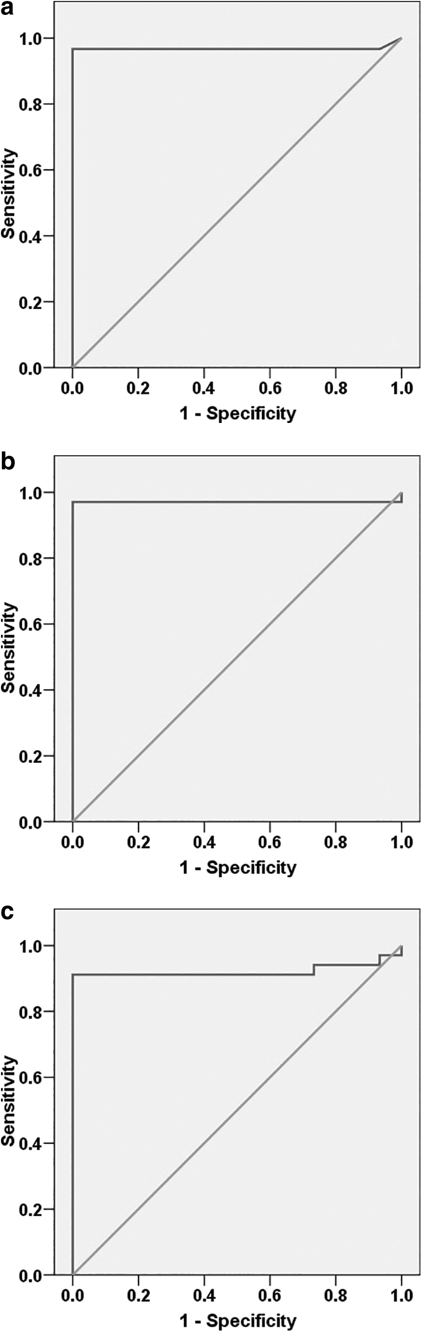
ROC analysis regarding miR-378a between
Discussion
Patients with IBD are predisposed to an assortment of problems owing to inadequate management and control of inflammation. This opens new avenues for exploring and scrutinizing novel candidate biomarkers for early IBD diagnosis. Accordingly, this study sought to demonstrate the differential expression of LINC00641 and miR-378 and their correlation in patients with IBD (UC and CD).
Although the exact pathogenicity of IBD forms remains unknown, it is well established that both arise as a consequence of genetic predisposition and immune gut flora dysregulation (Dalal and Chang, 2014). However, localization and endoscopic observations are not shared between both forms of IBD (Vespa et al., 2022).
In this study, white blood cell, platelet, ESR, and CRP levels were noticeably elevated in participants with UC and CD compared with healthy controls. Our results are consistent with those of Sayar et al. (2020) and Chen et al. (2020), who reported that patients with IBD exhibited a noticeable elevation in hematological profiles and CRP levels.
The blood expression of CRP is the most comprehensive biomarker of inflammatory disorders. The recurrence of IBD seems to be attributed to clinical signs and abnormally high CRP expression (Sproston and Ashworth, 2018). Furthermore, Karoui et al. (2011) explored the link between clinical and endoscopic activity and CRP level in patients with UC. However, high CRP levels are not disease specific, and they can occur in a variety of circumstances, including non-IBD enteritis, tissue damage, diabetes, malignancies, and cardiovascular disease (Fu et al., 2020; Hart et al., 2020; Jeong et al., 2019).
Our study revealed a significant decline in Hb and albumin levels in patients with UC and CD. Accordingly, previous studies have reported a noticeable decline in Hb and albumin levels in patients with UC and CD (Khan et al., 2017). In addition, Woźniak et al. (2019) stated that hypoalbuminemia has a significant impact on the onset of anemia in patients with IBD, and the severity of the inflammatory response is inversely correlated to albumin levels that arise from a decrease in albumin synthesis in the liver (Dalal and Chang, 2014). Moreover, Levitt and Levitt (2016) reported that hypoalbuminemia was correlated with disease activity, nutritional status, unresponsiveness to treatment, and an increased risk of colectomy in patients with acute severe UC.
This study demonstrated a statistically significant elevation in TNF-α and INF-γ levels in UC and CD patients compared with healthy participants. These data are consistent with their enhanced expression previously reported in patients with IBDs, which supports their role in the pathogenesis of IBDs (Eltayeb et al., 2020). Our findings are also supported by other studies investigating TNF-α levels in patients with IBD (Eltayeb et al., 2017). Increased TNF-α and INF-γ levels in the blood of UC and CD patients, released mainly from the intestinal colonic epithelium, have been proposed as markers of inflammation that contribute to disease severity (Eltayeb et al., 2020).
In this study, all participants with UC and CD showed elevated LINC00641 expression levels. In addition, patients with CD exhibited noticeably higher LINC00641 expression than those with UC. In concordance, Padua et al. (2016) and Chen et al. (2016), revealed that the expression of LncRNAs exhibited a noticeable overexpression in IBDs and was linked with the severity, intestinal permeability, and cell-intrinsic functions such as apoptosis and proliferation. Similarly, Hu et al. (2020) reported that LINC00641 is a new potential biomarker in patients with gastric adenocarcinoma, providing a predictive regimen for global survival.
Concerning the expression of miR-378a, all patients with UC and CD exhibited a notable elevation of miR-378a expression. Moreover, the differential expression of miR-378a was significantly higher in patients with CD than in those with UC. Consistent with our results, previous investigations have demonstrated an alteration in miR-378a expression in patients (Dubois-Camacho et al., 2019; Duttagupta et al., 2012). These alterations may be owing to the small number of patients included in previous studies or the use of pharmacological agents by the participants, which could reflect treatment rather than disease effects.
In our study, according to Pearson's correlations, serum LINC00641, miR-378a, CRP, TNF-α, INF-γ, and TLCs were positively associated. Moreover, the serum expression of LINC00641 and miR-378a was negatively associated with Hb levels in the study participants. Consistent with our results, Alper et al. (2017) and Solem et al. (2005) showed a positive correlation between CRP elevation and the TLC. Wang et al. (2016) reported a positive correlation between serum levels of miR-223, ESR, and CRP as indicators of disease activity in UC.
An ROC curve was constructed to test the diagnostic ability of LINC00641, producing a diagnostic accuracy (AUC = 92%) and a sensitivity = 88.24% and 100% specificity in discriminating patients with CD from healthy control participants. miR-378a had a high diagnostic accuracy (AUC = 97.1%) and a 97.06% sensitivity, specificity of 100% for distinguishing healthy controls from CD patients. It had a respectable capability to discriminate between CD and UC (AUC = 92.2%) and 91.18% sensitivity.
Conclusion
To our knowledge, this is the first study to report the differential expression of LINC00641 and miR-378a in individuals with IBDs and their correlation with CRP, hematological profile (ie, Hb and TLC), TNF-α, and INF-γ. This study showed that the elevation of LINC00641 and miR-378a coincided with the elevation of TNF-α and INF-γ in patients with IBD. This suggests that LINC00641 and miR-378a are prospective biomarkers of IBDs, which may help predict the progression of complications. However, this study has some limitations, including the sample size power analysis and the differential expression of miR-378a and LINC00641 in treatment response variability to TNF-α inhibitors. Larger and well-designed studies are required to further confirm the exact roles of LINC00641 and miR-378a in IBD development, progression, and treatment response to TNF-α inhibitors.
Footnotes
Authors' Contributions
N.A.H. and O.G.S. conceived and designed the research study; N.A.A-.H., O.G.S. contributed reagents/materials/analysis tools. N.A.A.-H. and N.A.H. analyzed the data. N.A.H. and N.A.A-.H. wrote the article. Finally, all authors reviewed the article.
Ethics Approval
This study was conducted in compliance with the Declaration of Helsinki, and the Ethics Committee at Faculty of Medicine, Beni-Suef University provided its approval (FMBSUREC/02102022/Abd-elhameed).
Informed Consent
Written informed consent was obtained from all subjects before the study.
Author Disclosure Statement
No competing financial interests exist.
Funding Information
No funding was received for this article.