
Abstract
The interleukin 1 (IL-1) family plays a significant role in the innate immune response. IL-1 receptor 2 (IL-1R2) is the decoy receptor of IL-1. It is a negative regulator that can be subdivided into membrane-bound and soluble types. IL-1R2 plays a role in the IL-1 family mainly through the following mechanisms: formation of inactive signaling complexes upon binding to the receptor auxiliary protein and inhibition of ligand IL-1 maturation. This review covers the roles of IL-1R2 in kidney disorders. Chronic kidney disease, acute kidney injury, lupus nephritis, IgA nephropathy, renal clear cell carcinoma, rhabdoid tumor of kidney, kidney transplantation, and kidney infection were all shown to have abnormal IL-1R2 expression. IL-1R2 may be a potential marker and a promising therapeutic target for kidney disease.
Introduction
Interleukin 1
Structure of IL-1R2
The first IL-1R2 clones were made from human and mouse B cells. Neutrophils, monocytes/macrophages, polymorphonuclear, dendritic cell, B cells (McMahan et al., 1991; Peters et al., 2013), and T cells (Subramaniam et al., 2004) all express IL-1R2. In humans, Il1r2 is located on the long arm of chromosome 2, band 2q12, and human chromosome 2q is the IL-1R cluster (Dale and Nicklin, 1999). The IL-1R2 protein is glycosylated and includes a long protein of 386 amino acids. It has a molecular weight of 68 kDa and is structurally similar to IL-1R1. In addition, like IL-1R1, IL-1R2 contains three extensively glycosylated immunoglobulin (Ig)-like extracellular structural domains as well as a helix-penetrating membrane region.
However, unlike other family members, IL-1R2 lacks the typical intracellular Toll-like receptors (TLRs)-IL-1 receptor (TIR) structural domain and is replaced by a tail of 29 amino acids. The absence of the TIR structural domain prevents IL-1R2 from transmembrane signal transduction (Peters et al., 2013; Supino et al., 2022). In addition, similar to IL-1R2, IL-18 binding protein (IL-18BP) is structurally incomplete with only one IgG domain. It functions as a decoy receptor by binding to IL-18 and blocking its biological activity (Novick et al., 1999).
IL-1R2 signaling pathway
TIRs are shared conserved regions and IL-1/TLRs are superfamily members of conserved proteins involved in immunity and inflammation (Garlanda et al., 2013). Both TLRs and IL-1Rs are receptors that play alarm roles in the natural immune system, with the former ligand being an exogenous (microbial mode of action on TLRs) alarm factor and the latter being an endogenous (IL-1 cytokines belonging to the IL-1R family) alarm mediator (Boraschi et al., 2018). IL-1 receptor accessory protein/IL-1 receptor 3 (IL-1RAcP/IL-1R3) is attracted when IL-1 binds to the membrane-associated IL-1R1. Myeloid differentiation factor 88 (MyD88), a linker protein that contains a TIR adapter, attracts linker proteins to the cytoplasmic TIR structural domains of IL-1R1 and IL-1RAcP to start a signaling cascade, which then activates IL-1R–associated protein kinase (IRAK). Next, phosphorylated IRAK binds to tumor necrosis factor (TNF) receptor–associated factor 6 (TRAF6) (Molgora et al., 2018; Schlüter et al., 2018; Supino et al., 2022).
MyD88, IRAK, and TRAF are central signal templates (Peters et al., 2013). This signal complex activates the downstream transforming growth factor kinase 1 (TAK1), and finally causes the transduction of various downstream signaling pathways such as nuclear factor-κB (NF-κB), activator protein-1 (AP-1), c-Jun amino-terminal kinase (JNK), p38 mitogen-activated protein kinase (p38 MAPK), extracellular signal–regulated kinases (ERKs) and interferon (IFN) regulatory factor (IRF) members. Eventually, various innate immune and inflammatory responses are activated (Molgora et al., 2018).
There are soluble and membrane-bound (mIL-1R2) versions of the IL-1R2 receptor. Soluble IL-1R2 is divided into secreted IL-1R2 (sIL-1R2) and shed IL-1R2 (shIL-1R2). The negative regulatory role IL-1R2 plays can be described as cis-regulation and trans-regulation (Mantovani et al., 2019; Zheng et al., 2013). In addition, there is an intracellular IL-1R2 that blocks the maturation of intracellular IL-1α precursor (pro-IL-1α), and this effect is lost by specific cleavage of IL-1R2 by activated IL-1 converting enzyme (caspase-1) (Zheng et al., 2013). Some researchers have pointed out that the breakdown of this negative regulatory effect is closely related to some sterile inflammation in the organism (Cavalli et al., 2021; Zheng et al., 2013).
For instance, the interaction of mIL-1R2 with IL-1RAcP/IL-1R3 prevents the formation of the IL-1R1-IL-1RAcP/IL-1R3 signaling receptor complex on the one hand and competes with the formed IL-1R1-IL-1RAcP/IL-1R3 for binding IL-1α and IL-1β on the other hand. This is a cis-regulatory function. Furthermore, mIL-1R2 has a higher affinity for IL-1β compared with IL-1α and IL-1Ra (which have a very low affinity for mIL-1R2), whereas IL-1β produces a richer and more intense inflammatory effect than IL-1α in the inflammatory response (Schlüter et al., 2018; Subramaniam et al., 2004). This may indicate that the negative regulatory role of IL-1R2 in the IL-1family is not negligible. In humans, the production of soluble IL-1R2 includes two pathways: alternative splicing and matrix metalloproteinases hydrolysis shedding (Peters et al., 2013).
It has been demonstrated that metalloproteinase inhibitors block all pathways of soluble IL-1R2 production, which implies that protein hydrolysis shedding is the main mechanism of soluble IL-1R2 production (Young et al., 2019). IL-1 must first undergo processing by caspase 1 to mature and become biologically active (Xia et al., 2021). The soluble IL-1R2 blocks the maturation of extracellular IL-1β mainly by binding to IL-1β precursor (pro-IL-1β) and blocking the action of caspase-1 enzyme. Of course, it also has a role in blocking IL-1α maturation, but does not have as high an affinity for pro-IL-1α as pro-IL-1β (Molgora et al., 2018; Schlüter et al., 2018; Symons et al., 1995). In addition, studies have demonstrated that soluble IL-1 receptor accessory protein (sIL-1RAcP) increases the stability of the soluble IL-1R2/IL-1 complex and enhances the ability of soluble IL-1R2 to inhibit the action of IL-1 (Schlüter et al., 2018; Smith et al., 2003).
Of note, soluble IL-1R2 barely binds to IL-1Ra, suggesting that the negative regulatory effect of soluble IL-1R2 on IL-1 is not disturbed by IL-1Ra (Boraschi et al., 2018). In addition, it was shown that IL-1 ligands do not bind both IL-1R1 and IL-1R2, and that ligand–receptor conjugation is independent (Colotta et al., 1994).
The macrophage polarization significantly influences how much IL-1R2 is expressed. Anti-inflammatory chemicals, which promote macrophage differentiation toward the M2 type, like glucocorticoids and IL-4, can upregulate IL-1R2 production. On the contrary, pro-inflammatory molecules that promote macrophage differentiation toward the M1 type, like lipopolysaccharide (LPS) and interferon α (IFN-α), cause downregulation of IL-1R2 production (Colotta et al., 1993; Supino et al., 2022). Furthermore, ectopic expression of IL-1R2 specifically blocks exogenous IL-1β signaling but increases the expression of IL-1α precursor forms and their downstream targets, such as IL-6, enhancing cell migration (Chang et al., 2009). Some researchers have found that IL-1R2 is highly expressed on tumor-infiltrating regulatory T lymphocytes (Treg), predicting that IL-1R2 may serve as a target for clearance of tumor Treg cells (De Simone et al., 2016). The pathways of pro-inflammatory responses involving IL-1 and the function of IL-1R2 are briefly given in Fig. 1 (created with BioRender.com).
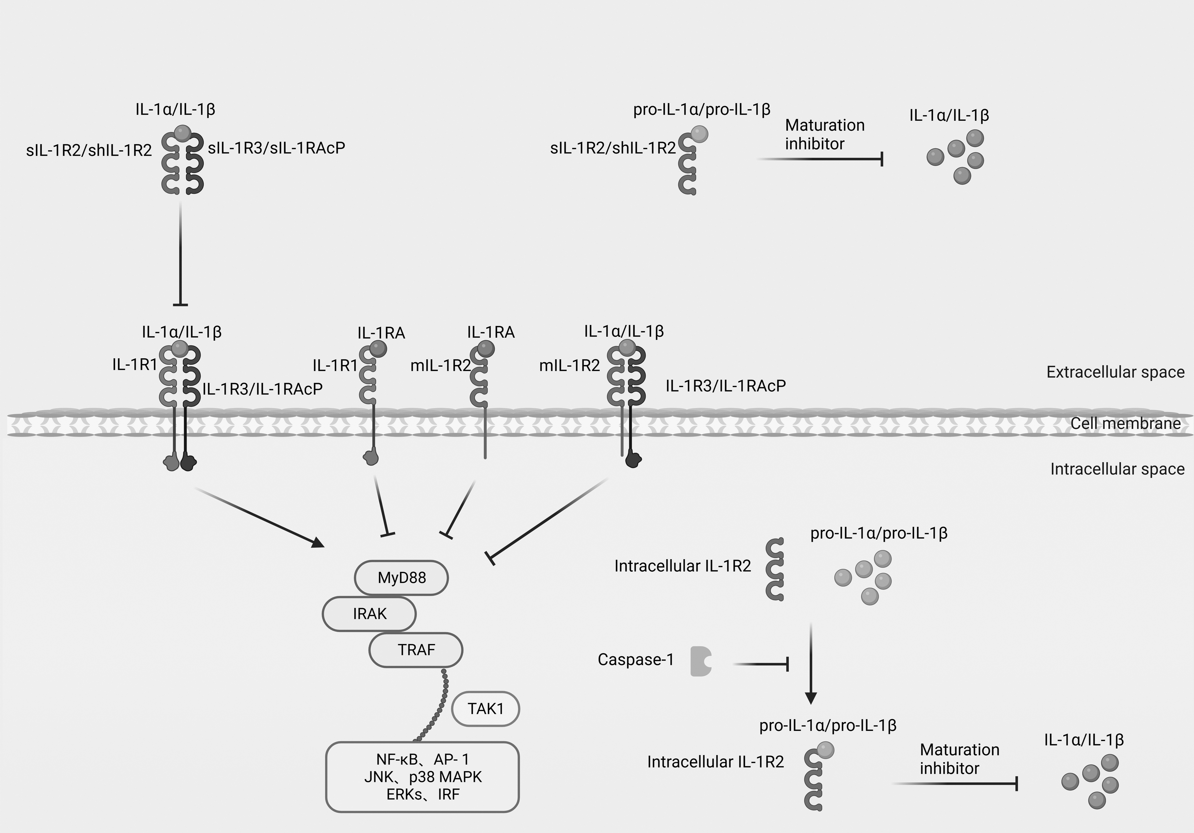
The mechanism of IL-1R2 in the IL-1 family. IL-1R2 participates in the IL-1–induced inflammatory response pathway by binding to IL-1AcP/IL-1R3 and inhibits the recruitment of downstream signaling. sIL-1RAcP increases the stability of the soluble IL-1R2/IL-1 complex. IL-1R2 inhibits the maturation of pro-IL-1α and pro-IL-1β. AP-1, activator protein-1; ERKs, extracellular signal-regulated kinases; IL-1, interleukin 1; IL-1Ra, IL-1 receptor antagonist; IL-1RAcP, IL-1 receptor accessory protein; IL-1R1, IL-1 receptor 1; IL-1R2, IL-1 receptor 2; IL-1R3, IL-1 receptor 3; IRF, interferon regulatory factor; JNK, c-Jun N-terminal kinase; mIL-1R2, membrane-bound IL-1 receptor 2; MyD88, myeloid differentiation factor; NF-κB, nuclear factor-κB; p38 MAPK, p38 mitogen-activated protein kinase; pro-IL-1α, IL-1α precursor; pro-IL-1β, IL-1β precursor; shIL-1R2, shed IL-1 receptor; sIL-1RAcP, soluble IL-1RAcP; sIL-1R2, secreted IL-1 receptor 2; sIL-1R3, soluble IL-1R3; IRAK, IL-1R-associated protein kinase; TAK1, transforming growth factor kinase 1; TRAF, tumor necrosis factor receptor–associated factor; VEGF-A, vascular endothelial growth factor A.
IL-1R2 in kidney disease
IL-1R2 is implicated in the regulation of both inflammation and immunity. The methylation levels of Il1r2 genes were significantly reduced in the systemic lupus erythematosus (SLE) patients (Lin et al., 2012), and the levels of sIL-1R2 were reduced considerably in endometriosis (Akoum et al., 2007), in organ transplantation, such as heart transplantation (Chen et al., 2013), small intestine transplantation (Asaoka et al., 2011), and facial transplantation (Kollar et al., 2018). An increased expression of Il1r2 has also been found in some neoplastic diseases, such as breast cancer (Zhang et al., 2020), Hodgkin's lymphoma (Oelmann et al., 2015), colon cancer (Spagnardi et al., 2022), gastric cancer (Yuan et al., 2021), and cutaneous melanoma (Torricelli et al., 2021), associated with proliferation, migration, and invasion of tumor cells.
The balance between cytokines and their antagonists is important for the control and regulation of immune responses. Previous study in Il1r2 gene knockout mice have found increased susceptibility to collagen-induced arthritis in knockout mice, and in models of arthritis, Il1r2 gene deletion plays an important role in inhibiting IL-1–regulated inflammation (Martin et al., 2017; Pinteaux et al., 2020; Shimizu et al., 2015). The balance between IL-18BP and IL-18 dictates pathology and in some cases correlates with disease severity (Novick et al., 2013). Therefore, an analogy can be made between IL-1R2 and IL-18BP, an IL-18 antagonist that is not a soluble receptor (Novick et al., 1999). Like the IL-1 receptor, also the type I interferon receptor (IFNAR2) exists in membrane-bound and soluble forms (Novick et al., 1994, 1995) and serve as decoy receptors that might affect pathology (Gilli, 2010).
Some investigators have recently addressed the potential role of IL-1R2 in kidney disease. The related studies are detailed in Table 1.
Studies Related to Interleukin 1 Receptor 2 in Kidney Disease
AKI, acute kidney injury; CACs, myeloid circulating angiogenic cells; CKD, chronic kidney disease; EPCs, endothelial progenitor cells; IgAN, IgA nephropathy; IL-1R2, interleukin 1 receptor 2; LN, lupus nephritis; LPS, lipopolysaccharide; PBMCs, peripheral blood mononuclear cells; RTK, rhabdoid tumor of kidney; SOD2, superoxide dismutase-2.
Chronic kidney disease
Genetic, metabolic, autoimmune, malignant, toxic, and environmental factors can cause different types of vascular, glomerular, and tubulointerstitial renal diseases and finally lead to the development of chronic kidney disease (CKD) and complications (Musgrove and Wolf, 2020). A previous study has shown that after LPS stimulation, the expression of superoxide dismutase-2 (Sod2), Il1α, Il1r1, and Il1r2 genes in peripheral blood neutrophils of the healthy control group was upregulated, whereas for peripheral blood neutrophils in CKD patients, Il1r1 and Il1r2 showed no change in gene transcription, Sod2 and ll1α were downregulated. Investigators found significant downregulation of Il1α, Il1r1, Il1r2, and IL-8 receptor antagonists (Il8ra) in HL60 cells with SOD2 transcriptional inhibition in vitro (Olsson et al., 2011). We can speculate that the expression of Il1r2 in CKD patients was influenced by SOD2 and the downstream IL-1 pathway.
It can be found that in CKD, neutrophil dysfunction and its resultant downregulation of Il1r2, causes a dysregulation of the body's immune inflammatory response. Combined with the role of IL-1R2 in the IL-1 family, the downregulation of Il1r2 enhances the IL-1–mediated inflammatory response in vivo, causing chronic and persistent kidney damage.
Acute kidney injury
Acute kidney injury (AKI) is a disease in which there is a sudden decrease in glomerular filtration rate and rapid loss of kidney function because of various causes (Levey and James, 2017). It was observed that Il1r2 expression was increased in kidney tissue during AKI. IL-1R2 interrupts the signaling cascade by sequestering ILs in serum and tissues, thereby preventing them from binding to the relevant functional receptors. Researchers proposed that the upregulation of Il1r2 expression is associated with the IL-10 and IL-6 pathways (Grigoryev et al., 2008). In acute kidney injury from various causes, IL-1 acts as an organism injury alarm as well as a pro-inflammatory cytokine, amplifying the inflammatory response of the body when the upregulation of IL-1R2 is supposed to play the role of a decoy receptor, balancing the intense inflammatory response of the body.
Lupus nephritis
SLE is a chronic inflammatory disease with lesions that can manifest in the kidneys. Lupus nephritis (LN) is a primary risk contributor to SLE's total morbidity and mortality (Almaani et al., 2017). The previous study showed that IFN-α promotes an antiangiogenic signature in SLE. The mRNA levels of Il1β, Il1r1, and Vegf-a in peripheral blood endothelial progenitor cells/myeloid circulating angiogenic cells of patients in the control group and the lupus group were significantly downregulated, whereas Il1ra and Il1r2 were significantly upregulated (Thacker et al., 2010). Although the study indicated that IL-1Ra has anti-angiogenic effects, it cannot be confirmed whether IL-1R2 plays a direct anti-angiogenic role or indirectly through its antagonism with IL-1.
IgA nephropathy
IgA nephropathy (IgAN) is the most common primary glomerular disease worldwide (Pattrapornpisut et al., 2021). In this study, the genotype “G/A” of Il1r2 gene rs4851527 significantly increased the risk of IgAN development in the hyper-dominant model, whereas Il1r2 gene rs3218977 was associated with a reduced risk of IgAN development in the dominant model. Il1r2 gene polymorphisms may play a critical role in IgAN (Xie et al., 2017). Il1r2 was found as one of the differentially methylated region–related differentially expressed genes in IgAN-discordant monozygotic twins, and functional enrichment analysis of these genes revealed the involvement of various inflammatory and immune pathways. Il1r2 was upregulated in peripheral blood mononuclear cells (PBMCs) of IgAN patients (Wei et al., 2021). The genetic polymorphism exhibited in IgAN can reflect the complexity of the etiology of IgAN, which is hereditary. IL-1R2, given its genetic polymorphism, can be attempted as a genetic signifier of IgAN and can play a significant role in the research and clinical diagnosis of IgAN.
Rhabdoid tumor of kidney
Rhabdoid tumor of kidney (RTK) is an uncommon pediatric kidney tumor that is extremely aggressive (Yamaoka et al., 2021). Immune system dysfunction is significantly correlated with RTK progression. Based on the TARGET database, the researchers collated and detected the expression profiles of immune-related genes (IRGs) in 65 patients with RTK, and suggested that inflammatory pathways were most commonly implicated in RTK. The investigators then built a prognostic model, and the Il1r2 gene was one of the seven IRGs (Chen et al., 2021). The results suggested that Il1r2 was correlated with RTK immunity disorder.
Renal clear cell carcinoma
Renal clear cell carcinoma is the main pathological subtype of renal cell carcinoma (Lucarelli et al., 2019). It is also a metabolic disease characterized by mutations in target genes involved in metabolic pathways (Ran et al., 2017). Il1r2 expression was significantly higher in renal clear cell carcinoma tissues with the tumor Furman pathological grade increasing. Compared with renal clear cell carcinoma patients with lower Il1r2 expression, the overall survival rate of patients with higher Il1r2 expression was better. The Janus kinase 2/signal transducer and activator of transcription 3 (JAK2/STAT3) pathway was shown to be engaged in IL-1R2–mediated regulation of cell function, and IL-1R2 may become a possible treatment target for renal clear cell carcinoma progression and metastasis by regulating the JAK2/STAT3 signaling pathway (Liu et al., 2022).
IL-1R2 is currently found to have great promise in the treatment of oncological diseases. It can be found to play a pro-cancer role in the majority of oncological diseases. Some researchers have found that IL-1R2 binds to the deubiquitinating enzyme USP15, which promotes the deubiquitinating enzyme activity of USP15 on breast cancer tumor stem cells, ultimately stabilizing stem cell expression in breast tumor–initiating cells (Zhang et al., 2020). More findings are the relationship between IL-1R2 and tumor infiltrating regulatory T cells (Tregs). For example, some research teams found that IL-1R2 can enhance the number and immunosuppressive function of Tregs in various tumor diseases and help tumor cells to undergo immune escape (Guo et al., 2018; Nikolouli et al., 2021; Zhou et al., 2021).
Targeting blockade of IL-1R2 would be a good strategy for treatment. The targeted blockade of IL-1R2 would be a good strategy for the treatment. In renal neoplastic diseases, both RTK and renal clear cell carcinoma, given the current results, IL-1R2 plays a pro-cancer role in both diseases. Therefore, targeted therapy with IL-1R2 neutralizing antibodies can be tried to eliminate IL-1R2 from tumor tissues by IL-1R2–specific neutralizing antibodies to slow down disease progression and prolong patient survival.
Kidney transplantation
Kidney transplantation is an alternative to kidney replacement therapy in end-stage renal disease (Augustine, 2018). For anti-rejection, long-term immunosuppressive therapy is required (Wojciechowski and Wiseman, 2021), and even the use of immunosuppressive agents cannot avoid the occurrence of the adverse effects of immune rejection and graft failure. In research involving recipients of allogeneic kidney transplants, researchers discovered that anti-rejection agents could effectively suppress the immune system by regulating several genes, including the upregulation of Il1r2 in peripheral blood lymphocytes. It is hypothesized that Il1r2 can interact via a specific, well-defined immunosuppressive pathway, although the exact mechanism has not been clarified (Wen et al., 2012). Researchers found that Il1r2 was upregulated in PBMCs of patients with kidney transplantation rejection. Il1r2 could be used as biomarkers for kidney transplant rejection, according to a hierarchical clustering analysis conducted on the cardiac transplant rejection profile (Chen et al., 2013).
A study has established a five-gene classifier (including Il1r2) based on the AlloMap molecular expression test to distinguish quiescence from rejection in kidney transplant rejection. Il1r2 is involved in the immune rejection pathway as a steroid response gene (Akalin et al., 2021). We can speculate that Il1r2 may be an assessable gene for the immune rejection status of kidney transplantation, and the expression level of Il1r2 may be able to predict the anti-rejection status of the organism. Studies have shown that anti-inflammatory chemicals that macrophage differentiation toward the M2 type can upregulate the production of IL-1R2 (Colotta et al., 1993; Supino et al., 2022), and we also speculate that the increased expression of Il1r2 in patients with anti-immune rejection after kidney transplantation may be related to this process. An attempt can be made to keep an eye on the expression of IL-1R2 in the peripheral blood of transplant patients to determine the effect of immunosuppressive drugs in patients and their long-term prognosis.
Extras
Low-dose hydrocortisone increased survival time in a lethal sepsis model. The amount of Escherichia coli bacteria in the kidney decreased following hydrocortisone treatment because it enhanced the expression of Tnfα, Il1r2, and histone deacetylase 4 (Hdac4) and lowered the expression of DNA methylation transferase (Dnmt3a) in both white blood cells and the liver (Doulias et al., 2018).
As mentioned previously, glucocorticoids are anti-inflammatory agents that promote the conversion of macrophages from M1 to M2. According to current findings, this process increases the expression of IL-1R2. At the same time, the role of IL-1 will not be missing in severe inflammatory responses that lead to organismal damage, when IL-1R2 may increase as a negative regulator, going to antagonize the binding of the functional receptor IL-1R1 to IL-1. However, we should also be concerned that excessive IL-1R2 expression may be detrimental to the clearance of exogenous microorganisms by the organism. Clinically, we can try to monitor the expression of IL-1R2 more perfectly to determine the course of the inflammatory response of the organism and even to guide the clinical use of drugs.
Limitations and prospects
IL-1R2 is being investigated in kidney disease, but little is known about the underlying pathological mechanisms or the cell types involved. It acts as a negative regulatory role in the family similar to IL-1Ra (Madej et al., 2017); IL-1Ra has been reported to have renal protective effects on amyloidosis in CKD (Afsar et al., 2018). However, studies have shown that IL-1R2 has a limited role in IL-1 regulation compared with IL-1Ra (Schlüter et al., 2018). As a primary antagonistic regulator of the IL-1 family, it may represent a prospective treatment target and a novel anti-IL-1 medication. In addition, IL-1R2 is induced under certain conditions and the relationship with prognosis or disease severity is variable; thus, as a molecular marker for specific kidney diseases, it is less stable and reliable. Future prospective studies are needed to reveal the link between IL-1R2 and kidney diseases.
Conclusion
In conclusion, we summarize the role of IL-1R2 as a decoy receptor in the IL-1 family and its involvement in immunity, inflammation, cell proliferation, and migration. Here we describe the clinical findings of IL-1R2 in kidney diseases, including CKD, AKI, LN, IgAN, RTK, renal clear cell carcinoma, kidney transplantation, and sepsis involving renal infection. IL-1R2 may be used as a biomarker of disease prognosis or severity and become a promising therapeutic target in the future.
Footnotes
Authors' Contributions
Conceptualization, H.Z. Writing—original draft preparation, H.H. Supervision, A.W. and X.M. Funding acquisition, H.Z. All authors have read and agreed to the published version of the article. All authors were involved in the study conception and design, acquisition of data, and analysis and interpretation of data.
Author Disclosure Statement
No competing financial interests exist.
Funding Information
This work was supported by grants from the National Natural Science Foundation of China (Grant No. 82000684), the Science and Technology Support (Social Development) Project of Bureau of Science and Technology of Changzhou (Grant No. CE20215024) and the Top Talent of Changzhou “The 14th Five-Year Plan” High-Level Health Talents Training Project (Grant No. 2022260).