
Abstract
Functional foods and/or their bioactive compounds playing a role in improving skeletal health have received considerable attention. The objective of the present study was to determine the extent to which certain functional foods as (1) whole, e.g., dried plum (DP), figs, dates, raisin, and blueberry, (2) fractionated, e.g., DP puree, DP juice, and DP pulp/skin, or (3) isolated, e.g., DP polyphenols, fructooligosaccharides (FOS), and β-hydroxy-β-methylbutyrate, forms reverse bone loss in an ovariectomized (Ovx) rat model of osteoporosis. Additionally, some of these components were tested in reversal of bone loss in combination. For this purpose, 180 3-month-old female Sprague-Dawley rats were divided into 15 groups (n = 12) and either Ovx (14 groups) or sham-operated (Sham, one group). Rats were maintained on a semipurified standard diet for 45 days after surgery to establish bone loss. Thereafter, rats were placed on one of the following dietary treatments for 60 days: casein-based diet (Sham and Ovx). The remaining 13 Ovx groups were placed on various treatment diets. Results showed that diets supplemented with 5% FOS + 7.5% DP was most effective in reversing both right femur and fourth lumbar bone mineral density and fourth lumbar calcium loss while significantly decreasing trabecular separation. There were no significant effects of treatment on serum or urine measures of bone turnover. Although other treatments were good at altering some bone parameters, none had the success in altering several bone health indicators as the diets supplemented with 5% FOS + 7.5% DP. The findings of this study suggest the combination of 5% FOS + 7.5% DP is capable of reversing Ovx-induced bone loss.
Introduction
O
In recent years, considerable attention has been given to exploring plant-based bioactive compounds and nutritional supplements as a substitute for conventional medicine. This attraction, in part, has been due to cross-sectional data supporting fruit and vegetable consumption yielding higher bone mineral density (BMD) prompted researchers to focus on the role of functional foods in bone health. 3,4 From the bone-preventive point of view, two comprehensive studies by Muhlbauer and colleagues 5,6 examined the bone-protective effects of numerous vegetables and herbs common in the human diet. Their findings indicated that among vegetables and herbs, onion had the most potent antiresorptive properties, followed by garlic and parsley. In terms of fruits, a follow-up study was conducted by the same group 7 that found prunes and oranges were effective in slowing bone resorption. Our earlier observations 8 –10 also showed the effectiveness of certain dietary components such as soy protein and prune in preventing bone loss in the ovariectomized (Ovx) rat model. According to the findings of a short-term study by our laboratory we were also able to demonstrate the positive effects of prune on bone in postmenopausal women and its influence on biomarkers of bone metabolism. 11
Fructooligosaccharides (FOS) have novel physiological functions within the small intestine and colon that may promote bone health. Dietary FOS have been shown to influence calcium and magnesium absorption in the colon and increase the bioavailability of isoflavones. 12 In 2006 13 we demonstrated that the combination of soy and FOS had the greatest effect in reversing the loss of tibial trabecular number, separation, and thickness. Our more recent studies have shown that adding FOS to either soy or prune improve their bone-protective efficacy through suppressed deoxypyridinoline (Dpd) and enhanced alkaline phosphatase activity. 13,14
Although the aforementioned studies and numerous other investigations show the ability of dietary supplements in preventing bone loss, hardly any studies have been conducted to show their bone-building properties. It is generally believed 15 that once bone loss, particularly in the trabecular area, occurred it would be difficult to bring bone back to normal. Hence, it would be of great interest to find natural compounds that can reverse this process. In a 1998 study 9 we were unable to show bone reversal effect of soy protein, whereas in a later study 16 we showed that dried plum (DP) was able to reverse bone loss in a very pronounced way. In that study incorporation of 15% DP was able to restore both femoral and tibial BMD to its original status.
DP contains several bioactive components that may provide bone-protective effects. Earlier studies from our laboratory 8,9 have shown a modest bone-preventive effect of genistein-rich soy protein in the Ovx rat model of bone loss in postmenopausal women. However, in another study we demonstrated that soy protein was unable to restore bone mass after its loss has already occurred. 9 Our positive results prompted the current investigation examining additional functional foods and nutritional supplements because they contain phytochemicals that aid in maintaining bone health and may prove to be pharmacologically active.
The present study was designed to investigate DP fractions, e.g., puree, pulp/skins, and juice, DP extract, e.g., polyphenols, whole foods such as raisins, figs, and dates, and β-hydroxy-β-methylbutyrate (HMB), a nutritional supplement on bone health. Several commonly consumed foods have had renowned “medicinal” properties due to their high antioxidant content and as such may be used to promote bone health. Each of these whole foods, food fractions, extracts, and nutritional supplements contains unique bioactive constituents that may enhance bone metabolism. For example, raisins contain high flavonol content and are considered one of the top 50 contributors of boron in the diet. 17 Boron is required to convert both estrogen and vitamin D to their active forms and may aid in promoting calcium absorption for skeletal maintenance. Figs and dates are also flavonoid-rich fruits high in soluble fiber and polyphenols. Evidence supports the role soluble fiber plays in enhancing calcium and magnesium absorption, 18,19 which is important in maintaining bone health. As a leucine metabolite HMB is linked to growth hormone, which stimulated the production of insulin-like growth factor-1 (IGF-1). Higher IGF-I concentrations reflect elevated bone formation rates because of its roles in stimulating osteoblasts and chondrocytes to increase bone production. 20 Furthermore, our previous research has provided sufficient evidence that DP can reverse bone loss; however, whether processed food fractions made from DP are as efficacious as the whole food is unknown.
Given the distinctive properties of these various functional foods and nutritional supplement it is not known whether combining some of these with FOS, which have been shown to enhance the activity of certain bioactive food components like soy and prune, 13,14 provides a synergistic effect on restoring bone.
Materials and Methods
Animals and diets
One hundred eighty female 3-month-old Sprague-Dawley rats (Harlan Sprague-Dawley, Indianapolis, IN, USA) were individually housed in an environmentally controlled facility. Guidelines for the ethical care and treatment of animals from the Animal Care and Use Committee at Oklahoma State University (Oklahoma City, OK, USA) were strictly followed. After 5 days of acclimation, rats were divided into 15 groups (n = 12 per group) and either Ovx (14 groups) or sham-operated (Sham, one group). Prior to surgery, whole-body bone mineral content (BMC) and BMD were assessed using dual energy x-ray absorptiometry (DXA) (QDR4500A Elite, Hologic Inc., Bedford, MA, USA). Thereafter, rats were maintained on a semipurified powdered casein-based diet, AIN-93M (Research Diets, New Brunswick, NJ, USA), for 45 days to induce bone loss. After bone loss was confirmed, the Sham group and one Ovx group continued to receive the same casein-based diet to serve as controls. The remaining 13 Ovx groups were fed one of the following diets: (1) 2% FOS; (2) 5% FOS + 7.5% DP; (3) 2% FOS + 5% DP; (4) 2% FOS + 2% DP polyphenol (equivalent to 7.5% DP powder); (5) 2% FOS + 7.5% DP juice; (6) 2% FOS + 7.5% DP puree; (7) 2% FOS + 7.5% DP pulp/skins; (8) 2% FOS + 7.5% raisin; (9) 2% FOS + 7.5% fig; (10) 2% FOS + 7.5% date; (11) 2% FOS + 7.5% blueberry; (12) 2% FOS + 0.25% HMB; and (13) 0.25% HMB. All diets were adjusted to have equivalent amount of carbohydrate, protein, fat, fiber, total energy, calcium, and phosphorus. Rats were pair-fed to the average food intake of the Sham group and had free access to deionized water. Food intake was recorded every 3 days, and body weight was measured weekly.
Animal necropsy and processing of samples
One day prior to necropsy, rats were placed in metabolic cages, and urine was collected. At the end of the 60-day treatment period, rats were anesthetized with a mixture of ketamine and xylazine (100 and 5 mg/kg of body weight, respectively) and bled from the abdominal aorta. Blood samples were collected, and serum was separated by centrifugation at 1,500 g for 20 minutes at 4°C. Aliquots of serum and urine were frozen and kept at −20°C for later analyses. The femurs, tibiae, and fourth lumbar vertebrae were removed, cleaned, and freed of surrounding soft tissues. The left tibiae were stored in 70% ethanol for histomorphometric analyses, whereas the other bones were frozen at −20°C until analyses. Uteri were collected, blotted, and weighed to confirm the success of ovariectomy.
BMD and BMC
BMD and BMC of the whole body, right femurs, and fourth lumbar vertebrae were assessed by DXA equipped with appropriate software for use with small laboratory animals and isolated bones. Whole-body BMD and BMC were assessed at the day of the surgery, 45 days after surgery to assure that bone loss had occurred due to ovariectomy, and at the end of 60 days of dietary treatment for evaluating treatment effects. To ash the bones, they were dried, weighed, and placed in a muffle furnace at 600°C for 24 hours. For assessing total mineral content, the amount of ash was weighed, and values were reported as percentage ash.
X-ray micro-computed tomography (μCT) assessment of right femur
The treatment effect on trabecular structure of the right femur was evaluated using x-ray μCT (μCT 40 scanner, Scanco Medical, Brüttisellen, Switzerland). The femur was scanned from the proximal growth plate in the distal direction (16 μm per slice). This region included 300 images obtained from each femur using a 1,024 × 1,024 matrix, resulting in an isotropic voxel resolution of 22 μm. An integration time of 70 milliseconds per projection was used, with a rotational step of 0.36°, resulting in a total acquisition time of 150 minutes per sample.
Bone morphometric parameters, including the bone volume to total volume ratio, trabecular number, separation, and thickness, connectivity density, and structure model index, were obtained by analyzing the volume of interest, which was selected from 25 slices away from the appearance of the growth plate at the proximal end of the femur to 125 slices. The three-dimensional images were also obtained using the image processing language option of the μCT 40 scanner for visualization and display.
Biochemical analyses of serum and urine
A commercially available radioimmunoassay kit was used to measure serum 17β-estradiol (E2) (Diagnostic Products Corp., Los Angeles, CA, USA). Serum osteocalcin (OC) and IGF-1 were measured as markers of bone formation. Serum OC was measure by double antibody immunoradiometric assay specific for rat OC (Immunotopics, Inc., San Clemente, CA, USA). Serum IGF-1, known to be involved in osteoblast proliferation, was measured using a radioactive immunoassay kit from the Nichols Diagnostics Institute (San Juan Capistrano, CA, USA). Serum and urinary calcium, magnesium, and phosphorus were determined colorimetrically using commercially available kits from Roche Diagnostics (Branchburg, NJ, USA). These colorimetric tests were performed on a Cobas-Fara II Clinical Analyzer (Roche Diagnostics Systems, Montclair, NJ, USA). Urinary Dpd was measured by competitive enzyme immunoassay in a microassay stripwell format (Quidel Corp., Mountain View, CA, USA).
Statistical analysis
Data analyses involved computation of means and SE of the means for each of the treatment groups using SAS (version 8.2, SAS Institute, Cary, NC, USA). Analysis of variance and least square means were calculated using the general linear model procedure, and the means were compared using Fisher's least significant difference for comparing groups. Differences were considered significant at P < .05.
Results
Food intake and body and uterine weights
The success of the surgical procedure was confirmed as the rats in Ovx groups experienced atrophy of uterine tissue (data not shown). Rats in the various treatment groups started with similar body weights and were pair-fed to the average food intake of the Sham group throughout the course of the study. However, in spite of pair feeding, the final mean body weight of rats in all Ovx groups was significantly higher than that of the Sham group (data not shown).
Whole-body BMD and BMC
Whole-body BMD and BMC prior to Ovx were not different among the groups. However, 45 days after ovariectomy, all Ovx rats on average had 3.5% lower (range, 1.63–6.62%) whole-body BMD compared to the Sham rats (Table 1). Whole-body BMD was slightly improved by addition of 2% FOS and 2% FOS + 2% DP polyphenol as their addition resulted in higher BMD values than the Ovx controls but not to the level of the Sham animals.
Within a column, values that do not share the same superscript letters are significantly (P < .05) different from each other.
Femoral and fourth lumbar BMD and BMC
Ovariectomy significantly reduced the right femoral BMC and BMD in comparison with intact rats. The loss of right femur BMD was reversed in the 2% FOS, 5% FOS + 7.5% DP, 2% FOS + 5% DP, 2% FOS + 2% DP polyphenol, 2% FOS + 7.5% DP juice, 2% FOS + 7.5% date, and 2% FOS + 0.25% HMB groups as the mean BMD of the animals in these groups were significantly higher than in the Ovx controls (Fig. 1).
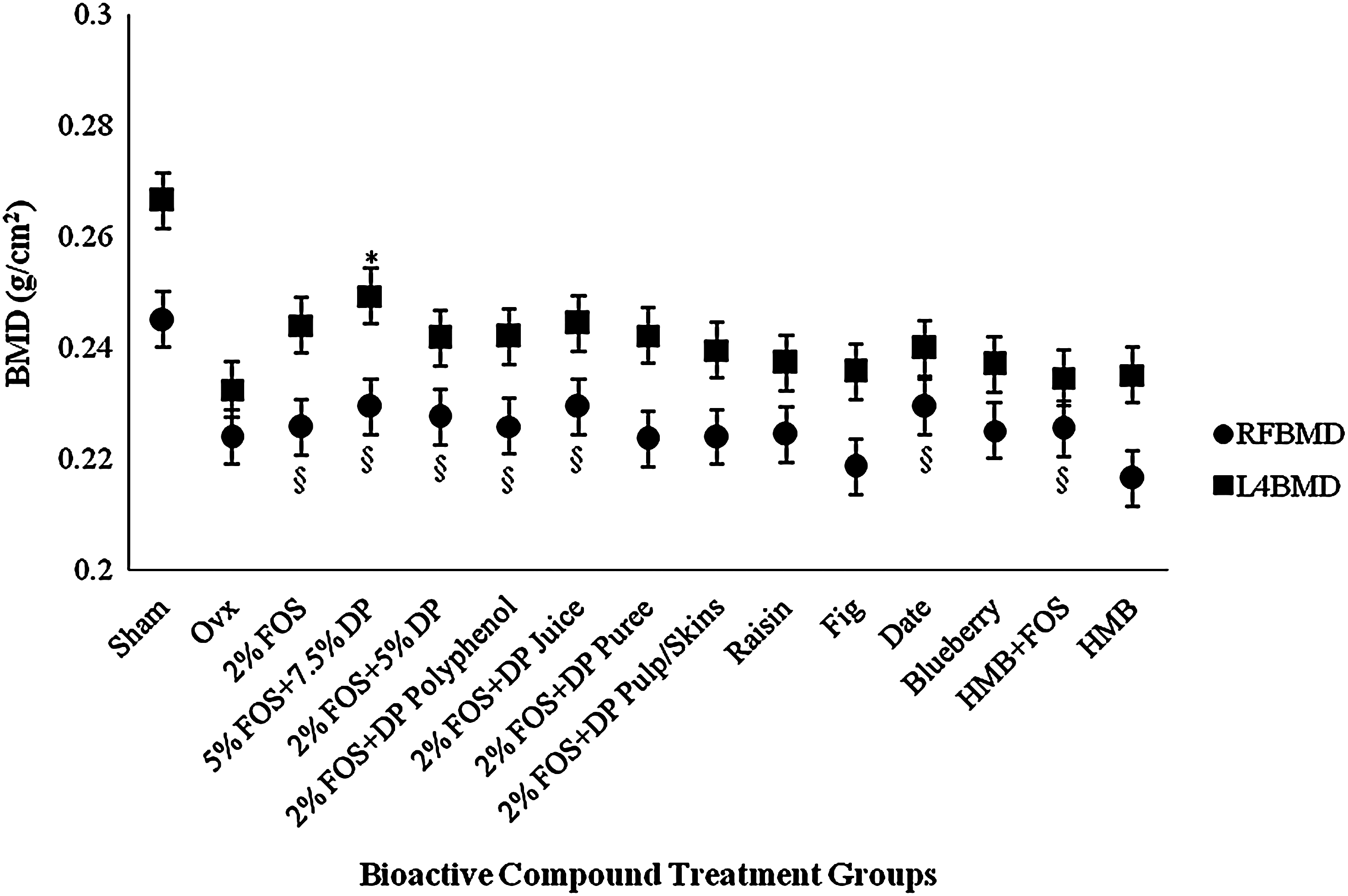
Right femur (∙) and fourth lumbar (▪) BMD. *P < .05, treatment that has a significantly improved BMD at the right femur compared to Ovx. Other bioactive treatments were found to be not significantly different from Ovx animals. § P < .05, treatment that had a significant effect on improving fourth lumbar BMD compared to Ovx. Other bioactive treatments were found to be not significantly different from Ovx animals.
In terms of fourth lumbar BMD 5% FOS + 7.5% DP had the best effect in reversing the loss of BMD as the mean BMD of this group was significantly higher than that in the Ovx controls (Fig. 1). However, the treatment was not able to bring the value of BMD up to the Sham level. All the other groups had somewhat of a modest effect as they had higher mean BMD values than the Ovx controls, albeit not significantly different (Fig. 1).
There were no significant differences in bone calcium, magnesium, and phosphorus contents of the right femur. Ovariectomy had significantly decreased the calcium content of the fourth lumbar vertebra. This loss was reversed in the 2% FOS, 5% FOS + 7.5% DP, 2% FOS + 5% DP, 2% FOS + 7.5% DP puree, 2% FOS + 7.5% raisin, 2% FOS + 7.5% fig, and 2% FOS + 7.5% date groups (Fig. 2), although the values were not significantly different from that of the Ovx controls. All the treatment groups did not show improvement in the fourth lumbar phosphorus content when compared to Sham animals. There were no differences in lumbar magnesium contents.
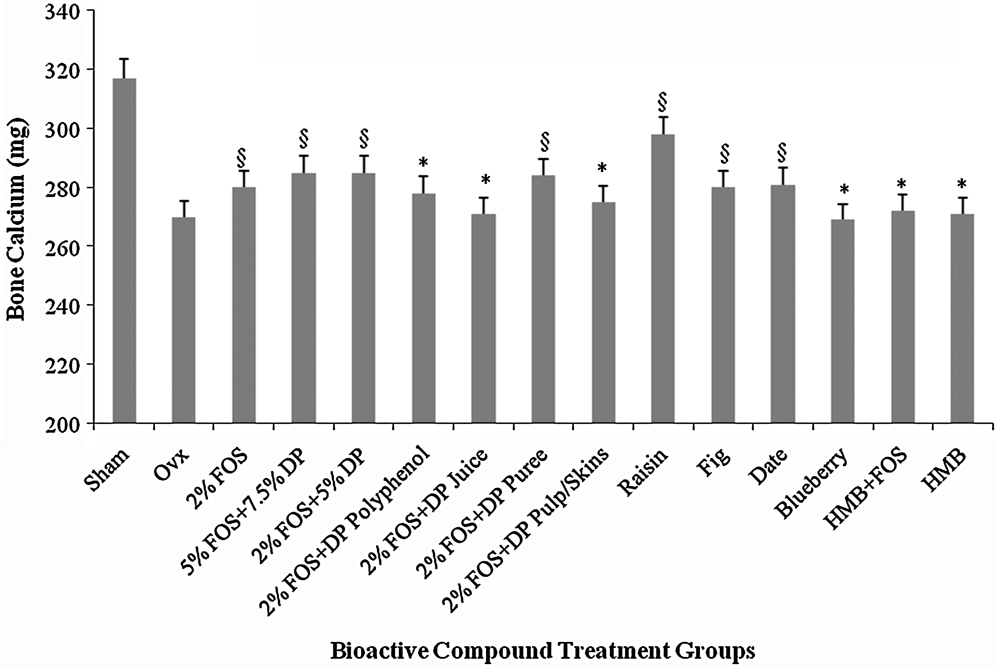
Fourth lumbar calcium content. *P < .05, treatment that is not significantly different from Ovx. § P < .05, treatment that is not significantly different from either Sham or Ovx.
Histomorphometric analyses of the femur
μCT analysis of the distal femur indicated that bone volume to total volume ratio, connectivity density, and trabecular thickness were significantly decreased and the structure model index and trabecular separation were increased by Ovx. None of the treatments altered trabecular thickness. The loss of bone volume to total volume ratio and connectivity density was not ameliorated by any of the dietary treatments. Trabecular number was significantly increased in the 2% FOS + 7.5% DP juice group only. Trabecular separation was significantly decreased by 5% FOS + 7.5% DP, 2% FOS + 2% DP polyphenol, and 2% FOS + 7.5% DP juice (data not shown).
Overall, data suggest that all the DP treatments are superior to raisin, fig, date, blueberry, and HMB treatments. Among the DP treatments 5% FOS + 7.5% DP seems to be most effective.
Serum and urine analyses (Table 2)
As expected, ovariectomy significantly reduced serum levels of E2, confirming the success of the surgery, and furthermore none of the treatments had any effect on serum estradiol levels, which indicate that the treatment do not exert true estrogenic effects. Serum OC, IGF-1, calcium, magnesium, and phosphorus were not altered by any of the treatments. Urinary excretion of Dpd (reported as nmol/mmol of creatinine), a marker of bone resorption, was significantly higher in all the Ovx groups, and none of the treatments was able to lower the Dpd excretion. There were no differences in urinary magnesium and phosphorus excretions. Urinary calcium excretion was highest in the 5% FOS + 7.5% DP and 2% FOS + 7.5% DP juice groups, whereas HMB-supplemented animals showed the lowest calcium excretion compared to the other groups.
Serum E2 is in pg/mL, OC in ng/L, IGF-1 in ng/mL, calcium, and phosphorus in mg/dL, and magnesium in mEq/L. Urinary Dpd is in nmol/mmol of creatinine, and calcium, magnesium, and phosphorus are in mg/mg of creatinine.
Within a column, values that do not share the same superscript letters are significantly (P < .05) different from each other.
Discussion
Osteoporosis-related fractures are enormous public health problems with immense socioeconomic impact. Therefore, a search for potential therapies for prevention of fractures is of vital concern. Osteoporotic fractures typically result from low BMD and BMC as well as poor bone quality. The purpose of the present study was to investigate the extent to which various bioactive constituents either alone or in combination reverse bone loss in an Ovx rat model of osteoporosis, to identify which food product of DP contained bone bioactivity, and to understand the level of FOS required to observe an effect.
Results from this study showed that diets supplemented with DP were able to improve right femur and fourth lumbar BMD in osteopenic Ovx rats while favorably modulating bone metabolism, resulting in improved BMD and biomechanical properties. Furthermore, among the DP treatment diets, supplementation with 5% FOS + 7.5% DP seemed to be most effective because of the significant improvements not only in right femur and fourth lumbar BMD but also by reversing the calcium loss of the fourth lumbar vertebrae associated with ovariectomy and lowering trabecular separation. These beneficial effects may be due to suppressing the rate of bone resorption, while allowing bone formation to continue. These findings, if confirmed in a clinical study, should have significant health implications for postmenopausal women at risk of osteoporosis.
In an attempt to understand the mechanism of action of DP, Smith's laboratory 21 showed that DP polyphenol enhances osteoblast alkaline phosphatase activity, calcified nodule formation, and type I collagen cross-linking in vitro. These alterations were suggested to be mediated by up-regulation of transcription and growth factors such as runt-related transcription factor, osterix, and IGF-1. In addition to stimulating bone formation, DP polyphenols decreased bone resporption by down-regulating receptor activator of nuclear factor κB ligand expression by osteoblasts. 21,22 DP polyphenols also suppressed osteoclast differentiation and activity under normal, oxidative stress, and inflammatory conditions in vitro. 23 DP was also able to decrease the level of high-sensitivity C-reactive protein, a marker of inflammation, in osteopenic postmenopausal women who have consumed 100 g of DP daily for 3 months (authors' unpublished data). Although antiresorptive properties of DP observed in vivo are in part mediated by polyphenols and their effects on osteoclast precursors and osteoblast-mediated signaling for osteoclastogenesis, the whole credit for bone-protective effects of DP yet cannot be given to polyphenols as other components of DP may also contribute to these positive effects.
Furthermore, our research has shown that DP is also efficacious in restoring the loss in trabecular microarchitectural properties of the tibia and the lumbar vertebra. 16 We have also shown that DP enhanced bone recovery during re-ambulation following skeletal unloading and had comparable effects to parathyroid hormone. 24,25 Our positive animal findings on the effect of DP on bone were confirmed by our short-term clinical trial 11 in which DP supplementation significantly increased indices of bone formation (alkaline phosphatase, bone-specific alkaline phosphatase levels, and IGF-1) in postmenopausal women while reducing bone resorption.
The present study also supports the proposal that certain DP fractions such as DP juice as well as DP polyphenols can reverse loss of BMD in the right femur. These findings are of importance because they can offer alternatives to individuals who may not be able to tolerate eating DP but may be able to drink its juice or take the polyphenols as a supplement. Although prune juice lacks fiber, compared to the whole food, because of filtration, it still contains similar concentrations of calcium, phosphorus, and magnesium. 26 Furthermore, the juice contains 80% of the recommended daily intake of boron, 26 which converts both estrogen and vitamin D to its active forms, making it possible that DP juice may exert bone-modulating effects similar to the whole food. It has been postulated that DP exerts its bone reversal properties because of its high polyphenol content, which is significantly lower in the juice compared to the whole food (184 mg/100 g of DP vs. 44 mg/100 g of DP juice) 26 ; however, in support of this, DP polyphenol was successful in reversing bone loss of the right femur. This combination was not successful in reversing bone loss at the fourth lumbar vertebrae, which was altered only by 5% FOS + 7.5% DP, suggesting that perhaps there may be more than the polyphenols responsible for the osteoporosis reversal effects. Interestingly, fourth lumbar BMD loss was also reversed by 2% FOS, 5% FOS + 7.5% DP, and 2% FOS + 5% DP, similar to that of the right femur; however, DP puree rather than the juice or polyphenol was able to reverse bone loss in the vertebrae, suggesting that there may be a site-specific mode of action for the DP fractions, which needs to be investigated further.
Evidence from this study has shown that there are other whole foods and a nutritional supplement that may be effective in reversing osteoporosis in addition to DP and its fractions. Date was shown to have significant bone reversal effects at both the right femur and fourth lumbar. Published literature, however, is scarce, and the use of date in bone health should be examined further because of the promising findings of this study.
HMB supplemented with 2% FOS was found to have bone loss reversal effects at the right femur, whereas raisin and fig exerted their effects at the fourth lumbar. These results again support a hypothesis that certain bioactive components of these whole foods or supplement may act in a site-specific manner, and this requires additional investigation.
Histomorphometric data support the proposal that DP treatments are efficacious in aiding in the restoration of bone and inhibition of further loss compared to the other bioactive compounds studied. This is evident by the significant reduction in trabecular separation by not only 2% FOS + 7.5% DP but the DP polyphenols and juice as well. Additionally, the DP juice showed significant increases in trabecular number, again leading to the conclusion that individuals who supplement their diet with DP as a whole food or fraction will show improvements in bone health not only through increases in BMD but also in its microarchitectural properties that contribute to improved bone strength. Together these improvements to bone will help to reduce the risk of osteoporotic fracture, and our results should be validated in a human trial to completely understand the full clinical potential of all these bioactive compounds.
The findings of this study suggest combining DP and FOS in the diet exerted benefits on right femur and fourth lumbar BMD and bone histomorphometric properties. FOS has been shown to increase absorption of minerals from the colon. 27 –29 In addition to increased mineral absorption, FOS has also been shown to increase calcium balance and bone mineralization, as well as decreasing bone turnover rate in Ovx rats 30 and mice. 31 Additionally, the results support the proposal that the 5% FOS + 7.5% DP supplementation had the greatest impact compared to the other test compounds, including other FOS + DP preparations, suggesting that there may be a critical dose of FOS and DP that is required for a positive bone outcome. This finding is important especially when trying to extrapolate the preclinical learning to recommendations for an effective dose for women trying to optimize bone health through nutritional intervention.
Footnotes
Author Disclosure Statement
C.D.J. is an employee of Abbott Nutrition. B.H.A., S.C.C., S.H., S.C.C., and M.P.A. declare no competing financial interests.