
Abstract
Genotoxic data of medicinal plants and functional foods are required as part of the risk assessment by international regulatory agencies. Due to its food consumption and ethnopharmacological relevance, pequi oil (Caryocar brasiliense Camb.) is one of these compounds to be studied. The aim of this study was to evaluate the cytotoxic, genotoxic, and clastogenic effects of the oil from the pulp of C. brasiliense (OPCB) in vivo and in vitro. Initially, the Artemia salina in vitro assay was conducted to determine the cells viability rate of different doses of the OPCB. Subsequently, comet assay (Organization for Economic Cooperation and Development, OECD 489) and micronucleus test (OECD 474) were performed in blood and bone marrow of Wistar rats treated orally with a 125, 250, 500, or 1000 mg/kg/bw of the OPCB for 4 weeks. The chemical analysis indicated the presence of β-carotene and lycopene in the oil. In the A. salina test, all OPCB doses maintained cell viability rates statistically similar to the negative control. The in vivo tests performed showed that OPCB did not show significant genotoxic or clastogenic effects in cells analyzed with the four doses tested. Altogether, these results indicate that, under our experimental conditions, C. brasiliense fruit oil did not reveal genetic toxicity in rat cells.
Introduction
M
Caryocar brasiliense Camb., popularly known as pequi or piqui, is a distributed, cultivated, and consumed plant in the Brazilian Midwest. 2 Chemical analysis of the C. brasiliense fruit showed high levels of antioxidants such as carotenoids, total phenols, 3 –5 and unsaturated fatty acids, especially oleic acid. 2,6 Antioxidants are important in preventing degenerative diseases, while oleic acid plays a fundamental role in the prevention of cardiovascular diseases. 7
C. brasiliense plays an important role in the local economy and is widely applied for vegetable oil production, in the cosmetic industry, and in traditional cuisine due to its characteristic flavor. 8 The plant is also used in the treatment of respiratory diseases such as influenza, asthma, 9 as well as to treat gastric lesions. 10 Regarding the pharmacological effects of the species, there are reports in the literature of anti-inflammatory, hypocholesterolemic, 11 antifungals, 12 and chemopreventive effects on precancerous lesions in animal models. 13,14
Several toxicological studies were conducted for this species. Fonseca et al. reports that the aqueous extract from barks and leaves caused toxic effects after acute administration in Swiss mice. 15 Castro et al. considered the fruit aqueous extract as genotoxic in Drosophila melanogaster studies. 16 Traesel et al. demonstrated the absence of toxicity of the pequi fruit pulp oil after chronic treatment in Wistar rats. 17 However, to our knowledge, safety studies on the genotoxicity of pequi fruit pulp oil have not yet been critically conducted and published.
The toxicity test against Artemia salina is a biological assay, which determines the cytotoxic effect of a substance and is used for initial screening of toxicity. 18 The comet assay is used to detect DNA strand breaks in cells of animals that have been exposed to a test substance. 19 The micronucleus test is used for detection of chromosome damage in rodent bone marrow. 20 The aforementioned assays are routinely used for an assessment of toxicological spectrum of chemicals, plants, and medicines and are required by international regulatory agencies for the production of new medicines.
Considering the food and pharmaceutical potential of C. brasiliense oil and the lack of information on the toxicity genetic of the species, the objective of this study was to evaluate the cytotoxic, genotoxic, and clastogenic potential of pequi oil in in vivo and in vitro experimental models.
Materials and Methods
Plant collection and identification
C. brasiliense fruits were collected in a Cerrado area from Campo Grande, Mato Grosso do Sul-Brazil (latitude 20° 26′ 34″ South and longitude 54° 38′ 47″ West) according to a permit issued by the Brazilian Environmental Agency (No. 54442-1—MMA/ICMBio/SISBIO). The fruits were selected according to their state of maturation. A voucher specimen was authenticated by Dra. Zefa Valdivina Pereira and deposited (No. 4752) in the Herbarium of the Federal University of Grande Dourados (UFGD).
Preparation of oil
The fruits were transported to the Laboratory of Food Technology of the Faculty of Exact Sciences and Technology (UFGD) and were sanitized with water and sodium dichloroisocyanurate dihydrate 0.66% (SumavegR) for 5 min. The pequi fruits were manually depulped and dehydrated in an air circulation oven at 40°C for 72 h. The dried pulp was cold-pressed in an “expeller” press to obtain the oil. The oil was centrifuged at 15,000 g for 15 min for complete separation of sediment. The oil was then packed in amber glass containers and kept at −8°C for further analysis.
Carotenoids characterization: high performance liquid chromatography
The sample (1 g) used for total carotenoids was solubilized with 5 mL of ethyl acetate:methanol (1:1 v:v) in ultrasonic for 15 min. The mixture was then filtered through a 0.45-μm Millex filter and directly analyzed by high-performance liquid chromatography (HPLC). The sample was analyzed using an analytical HPLC (LC-6AD; Shimadzu, Kyoto, Japan). The column ODS HYPERSIL C-18, 150 mm length × 4,6 mm inside diameter. The elution was carried out using 85% acetonitrile, 15% ethylacetate, in 15 min 50% acetonitrile, 50% ethylacetate, in 25 min returning to the initial condition. The flow rate was 0.8 mL min−1 and injected volume was 10 μL. All chromatographic analyses were performed at 22°C. The β-carotene (≥97%; Sigma-Aldrich) and lycopene (≥97%; Sigma-Aldrich) were used as standards. The compounds were identified with the aid of diode-array detector scanning in the spectral range of 200–800 nm. Compounds were easily identified based on their absorption spectra in the ultraviolet region and in retention time of standards and did not reveal interferences in retention time of the sample in HPLC. Compounds found in sample were unambiguously identified by performing coinjection experiments, in which aliquots of the sample and standard were mixed and diluted to a known volume, and analyzed through HPLC. The relative standard deviation for the retention times of replicated injections (n = 5) were <2%, there by demonstrating good repeatability. The contents estimation of the β-carotene and lycopene of the C. brasiliense was performed by external calibration using HPLC. A linear least square regression of the peak areas as function of the concentrations was performed to determine the correlation coefficients. The equation parameters (slope and intercept) of the standard curve were used to obtain the concentration values for the samples.
A. salina assay
The C. brasiliense lethality potential was evaluated using the brine shrimp larvae lethality bioassay (A. salina). Initially, A. salina eggs were immersed in a sea-salt solution and exposed to artificial light for 48 h until larvae hatch. Then, the nauplii (n = 10) were placed into wells containing different doses of the C. brasiliense pulp oil (OPCB) (10, 50, 100, 250, and 500 μg/mL). A negative control group was prepared and exposed to saline, and a positive control group was exposed to potassium dichromate (K2Cr2O7). The assays were performed in quadruplicates. The wells were maintained in artificial light for 24 h, 18 the evaluation of the nauplii survivors was performed, and the cells viability rate (%) for each treatment was calculated.
Animals
Sixty Wistar rats (Rattus norvegicus) from both sexes (30 female and 30 male) of the same age (8–10 weeks) and weight (200 g ± 15%) were used in this study. The animals were obtained from the State University of Maringa and were housed in polypropylene rodent cages (5 animals each), under standard conditions (23 ± 2°C, 40–60% humidity and 12 h light and dark cycle) and had free access to water and food. The experimental procedures were performed in accordance with the Ethical Principles in Animal Research and approved by the Ethics Committee in Animal Experimentation from the UFGD (protocol: 17/2015).
The rats were divided into experimental groups (n = 10 animals per group, 5 males and 5 females) as follows: negative control group (treated for 4 weeks with saline, orally); positive control group (treated with 50 mg/kg/bw of cyclophosphamide monohydrate, i.p.); and test groups (treated for 4 weeks with different doses of the oil: 125, 250, 500, or 1000 mg/kg/bw, orally). After the treatment period, the animals were euthanized by anesthetic overdose (isoflurane inhalation) followed by exsanguination.
Comet assay
The experimental design of the comet assay was established taking into account the guidelines of the Organization for Economic Cooperation and Development (OECD) 19 for chronic exposure. The analyses were performed on whole blood of the animals after 4 consecutive weeks of treatment.
Blood samples were collected from caudal puncture, and 40 μL were transferred to wells containing 120 μL of low-melting point agarose (1.5%) at 37°C. The mixture was homogenized and transferred to plates precoated with normal agarose (5%). After solidification of the agarose (3°C 20 min), the slides were transferred to final lysis solution for 2 h at 3°C.
After lysis, the slides were placed in electrophoresis tank filled with alkaline buffer and were left at rest for 20 min to denature the DNA. After the denaturation step, the electrophoretic run was started (20 min at 4°C with 300 mA and 25 V). Subsequently, the slides were removed from the chamber and covered by neutralization solution in three cycles of 5 min each. The slides were fixed in absolute ethanol for 10 min and then transferred to the refrigerator until the time of analysis. Slides were stained with ethidium bromide solution (0.002 mg/mL) and analyzed with a fluorescence microscope equipped with excitation filter (420–490 nm) and barrier filter (520 nm).
Each slide was identified and examined in a blind test, in which 100 cells were analyzed per animal. The comet findings were classified as follows: class 0 (no damage); class 1 (comet tail shorter than the diameter of the nucleoid); class 2 (comet tail once or twice the diameter of the nucleoid); and class 3 (comet tail greater than twice the size of the nucleoid). From the readings, two parameters were calculated for each animal: damage index and damage frequency. The damage index refers to the extent and severity of the damage observed in the cells. Damage frequency shows the amount of cells, out of the 100 analyzed cells, which have some kind of damage.
Micronucleus assay
The experimental design of the micronucleus test was established in accordance with the OECD guidelines. 20 The analyses were performed with polychromatic erythrocytes (PCEs) from the bone marrow of animals after 4 consecutive weeks of treatment to obtain chronic data.
Immediately after euthanasia, each animal had its right femur removed and separated from adjacent muscles. The two ends of the femur were cut to expose the bone marrow. The marrow canal was washed with 1 mL of fetal bovine serum so as to push the marrow into a centrifuge microtube. The suspension was centrifuged for 5 min at 1000 g. The supernatant was discarded, and the pellets were used to make smears on slides. The slides were fixed with methanol for 10 min and stained with Giemsa for 15 min.
Each slide was identified and analyzed under blind conditions, and 2000 PCEs were analyzed per animal. Each erythrocyte was identified by the presence or absence of micronuclei. To evaluate the possible cytotoxic effects, the ratio of polychromatic and normochromatic erythrocytes (PCEs/NCEs) was calculated by analyzing 200 random erythrocytes per animal.
Statistical analyses
The results are reported as mean ± standard error of the mean. The normality of the samples was checked using the Kolmogorov–Smirnov test. The differences between groups were determined by analysis of variance (one-way) followed by Tukey test. P-values <.05 were set as the level of significance. The statistical analyses were performed by GraphPad Prism software version 5.00 for Windows.
Results
Characterization of the oil
The analysis of the compounds present in the OPCB was performed by HPLC with diode-array detector. The data are presented in Table 1 and Figure 1. The chromatographic analysis indicated the presence of β-carotene and lycopene in the oil.
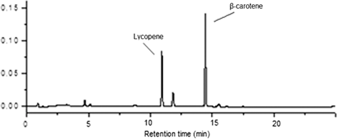
Composition of carotenoides (lycopene and β-carotene) in the OPCB. OPCB, Caryocar brasiliense pulp oil.
Linear regression, formula: y = a + bx, where y = ratio of peak areas; x = concentration (μg); a = intercept; and b = slope.
A. salina assay
The results of this test (Fig. 2) show the cell of the OPCB when tested in A. salina nauplii in doses of 10, 50, 100, 250, and 500 μg/mL when compared with the negative and positive controls. The test was performed in quadruplicate and the results demonstrate the low lethality of C. brasiliense oil in this experimental model.
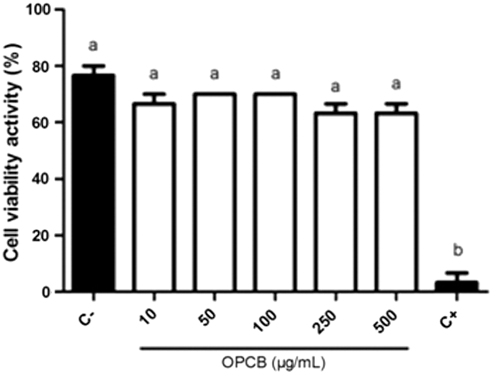
Effects of treatment with OPCB and K2Cr2O7 (positive control) on the Artemia salina assay. Results are presented as mean ± SEM, n = 10, in quadruplicate. One-way ANOVA followed by the Tukey test was used (P < .05). Different letters (a and b) indicate statistically significant differences. Cell viability activity: percentage of surviving nauplii. ANOVA, analysis of variance; SEM, standard error of the mean.
Comet assay
The results of the comet assay in this study were divided into damage index (Table 2) and frequency of damage (Fig. 3). None of the tested doses of the OPCB (125, 250, 500, and 1000 mg/kg/bw) induced any significant damage and was statistically similar to animals from the negative control.
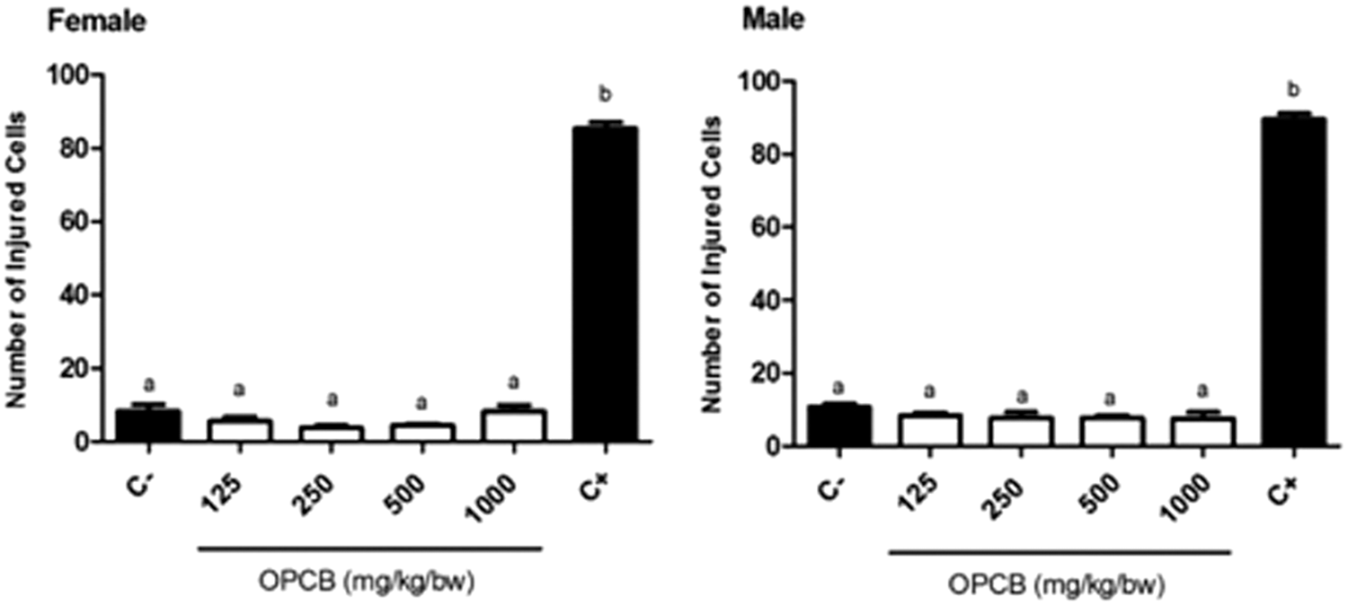
Effects of treatment with OPCB and cyclophosphamide (positive control) on the DNA damage frequency using the peripheral blood of female and male Wistar rats. Results are presented as mean ± SEM, n = 5 animals per group. One-way ANOVA followed by the Tukey test was used (P < .05). Different letters (a and b) indicate statistically significant differences. Damage frequency: number of cells with damage in 100 cells analyzed.
Results are presented as mean ± standard error of the mean, n = 5 animals per group. One-way analysis of variance followed by the Tukey test was used (P < .05). Different letters indicate statistically significant differences. Damage index: type of damage × number of cells with damage.
Micronucleus assay
Figure 4 shows the difference between the means of micronucleated PCEs (MN-PCEs) in all experimental groups. All OPCB-treated animals (125, 250, 500, and 1000 mg/kg/bw) showed the number of MN-PCEs in a similar manner to the control negative group. Similarly, the treatment with OPCB did not cause any significant change in the PCEs/NCEs ratio compared with negative control group (Fig. 5).
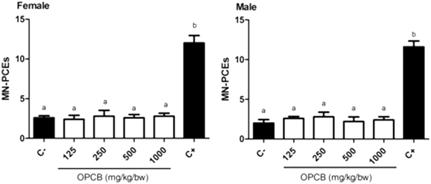
Effects of treatment with OPCB and cyclophosphamide (positive control) on the counts of MN-PCEs using the bone marrow of female and male Wistar rats. Results are presented as mean ± SEM, n = 5 animals per group. One-way ANOVA followed by the Tukey test was used (P < .05). Different letters (a and b) indicate statistically significant differences. A total of 2000 cells were analyzed in each animal. MN-PCEs, micronucleated polychromatic erythrocytes.
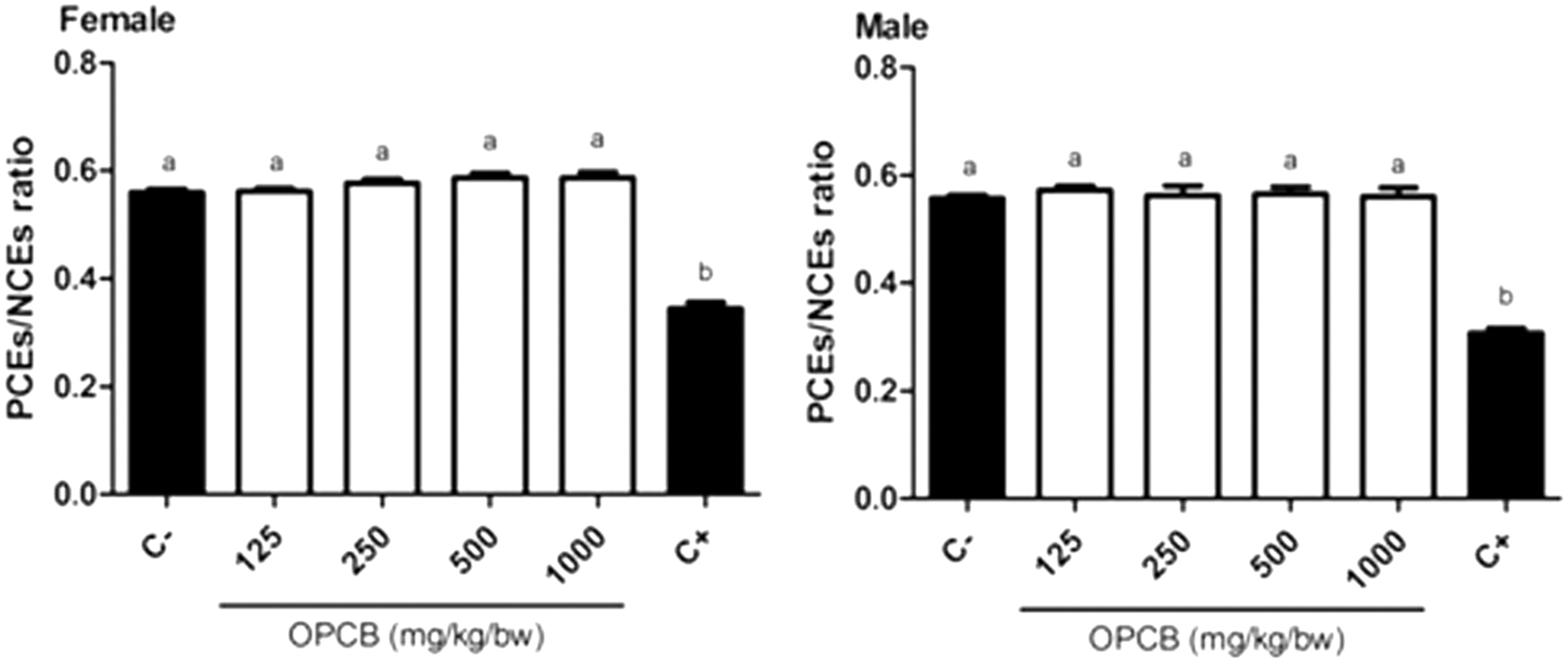
Effects of treatment with OPCB and cyclophosphamide (positive control) on the PCEs/NCEs ratio using the bone marrow of female and male Wistar rats. Results are presented as mean ± SEM, n = 5 animals per group. One-way ANOVA followed by the Tukey test was used (P < .05). Different letters (a and b) indicate statistically significant differences. A total of 100 cells were analyzed in each animal. PCEs/NCEs, polychromatic to normochromatic erythrocytes.
Discussion and Conclusion
Plant-derived products have been widely used throughout the world for the production of food and drugs. The consumption of medicinal plants and functional foods are being accepted as an alternative to conventional allopathic medicine in developing and developed countries. 21 While the widespread use of herbal compounds increases, there are concerns regarding the safety of these products. 22 The genetic toxicity study is crucial in the safety assessment of any product of plant origin. In this study, for the first time, we present the genotoxicity parameters of an oil (OPCB) routinely consumed in several regions of Brazil.
Chemical studies performed with pequi showed that this fruit contains bioactive components with important antioxidant properties. There are reports on the phenolic compounds such as gallic acid, quinic acid, 23 and some specific flavonoids such as quercetin, 24 besides tannins, saponins, 25 and oleic acid. 17 Other authors presented data on the carotenoid composition of the oil, highlighting the presence of β-carotene and lycopene. 26,27 In this study, the chromatographic analysis of carotenoids confirmed the compounds described in the literature. These substances are of large importance to the ethnopharmacology due to of its biological properties, in particular, at maintenance of the immune system and prevention of chronic diseases.
The relationship between carotenoids intake and health benefits are well established, since carotenoids act as vitamin A precursors, performing antioxidant function. 28 The antioxidant potential of medicinal plants plays an important role in genetic toxicology, since reactive oxygen species can cause DNA strand breakage. DNA damage caused by oxidation is considered as the most relevant type of damage to cell metabolism. 29 Evidences suggest that cumulative damage to DNA caused by reactive oxygen species contributes to various adverse clinical conditions, such as cancer. 30 For this reason, the consumption of antioxidants compounds is directly associated with the prevention of the development of diseases such as cancer.
Several biological assays can be used for monitoring and initial screening of the toxicological potential of plant extracts. The lethality test with A. salina is a simple, quick, and inexpensive method used in natural product research. The procedure determines viability rates of compounds in extracts and can be correlated with acute oral toxicity data in animals. 18 In this study, none of the OPCB doses was able to promote significant lethality in the A. salina nauplii, indicating the low toxicity of the oil in this experimental model. These results correlate with the high LD50 reported for OPCB in the literature (above 2000 mg/kg/bw), 17 corroborating the hypothesis of low toxicity of pequi oil.
Genotoxicity studies are in vitro, and in vivo assays are designed to detect the potential of substances under investigation to cause gene mutations and chromosomal abnormalities, 31 since these events are considered important prerequisites for the development of adverse health effects such as cancer. Genotoxicity is not a measurement for carcinogenicity, but is often used as an indicator for cancer, once this test measures an initial or intermediate event in tumorigenesis.
The comet assay is a sensitive and reliable method to detect lesions in DNA strands. This test is considered to be a genotoxic test since these lesions are still amenable to repair. 19 In this study, regarding the two parameters evaluated in the comet assay, the negative control group showed lower values of lesions in analyzed cells when compared with the positive control group. This confirms the low rates of genetic damage in healthy rats and confirms that the administration of cyclophosphamide in the positive control group resulted in the expected effect. All doses of the oil administered chronically to the animals showed damage index and frequency of damage similar to the negative control and different to the positive control. In all groups treated with the oil, most of the observed cells showed no damage to the DNA, confirming that the administration of the OPCB resulted in no genotoxic effect in treated animals.
The second cytogenetic assay performed in the present study to verify the genotoxic potential of OPCB was the micronucleus test. The micronucleus test is a biological assay performed to detect clastogenic and aneugenic agents. It is one of the most well-established tests in the field of genetic toxicology, being widely accepted by international regulatory agencies and providing important mutagenic data. 20 The increased MN-PCEs frequency may play an important role in the neoplastic development of certain tumors. In this study, there was no difference in the number of MN-PCEs among the negative control and groups treated with four doses of the OPCB. As expected, the animals treated with cyclophosphamide showed a frequency of MN-PCEs higher when compared with all other groups. These results are in accordance with the comet assay and indicate that the oil of C. brasiliense pulp showed no clastogenic or aneugenic effect on the bone marrow cells of the rats, treated with OPCB for 28 consecutive days.
Besides the clastogenic effects, the micronucleus test also allows the determination of the cytotoxic effects of the test substance by analyzing the relationship between PCEs/NCEs in bone marrow samples. Compounds that reduce the production of young erythrocytes (polychromatic) are considered cytotoxic. 32 The values of the ratios between the two cell maturation were statistically similar among the four groups treated with the OPCB and the negative control group. These results indicate that pequi oil is not able to affect the production of new erythrocytes at cord level and, therefore, is not cytotoxic for this cell type.
The results of our in vivo tests are in accordance with data obtained from tests of the antimutagenic effect of pequi pulp. In 2008, two studies evaluated the efficacy of the aqueous extract of the fruit pulp against the mutagenicity induced by the cyclophosphamide and bleomycin agents in bone marrow of mice 9 and rats. 33 The results of both studies show the antimutagenic potential of the extract, which protected the DNA of the animals investigated against the genotoxic action of the drugs. The authors related these results to the antioxidant properties of the fruit.
Several plants with important ethnopharmacological and food use were assessed for genotoxic potential in recent years. The oils obtained from the fruits of Acrocomia aculeata 34 and Attalea phalerata, 35 for instance, demonstrated low genotoxicity through the comet and micronucleus assays. However, other extracts showed significant genetic toxicity. In the last two decades, 458 studies evaluated the genotoxic potential of medicinal plants, to which the genotoxic activity was reported in 28.4% of them. 36 This high incidence of positive results reinforces the need for toxicological evaluations of plants and their derivatives with nutritional and medicinal interest.
In conclusion, under the experimental conditions used in the present work, our findings provide the first evidence that the C. brasiliense fruit oil, administered by gavage, is not genotoxic in the leukocytes of rats, and by the micronucleus test, that it has no clastogenic/aneugenic effect on the bone marrow either. Chemical analysis indicated that the oil is rich in carotenoids, with marked presence of lycopene and β-carotene. The data obtained are important since they support the use of a species of economic, nutritional, and pharmacological importance.
Footnotes
Acknowledgment
The authors thank Dr. Ghazal Nabi Edavile for providing language help.
Author Disclosure Statement
No competing financial interests exist.