
Abstract
Although leaves of Anchietea salutaris are used in Brazilian traditional medicine, there is no available data in the literature proving its efficacy and safety. Thus, the aim of the study was to perform a meticulous botanical, phytochemical, toxicological, and pharmacological investigation of A. salutaris in Wistar rats. At first, a morphoanatomical characterization of Anchietea pyrifolia leaves and stems was performed. Then, a purified infusion (ethanol-soluble fraction obtained from A. pyrifolia [ESAP]) was obtained followed by its chemical profile elucidation. Furthermore, an acute toxicity test was performed, and the acute and prolonged diuretic and hypotensive effects were also evaluated in Wistar rats. Finally, the vasodilatory responses of ESAP in mesenteric vascular beds were investigated. The main secondary metabolites identified from ESAP were O-glycosylated flavonoids, chlorogenic acids, and phenylpropanoic acid derivatives. ESAP did not promote any toxic effects in female rats nor increased urinary excretion in male rats after a single exposure. However, ESAP significantly reduced renal elimination of sodium, potassium, and chloride after prolonged treatment. An ESAP highest dose promoted significant acute hypotension without affecting blood pressure levels after prolonged use. Furthermore, its cardiovascular effects seem to be related with the calcium-activated potassium channel activation in resistance vessels.
Introduction
Hypertension is a medical condition characterized by sustained blood pressure elevation (≥140 and/or 90 mm Hg). Known as the most recurrent noncommunicable chronic diseases in the world, hypertension affects people of all ages, reaching about 32.5% of adults and over 60% of the Brazilian elderly. 1,2
In an attempt to reduce the incidence and minimize the effects of hypertension, drug and nondrug therapies are routinely employed. In pharmacological treatment, renin/angiotensin system inhibitors, betablockers, calcium channel blockers, and diuretics are used as first-line agents. 2 However, patients’ adherence to conventional treatments is still low and about one-third of the population present controlled blood pressure levels. This fact can be explained by the number of collateral effects that occur to some patients or difficulties of access to antihypertensive drugs by the low-income population that uses the Brazilian Health System. Thus, even though some classes of antihypertensive drugs are distributed free of charge to the population by the government, these drugs are not always available. 3
Therefore, the use of medicinal plants can be seen as an alternative to reduce unwanted side effects, avoid irrational consumption of manufactured drugs, and solve problems in primary care. 2 The desire for a more natural treatment is intensifying among people and, therefore, medicinal species have become the target of numerous studies to demonstrate its efficacy and safety, aiming at the development of new herbal medicines. 4 For this reason, the Brazilian cerrado was chosen as the main focus of this research, since it is the second largest biome in the area, representing around 23% of the country's surface. 5
Anchietea pyrifolia (Mart.)
Although leaf infusion of A. pyrifolia is used in folk medicine as it presents diuretic effects, there are no reports in the literature demonstrating its efficacy and safety. Therefore, we performed a meticulous botanical, phytochemical, toxicological, and pharmacological investigation of A. pyrifolia in Wistar rats after acute and prolonged exposure. Furthermore, we investigated the molecular mechanisms adjacent to their pharmacological effects using isolated mesenteric vascular beds (MVBs).
Materials and Methods
Chemicals
Xylazine, ketamine hydrochloride (Syntec, São Paulo, SP, Brazil), heparin (Hipolabor, Belo Horizonte, MG, Brazil), 4-aminopyridine (4-AP), acetylcholine, dextrose, ethylenediaminetetraacetic acid, indomethacin, hydrochlorothiazide (HCTZ), Nω-Nitro-L-arginine methyl ester, phenylephrine, sodium deoxycholate, NaCl, KCl, NaHCO3, MgSO4, CaCl2, tetraethylammonium (TEA), and KH2PO4 (Sigma-Aldrich, St. Louis, MO, USA). All other reagents were obtained in analytical grade.
Plant collection and identification
A. pyrifolia (Mart.)
Extract preparation
Leaves were sanitized with tap water and dried in a circulating air oven at 48°C for 2 days. Dried and pulverized leaves of A. pyrifolia (60 g) were suspended in 1 L of boiling water (97°C) and the mixture obtained was then cooled to room temperature for ∼5 h. Subsequently, the plant material was filtered and the infusion was treated with three volumes of ethanol, originating a precipitate and an ethanol-soluble fraction (ethanol-soluble fraction obtained from A. pyrifolia [ESAP]). In the end, ESAP samples were lyophilized, stored in a freezer at −18°C, and used for further studies.
Pharmacobotanical assays
Anatomical study
A. pyrifolia leaves and stems were fixed in formalin–acetic acid–alcohol (FAA) solution 11 for 3 days, sanitized with distilled water, and then stored in 70% ethanol (v/v). 12 The fixed samples of mature leaves and stems from the third node were hydrated and cut by hand using razor blades. The sections were double stained with basic Fuchsin and Astra Blue. 13 For detachment of epidermis, fixed leaves were washed in distilled water, immersed in a solution of commercial bleach (5.25% sodium hypochlorite) until translucent. Then, the samples were immersed briefly in a diluted acetic acid solution, washed with water, and stained in 1% Safranin in ethanol 50%. Slides were mounted in glycerin 50% solution and sealed with transparent nail polish. 14
The histochemical analysis was performed in sections of fresh leaves and stems with razor blades for the detection of lignin, lipids, starch, and phenolic compounds. Standard solution of phloroglucinol/HCl was used to identify lignified tissues, 15 Sudan III to detect the occurrence of lipophilic compounds, 16 iodine solution (iodine potassium iodide) to stain starch, 12 and ferric chloride 11 and potassium dichromate 17 to detect the presence of phenolics. Blank sections were used for comparative analysis. Samples were analyzed and photomicrographs were prepared using an Olympus CX31 microscope equipped with Olympus C-7070 digital camera.
Field emission scanning electron microscopy and energy-dispersive X-ray spectroscopy analyses
The plant material fixed in FAA was sectioned and passed through a series of ethanol solutions (80%, 90%, and 100%). The fully dried sections were mounted on aluminum stubs with double-sided adhesive tapes and coated with gold using a sputter coater (Quorum SC7620). The sections were analyzed and imaged using a Mira 3 Tescan field emission scanning electron microscopy (FESEM; Oxford Instruments, Oxford, United Kingdom) in high vacuum mode at 15 kV accelerating voltage. Qualitative and quantitative X-ray microanalyses were done for certain crystals using an energy-dispersive X-ray spectroscopy (EDS) detector attached to the FESEM. The FESEM and EDS analyses were performed in the State University of Ponta Grossa at the multiuser laboratory (c-LABMU).
LC-DAD-MS analyses
ESAP was analyzed by LC-DAD-MS using a UFLC Shimadzu Prominence coupled to diode array detector and mass spectrometry (MicroOTOF-Q III Bruker Daltonics). The analyses were performed on Kinetex column chromatographic C18 (2.6 μ, 100A, 150 × 2.1 mm; Phenomenex), applying the same parameters described by Dembogurski et al. 18 ESAP was prepared with methanol and deionized water (7:3 v/v) at concentration 1 mg/mL, filtered on Millex filters (0.22 × 13 mm PTFE; Millipore), and 2 μL was injected in the chromatographic system. The mass spectrometry (MS) analyses were performed in negative and positive ion modes, and the constituents were identified according to the spectral data (ultraviolet [UV], MS, and MS/MS) compared with data obtained in the literature.
Safety and efficacy assessments
Animals
Male and female Wistar rats (8–12 weeks) were housed at the bioterium under standard and controlled conditions (22°C ± 2°C; 12-h light/12-h dark cycle, 50% ± 20% of relative humidity) and had free access to water and commercial food. Before the onset of the experiments, rats were left for 10 days to acclimatize to new laboratory conditions. All animal experiments presented in this work were previously approved by the Ethics Committee in Animal Experimentation from the Federal University of Paraná (protocol: 05/2017) and UFGD (protocol: 21/2017).
Study design
All animals were randomly divided into four groups (n = 8) for the acute toxicity test and into five groups (n = 6) for the diuretic and blood pressure investigations. Rats were treated with a single dose (acute toxicity and acute diuretic activity) or once a day, for 7 days (prolonged investigations), with ESAP (30, 100, and 300 mg/kg), HCTZ (25 mg/kg), or vehicle (filtered water 1 mL/100 g) by oral gavage.
Safety assessment
Acute toxicity test
ESAP oral acute toxicity was evaluated in female rats according to the protocol 425 established by the Organization for Economic Co-operation and Development (OECD) in 2008. 19 After a 12-h period of fasting (only food was withheld overnight), three different doses (30, 300, and 2000 mg/kg) of ESAP were administered to rats (n = 8) by oral gavage. Vehicle (filtered water) was administered to the control group (1 mL/100 g). Food was only given to rats 1 h after treatment. Animals were carefully observed during the first 24 h for any signs of death or toxicity and thereafter, for 14 consecutive days. Body weight, food, and water intake were evaluated daily and registered. To observe possible changes in behavior induced by ESAP, rats were closely examined for any changes in activities on the central nervous system, consciousness, activity and coordination of motor system and muscle toning, corneal and headset reflexes, and activities on the autonomic nervous system. 20 Such parameters were previously described by Malone and Robichaud in 1962 and are widely know as the “hippocratic screening.”
The night before euthanasia, animals were kept under fasting conditions (10 h) and had free access to water. On day 15 after treatment, animals were euthanized by isoflurane anesthesia (inhalation) followed by exsanguination. Heart, lung, spleen, liver, kidney, uterus, and ovaries were removed, properly cleaned, weighed, macroscopically examined, and the relative organ weight calculated. Heart, liver, and kidney samples were sent to histopathological analyses that were performed by two veterinary pathologists from UFGD.
Pharmacological investigations
Acute diuresis
The diuretic activity of ESAP was investigated according to a methodology previously described by Gasparotto Junior et al. 21 Fasted male rats were divided into five groups (n = 6) and received, by oral gavage, a single dose of 5-mL/100 g of physiological saline solution (0.9% NaCl). Such procedure is of utmost importance to impose controlled water and salt balance. Afterward, each group received one single administration of ESAP (30, 100, and 300 mg/kg), HCTZ (25 mg/kg), or vehicle (filtered water 1 mL/100 g) by oral gavage. Immediately after treatments, animals were placed in individual metabolic cages for 24 h with free access to commercial feed and filtered water. Urine was collected and the volume recorded at 8 and 24 h (expressed as mL/100 g of body weight). Urinary levels of potassium (K+), sodium (Na+), and chloride (Cl−) were quantified with the aid of an ion selective meter (COBAS INTEGRA 400 plus; Roche). Urinary excretion load of K+, Na+, and Cl− was obtained by multiplying the concentration of electrolytes (mEq/L) by the urinary flow (mL/min). Data are expressed as μEq/min/100 g. Density and pH were determined on fresh urine samples using a handheld refractometer (NO107; Nova Instruments, Brazil) and a digital pH meter (Q400MT; Quimis Instruments, Brazil), respectively.
Prolonged diuretic activity
Rats (n = 6) were treated with ESAP (30, 100, and 300 mg/kg), HCTZ (25 mg/kg), or vehicle (filtered water 1 mL/100 g) once daily, for 7 days. Each animal was individually placed in metabolic cages during the experimental period. The total amount of urine was collected every 24 h and the following parameters were analyzed: pH, density, volume, and electrolyte concentrations (Na+, K+, and Cl−).
Arterial pressure and heart rate evaluation
Thirty normotensive male rats, equally divided into five groups (n = 6), were intramuscularly anesthetised with ketamine and xylazine (100 mg/kg; 20 mg/kg) followed by a subcutaneous bolus injection of heparin (50 IU). Tracheotomy was performed to allow animals to spontaneously breathe and the left carotid artery was isolated, cannulated, and connected to a pressure transducer coupled to a PowerLab® recording system. An application program (Chart, v 4.1; all from ADI Instruments, Castle Hill, Australia) recorded the systolic blood pressure (SBP), diastolic blood pressure (DBP), mean arterial pressure (MAP), and heart rate (HR). After that, rats received ESAP (30, 100, or 300 mg/kg), HCTZ (25 mg/kg), or water (1 mL/100 g of body weight) intraduodenally and changes in SBP, DBP, MAP, and HR were recorded for 20 min.
For measurement after prolonged treatment, normotensive rats (n = 6) were treated with ESAP (30, 100, and 300 mg/kg), HCTZ (25 mg/kg), or vehicle (filtered water 1 mL/100 g) by oral gavage for 7 days. After the treatments, rats were anesthetized and prepared for the measurement of the hemodynamic parameters as previously described. After the stabilization period (15 min), changes in SBP, DBP, MAP, and HR were recorded for 5 min. At the end of the experimental period, blood samples from the left carotid artery were collected and serum levels of urea, creatinine, Na+, and K+ were quantified. Animals were then euthanized with an overdose of isoflurane anesthetic (inhalation, two to three times of the anesthesic dose).
Effects on peripheral vascular resistance
First, normotensive male rats were anesthetized with ketamine and xylazine (100 and 20 mg/kg) by intraperitoneal route. MVBs were isolated and prepared according to methods previously described by McGregor. 22 MVBs (n = 5) were put in a water-jacketed organ bath and perfused (at 4 mL/min) with physiological saline solution (PSS) (composition in mM: NaCl 119; KCl 4.7; CaCl2 2.4; MgSO4 1.2; NaHCO3 25.0; KH2PO4 1.2; dextrose 11.1; and EDTA 0.03) at 37°C and gassed with 95% O2/5% CO2. With the aid of a pressure transducer coupled to a PowerLab recording system and an application program (Chart, v 4 .1), changes in perfusion pressure (PP, mm Hg) were identified. After equilibration (45 min), its integrity was confirmed by a bolus injection of KCl (120 mmol). To observe the endothelial viability of preparations, different MVBs were continuously perfused with PSS plus Phe (3 μM) to promote a prolonged increase in PP. Afterward, a bolus injection of ACh (1 nmol) was performed and the PP reduction was measured. Some preparations were perfused with PSS containing sodium deoxycholate (1.8 mg/mL) for 30 sec to remove chemically the endothelium of MVBs. Then, aiming to prove the loss of endothelial responsiveness, preparations were continuously perfused with PSS plus Phe (3 μM), and following sustained PP increase, a dose of ACh (1 nmol) was applied into the perfusion system.
MVBs containing or not functional endothelium were continuously perfused with PSS plus Phe (3 μM) and, right after the stabilization period, different preparations received bolus injections of ESAP (0.1, 0.3, and 1 mg) and the PP reduction was measured. The following doses were only administered after the return of the PP to the same level previously recorded, with a minimal interval of 3 min between doses. Then, MVBs were perfused with PSS containing Phe (3 μM) plus indomethacin (1 μM; a nonselective cyclooxygenase inhibitor), L-NAME (100 μM; a nonselective nitric oxide [NO] synthase inhibitor), KCl (40 mM), TEA (5 mM; a nonselective calcium-sensitive [KCa] K+ channel blocker), 4-AP (10 μM; a voltage-dependent [KV] K+ channels blocker), and glibenclamide (GLB; 10 μM; a selective Kir6.1 ATP-sensitive [KATP] K+ channels blocker), used combined or alone. After 15 min of continuous perfusion, ESAP (0.1, 0.3, and 1 mg) was once again injected into the perfusion system and its ability to reduce PP in the presence and absence of different inhibitors was evaluated.
Statistical analyses
Analyses were performed using one-way analysis of variance followed by Dunnett's test or Student's t-test. Results are expressed as mean ± standard error of the mean. P-value of less than .05 were set as the level of significance. All graphs and analyses were performed using GraphPad Prism Mac 6.0h.
Results
Anatomical profile and histochemical characterization
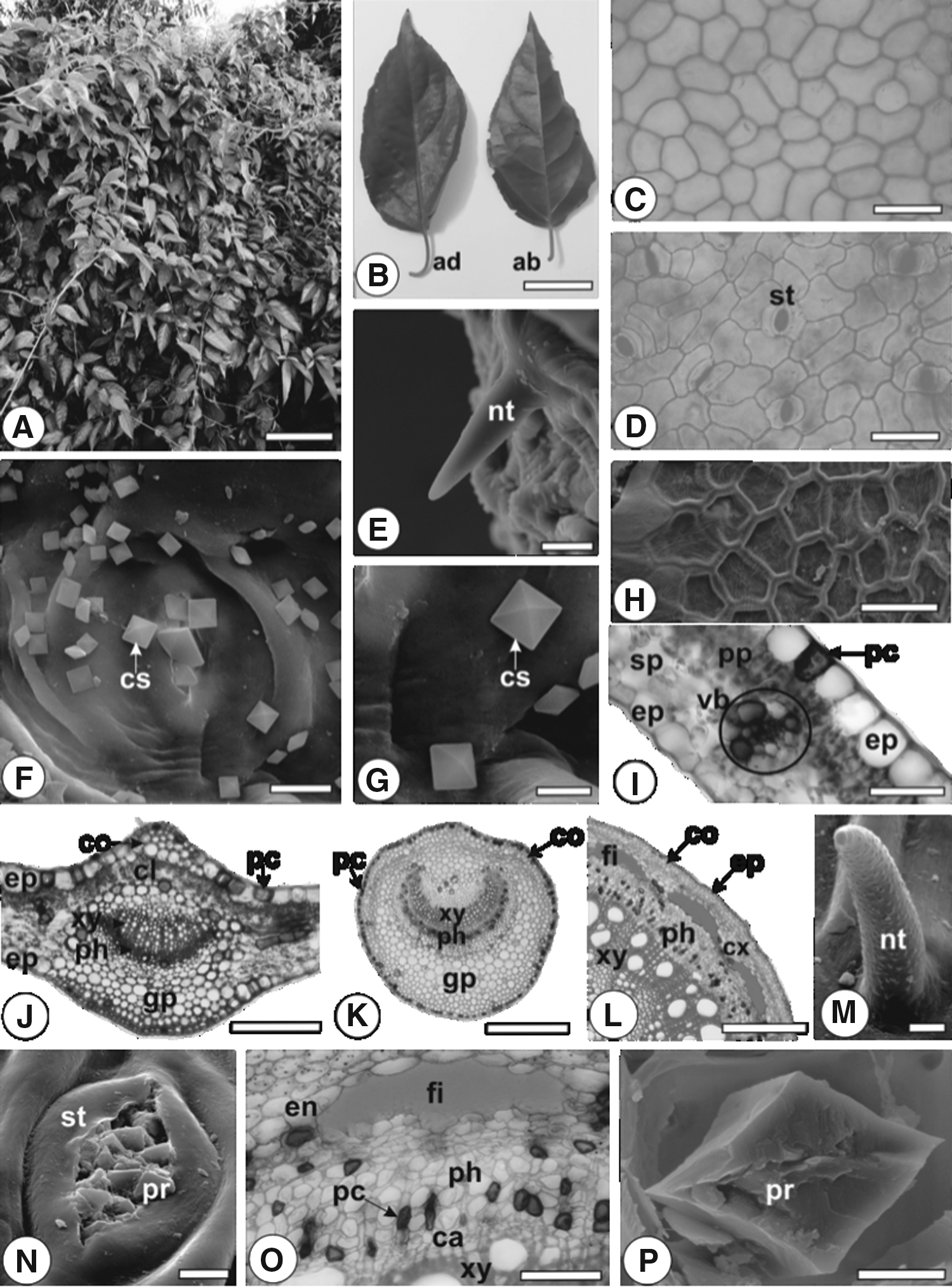
Anatomically, the leaves of A. pyrifolia (Mart.)
Morphoanatomy of Anchietea pyrifolia (
The leaf, in cross-section, shows one-layered epidermis covered by thin and slightly striate cuticle (Fig. 1I). The cells located in the upper surface are bigger than in the lower surface and anticlinally elongated (Fig. 1I, J). The mesophyll is dorsiventral and is formed by one to two layers of the palisade and about four layers of spongy parenchyma (Fig. 1I, J). The veinlets traversing the mesophyll region are represented by small collateral vascular bundles enclosed by parenchymatic sheath (Fig. 1I).
The midrib, in transection, has biconvex shape (Fig. 1J). The epidermis is uniseriate and covered by slightly thick and striate cuticle. Crystal sand with the same features as previous described for the leaf blade is found on the epidermis. Nonglandular trichomes are also observed. Beneath the epidermis, up to four layers of angular collenchyma is found in the adaxial side and one layer in the abaxial side. In the adaxial side, chlorenchyma is continuous. Some crystalliferous idioblasts containing prismatic crystals or druses and tiny starch grains are found in the ground parenchyma. The vascular system is represented by unique and central collateral vascular bundle arranged in open arc (Fig. 1J).
In cross-section, the petiole is rounded in shape with a slight raise on the adaxial side. A thin and striated cuticle covers the single-layered epidermis that also shows crystal sand on the outer surface. Beneath the epidermis, a continuous angular collenchyma formed by up to three layers is found. The stele is represented by one collateral vascular bundle in open arc. Cambia can be observed. Starch grains and crystalliferous idioblasts (Fig. 2F), as described in the midrib, are found in the ground parenchyma.

Histochemistry of A. pyrifolia (
The stem is circular in shape. The epidermis has the same features described for leaves, showing nonglandular trichomes (Fig. 1M) and crystal sand on outer surface that seem to come from stomata (Fig. 1N). Beneath the epidermis, three-layered angular collenchyma occurs as an uninterrupted ring. Crystalliferous idioblasts containing druses and prisms (Fig. 1P) are found in the cortex. An endodermis bounds the internal part of the cortex (Fig. 1O). The vascular cylinder shows cambia that produces inward xylem and outward phloem, and perivascular fiber caps abutting the phloem (Fig. 1L, O). The pith is made up of thin-walled parenchymatous cells and some of them contain phenolic compounds (Fig. 2J).
Phenolic compounds are present in epidermal cells in the leaf blade (Figs. 1I, 2A, B), midrib (Figs. 1J, 2C), petiole (Fig. 1K), and stem, beyond in the cells near the minor vascular bundles in the leaves (Fig. 1I), gathered in the ground parenchyma (Fig. 1J) and in the phloem (Fig. 2D) of midrib and petiole (Fig. 1K). Tanniniferous cells were found in the collenchymatous tissue in the petiole of species of genera Rinorea and Fusispermum, both belonging to the Violaceae family. 23
Lipophilic compounds are observed in the cuticle of leaves, petioles (Fig. 2F), and stems (Fig. 2K); lignified elements were seen in the fibers of petioles (Fig. 2E) and stems (Fig. 2H), beyond in the vessel elements of leaves, petioles (Fig. 2E), and stems (Fig. 2H), and in the cells of perimedullary region (Fig. 2I); tiny, isolated or aggregated starch grains are found in the ground parenchyma of midrib, petiole, stem, and in the perimedullary region of stem (Fig. 2G).
The EDS spectra of the crystals present in A. pyrifolia show large peaks of calcium, carbon, and oxygen (Fig. 3). These results confirm that the chemical composition of these crystals is calcium oxalate. The EDS spectrum of bipyramidal crystal sand on the epidermis (Fig. 3A) shows prominent peaks of calcium (53.67%), carbon (24.43%), and oxygen (21.90%). Whereas, the EDS spectrum of a prismatic crystal (Fig. 3B) shows major peaks of calcium (40.33%), carbon (14.55%), and oxygen (45.12%) and of a druse (Fig. 3C) presents major peaks of calcium (47.49%), carbon (12.26%), and oxygen (40.25%). The unlabeled peaks in the spectra characterize conductive metal used for covering the samples for FESEM analysis. Hoyos-Gómez 23 in 2015 studied 10 species of the Violaceae (Rinorea and Fusispermum genus) and found calcium oxalate crystals, as rhombic crystals and druses, in the collenchyma tissue.
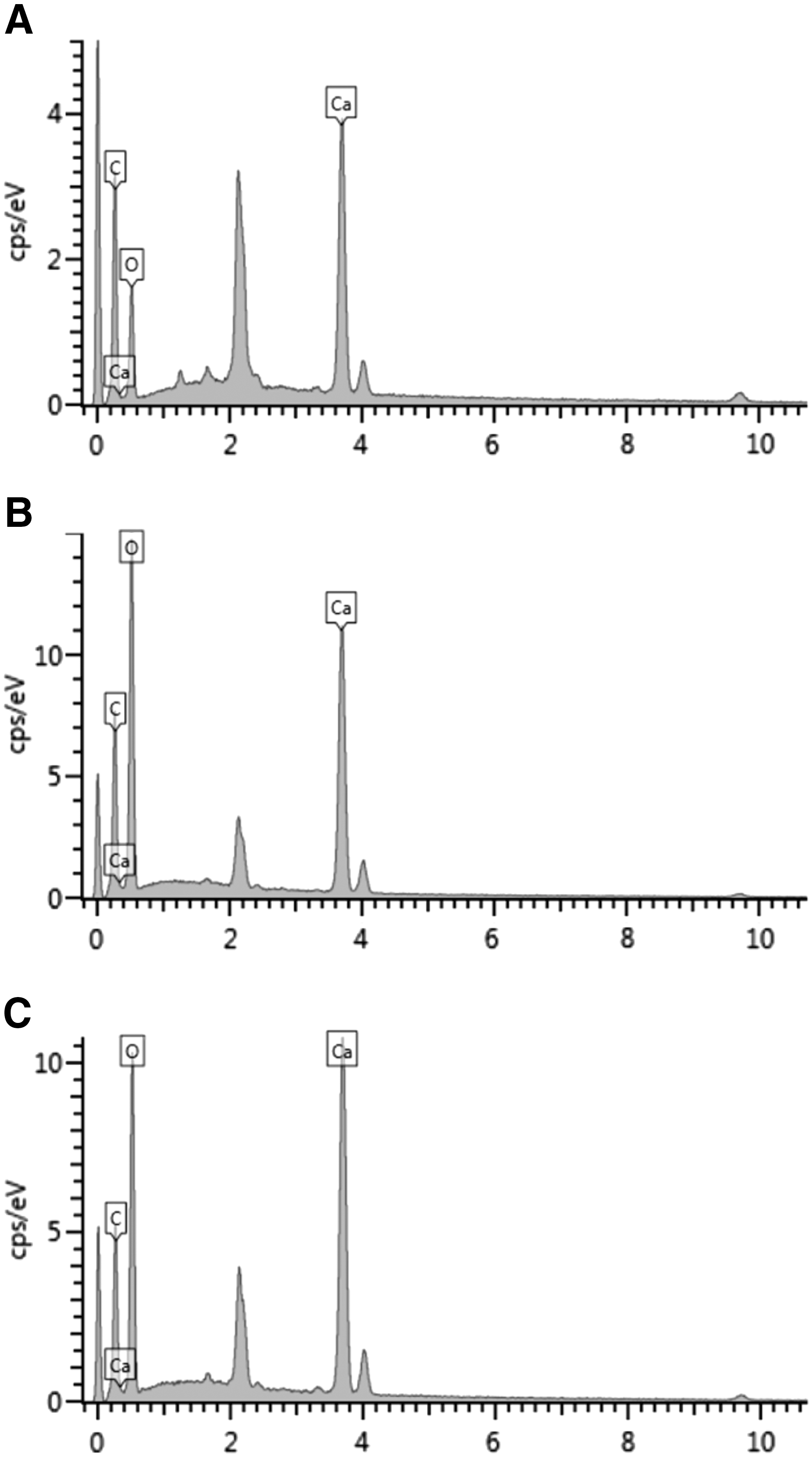
EDS spectrum of crystals of A. pyrifolia. Bipyramidal crystal sand on the epidermis
Identification of the constituents from ESAP by LC-DAD-MS
ESAP extract was analyzed by LC-DAD-MS and 19 compounds could be detected and identified (Table 1, Fig. 4). The compounds
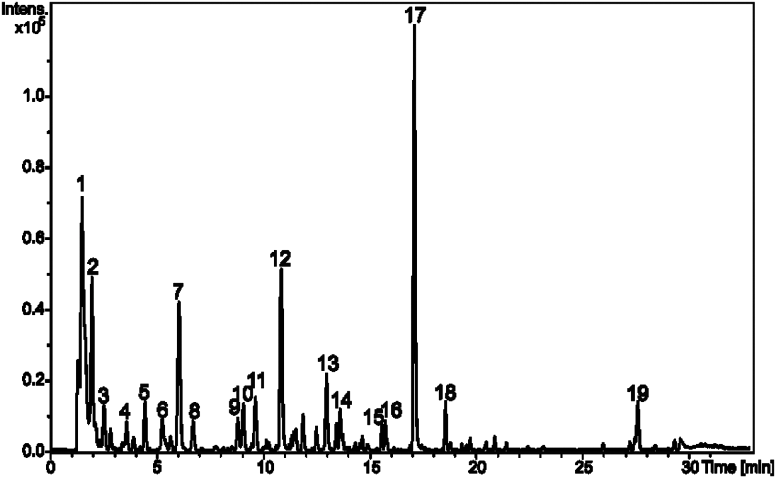
Base peak chromatogram (negative ion mode) of the extract from A. pyrifolia.
Identification of the Constituents from Anchietea pyrifolia Extract by Liquid Chromatography-Diode Array Detection-Tandem Mass Spectrometry
MF, molecular formula; MS, mass spectrometry; NI, nonidentified; RT, retention time; UV, ultraviolet.
The compounds
The metabolites
Safety evaluation
Neither deaths nor changes in behavior were observed in animals after acute exposure to ESAP (30, 300, and 2000 mg/kg) (data not shown). Female rats did not present any significant changes in food and water intake as well as in body weight gain (Table 2). However, the relative body weight gain of rats treated with ESAP 300 mg/kg differed significantly when compared with the control group. Regarding the relative organ weight, no significant changes were observed among groups treated with all doses of ESAP when compared with the control (Table 2). Furthermore, no gross signs of toxicity were observed in vital and reproductive organs (data not shown) as well as no histopathological changes in heart, liver, and kidney samples (Fig. 5). Therefore, ESAP median lethal dose (LD50) can be considered higher than 2000 mg/kg.
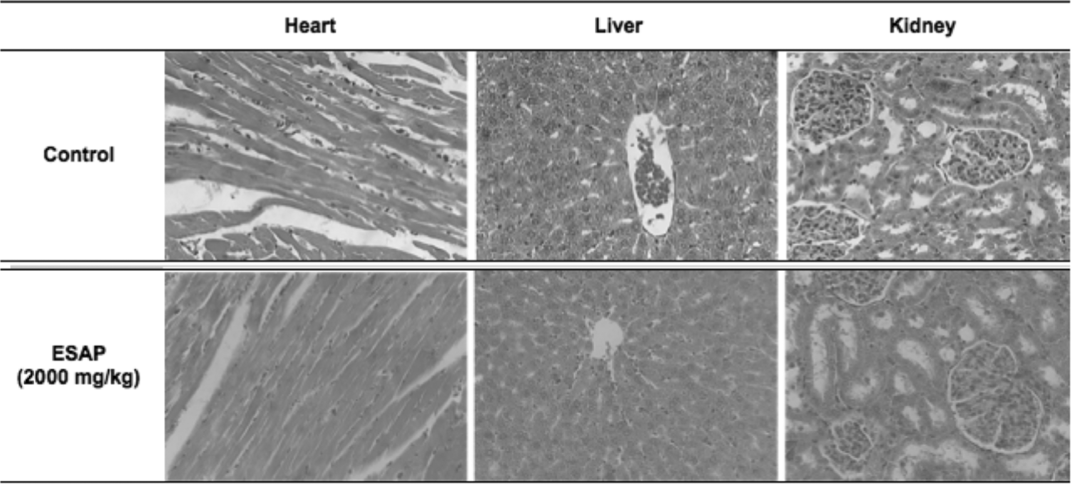
Histopathological assessment of heart, liver, and kidney from rats orally treated with the vehicle (control) or with the highest dosage of ESAP (2000 mg/kg) in the acute toxicity test. HE (40 X). ESAP, ethanol-soluble fraction obtained from Anchietea pyrifolia.
Body Weight Gain, Relative Organ Weight, Food and Water Intake of Rats Treated Orally with Ethanol-Soluble Fraction from Anchietea pyrifolia
Statistical analysis was performed using one-way ANOVA followed by Dunnett's test. Values are expressed as mean ± SEM (n = 8) in comparison to the control group.
P ≤ .05.
ANOVA, analysis of variance; bw, body weight; ESAP, ethanol-soluble fraction obtained from Anchietea pyrifolia; SEM, standard error of the mean.
Diuretic effects
Acute diuretic activity
In this experiment, no significant increase in diuresis was observed after acute administration of three different doses (30, 100, and 300 mg/kg) of ESAP in 8 and 24-h samples. HCTZ, as expected, increased diuresis in 8-h samples. However, this effect could not be sustained 24 h after drug administration (Table 3). Although ESAP did not induce diuresis, the highest dose (300 mg/kg) significantly increased pH levels in 8 and 24-h samples. Moreover, ESAP 30 and 300 mg/kg promoted a significant decrease in urine density in 8 and 24-h samples (Table 3). Regarding the urinary electrolyte analysis, HCTZ, as expected, increased Na+, K+, and Cl− excretion after 8 h and returned to values similar to the control after 24 h. None of ESAP doses increased the amounts of Na+, K+, and Cl− in 8 or 24-h urine samples (Table 4).
Effect of Acute Oral Administration of Ethanol-Soluble Fraction Obtained from Anchietea pyrifolia on the Urinary Volume, pH, and Density in 8 and 24-h Urine Samples
For ESAP treatments, statistical analysis was performed using one-way ANOVA followed by Dunnett's test. HCTZ treatments were compared with the control using Student's t-test. Values are expressed as mean ± SEM (n = 6).
P ≤ .05 when compared with the control group.
HCTZ, hydrochlorothiazide.
Effect of Acute Oral Administration of Ethanol-Soluble Fraction Obtained from Anchietea pyrifolia on Urinary Electrolyte Excretion in Urine 8 and 24 h
For ESAP treatments, statistical analysis was performed using one-way ANOVA followed by Dunnett's test. HCTZ treatments were compared with the control using Student's t-test. Values are expressed as mean ± SEM (n = 6).
P ≤ .05 when compared with the control group.
Saluretic index = μEq/min/100 g problem group/μEq/min/100 g control group.
El, excreted load.
Prolonged diuretic activity
Prolonged treatment with ESAP (30, 100, and 300 mg/kg) did not promote significant increase in diuresis after a 7-day treatment. Only ESAP lowest dose (30 mg/kg) was able to significantly increase diuresis on the seventh day after administration as it statistically differed from the control (Table 5). However, daily treatment with ESAP (30, 100, and 300 mg/kg) significantly decreased urine excretion of Na+, K+, and Cl− on days 3 and 7 after treatments (Table 5). HCTZ, as expected, increased urine volumes and electrolyte excretion on days 3 and 7 after treatments (Table 5). pH and density values were not altered by any treatment (data not shown).
Effects of Prolonged Oral Administration of Ethanol-Soluble Fraction Obtained from Anchietea pyrifolia on Cumulative Urine Volume, Na+, K+, and Cl− Excretion
For ESAP treatments, statistical analysis was performed using one-way ANOVA followed by Dunnett's test. HCTZ treatments were compared with the control using Student's t-test. Values are expressed as mean ± SEM (n = 6).
P ≤ .05 when compared with the control group.
Acute treatment with ESAP reduces SBP and DBP values in normotensive male rats
Basal SBP, DBP, MAP, and HR recorded after the 15-minute stabilization period and before extract administration were 103.21 ± 3.31 mm Hg, 65.12 ± 2.13 mm Hg, 84.25 ± 2.53 mm Hg, and 249.16 ± 8.50 beats per minute (bpm), respectively. The positive control (HCTZ), as expected, significantly reduced basal SBP, DBP, and MAP and HR to 88.35 ± 3.09, 57.71 ± 3.42, and 71.04 ± 2.36 mm Hg, whereas the control group (vehicle) barely changed these values, as follows: 104.53 ± 8.64, 66.85 ± 3.52, and 81.19 ± 5.86 mm Hg. Only the acute administration of ESAP highest dose (300 mg/kg) significantly reduced SBP and DBP values to 87.64 ± 2.54 and 51.72 ± 3.72, respectively (Table 6).
Effect of Acute and Prolonged Oral Administration of Ethanol-Soluble Fraction Obtained from Anchietea pyrifolia on Arterial Pressure and Heart Rate of Male Wistar Rats
For ESAP treatments, statistical analysis was performed using one-way ANOVA followed by Dunnett's test. HCTZ treatments were compared with the control using Student's t-test. Values are expressed as mean ± SEM (n = 6).
P ≤ .05 when compared with the control group.
bpm, beats per minute; DBP, diastolic blood pressure; HR, heart rate; MAP, mean arterial pressure; SBP, systolic blood pressure
Prolonged treatment with ESAP was not able to significantly change arterial blood pressure and HR values in normotensive rats when compared with animals from the negative control group. On the other hand, HCTZ significantly reduced SBP, DBP, and MAP levels (Table 6). Regarding serum urea, creatinine, K+, and Na+ levels, only ESAP highest dose (300 mg/kg) significantly decreased urea values when compared with the control (Table 7).
Effects of Prolonged Oral Administration of Ethanol-Soluble Fraction Obtained from Anchietea pyrifolia on the Biochemical Parameters of Male Wistar Rats After 7 Days of Treatment
For ESAP treatments, statistical analysis was performed using one-way ANOVA followed by Dunnett's test. HCTZ treatments were compared with the control using Student's t-test. Values are expressed as mean ± SEM (n = 6).
P ≤ .05 when compared with the control group.
Effects on peripheral vascular resistance
In this experiment, a sustained increase in vascular PP was observed after continuous perfusion of MVBs with Phe. Such perfusion was dose dependently reduced after administration of ESAP into the perfusion apparatus. Also, an expressive dose-dependant vasodilator response in MVBs was induced by ESAP, followed by a reduction in PP values for doses of 0.1, 0.3, and 1 mg to 1.46 ± 0.84, 5.62 ± 1.98, and 12.44 ± 2.19 mm Hg, respectively (Fig. 6A).
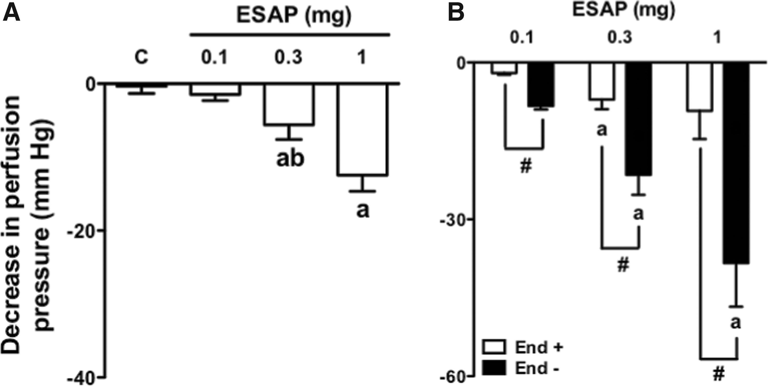
Vasorelaxant effect of ESAP does not depend on endothelium mediators in the MVBs of rats. MVBs were perfused with PSS containing Phe (3 μM) on intact
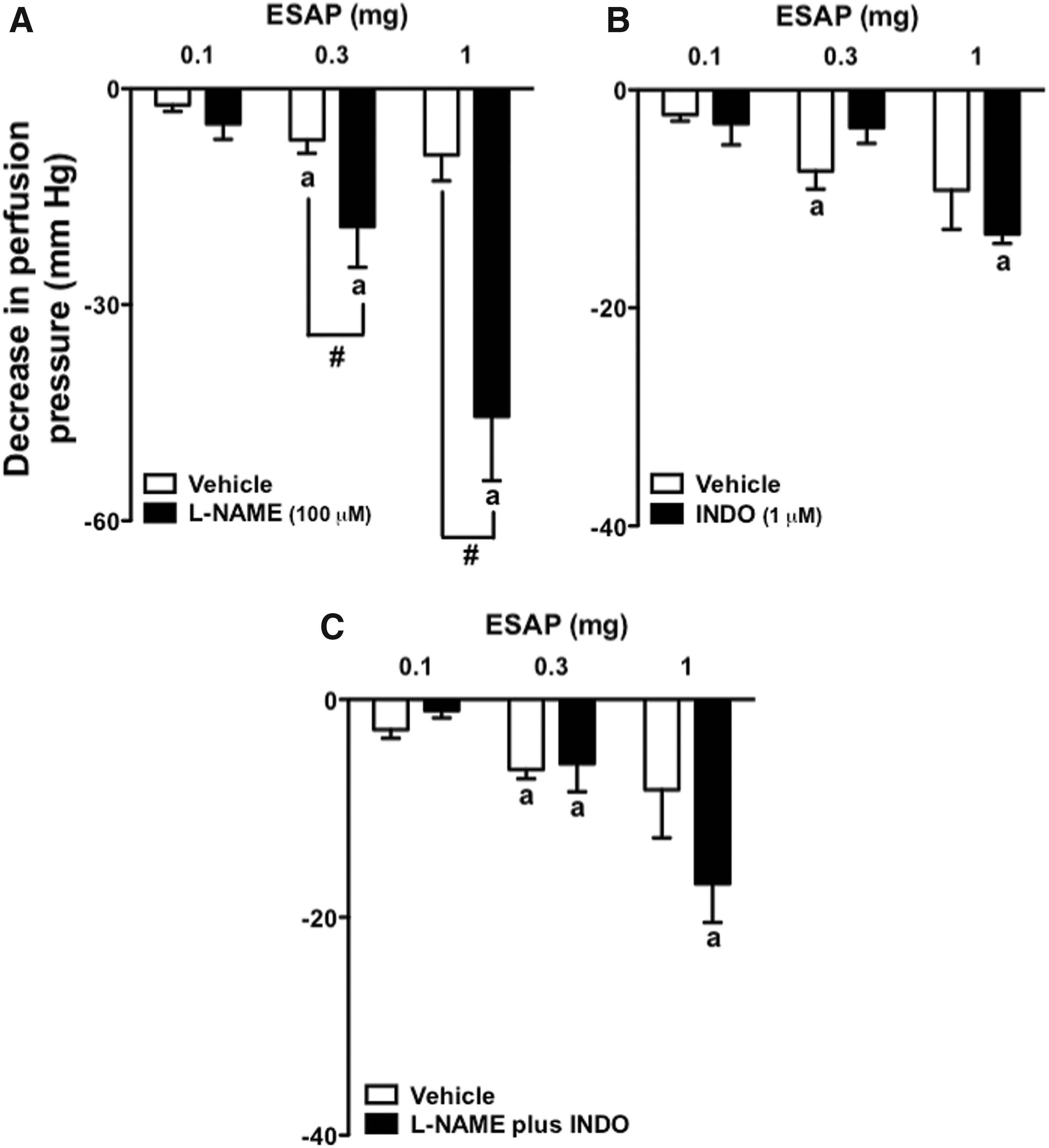
Treatment with sodium deoxycholate was able to reduce the effects of ACh on MVBs by ∼80% (data no shown), confirming the efficacy of chemical removal of the endothelium. The vasodilatory effect of all doses of ESAP was significantly increased in the absence of endothelium (Fig. 6B) or in preparations with intact endothelium perfused with L-NAME (Fig. 7A). The vasodilatory effects of ESAP remained unaltered in preparations with intact endothelium perfused with indomethacin (Fig. 7B), or L-NAME plus indomethacin (Fig. 7C).
Vasorelaxant effect of ESAP does not depend on nitric oxide or prostaglandins in the MVBs of rats. MVBs were perfused with PSS containing Phe (3 μM) plus L-NAME
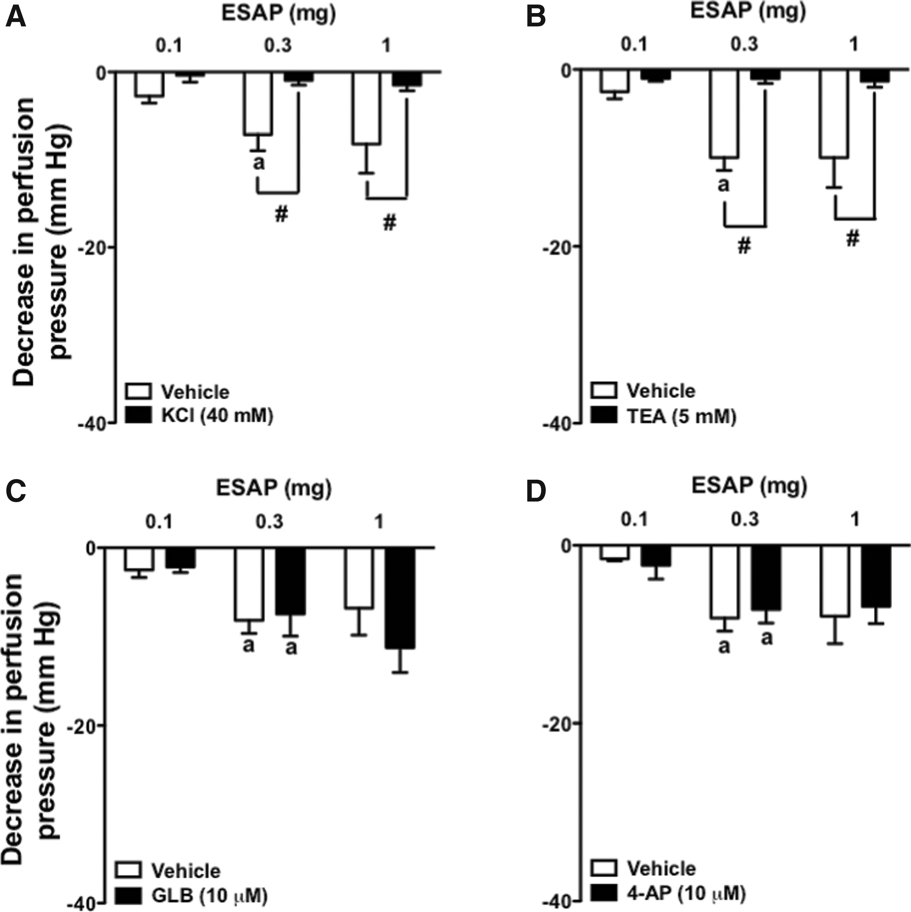
The effects of ESAP were abolished by the perfusion of MVBs with nutritive solution added of 40 mM KCl (Fig. 8A). Contrarily, only minor effects could be noted after infusion of GLB or 4-AP (Fig. 8C, D). Curiously, treatment with TEA vanished the vasorelaxant effect induced by all ESAP doses (Fig. 8B).
Vasorelaxant effect of ESAP depends on calcium-activated potassium channels in the MVBs of rats. MVBs were perfused with PSS containing Phe (3 μM) plus KCl
Discussion
In Brazil, the Cerrado represents about 23% of the land surface and possesses a huge diversity of species. For this reason, this area has become the target of a broad number of scientific studies to discover new molecules with powerful biological effects.
27
A. pyrifolia (Mart.)
In the first stage of this work, we performed a pharmacognostic study using A. pyrifolia leaves and stems willing to broaden our knowledge on the Brazilian Cerrado. Thus, we present the main pharmacognostic characteristics of this species, offering a unique pattern for its morphological and microchemical characterization. In fact, this gives us confidence about the identification of the species studied and provides important data regarding the quality control of the plant drug. Morphoanatomical studies are the most accurate possible means of identifying plant species to prevent tampering and ensure quality standard of the species under study. 28,29
In the meantime, a detailed characterization of the main secondary metabolites from ESAP was performed by LC-DAD-MS and 19 compounds were observed. The chemistry composition of A. pyrifolia is underexplored, and nonpolar metabolites have been reported, such as triterpenoids and fatty acid derivatives. 30 However, in our study O-glycosylated flavonoids, chlorogenic acids, and other phenylpropanoid acid derivatives were identified from ESAP. These compounds have not been described from A. pyrifolia and they probably are related to pharmacological properties, since the aqueous extraction prevails polar metabolites extraction.
Medicinal plants, as well as drugs of synthetic origin, have the same potential to cause serious toxic effects. Thus, toxicological studies are of utmost importance to assess safety and possible toxic effects that may occur during therapeutic use. For this reason, the next stage of this research was destined to investigate the possible toxic effects of this species after acute exposure. In this experiment, female Wistar rats were used, as they are considered more sensitive than male rats. 19 As known, animal death is a clear sign of toxicity. However, other characteristics may indicate less serious toxic effects such as body weight loss and behavior change. 31 In this work, no deaths nor weight loss were observed in all rats treated with single doses of ESAP. Daily observations of changes in behavior were performed according to the Hippocratic screening, which estimates the pharmacological and toxicological nature of the test substance. 20 Besides, such parameters are fundamental for evaluating animal general health status. 32 According to data obtained in the safety assessment, animals showed no changes in behavior, in food and water intake, as well as no gross nor histopathological changes in organs examined. Therefore, ESAP can be considered safe in this animal model at all doses tested.
Since the acute toxicity test proved ESAP safety in rats, we then performed an ethnopharmacological investigation of this species in male Wistar rats. First, the diuretic potential of this species was accessed by performing acute and prolonged tests. Despite its clear indication as a diuretic agent by Brazilian popular healers, no diuretic effects were observed in rats after acute administration and only ESAP lowest dose (30 mg/kg) increased diuresis on the seventh day of treatment. Although ESAP 30 mg/kg values on cumulative urine were statistically different from the control group, this volume is still low as it significantly differed from HCTZ, demonstrating a not so potent diuretic effect. In addition, the small variations found in the urinary pH after ESAP treatments can be attributed to the extract itself, which, because of its alkaline pH, may have slightly raised pH since this effect did not remain after prolonged treatment. In addition, the small changes in urinary density observed after acute treatments were also considered to be incidental and of no significant clinical relevance. On the other hand, prolonged treatment with all doses of ESAP decreased electrolyte (Na+, K+, and Cl−) excretion on days 3 and 7 after administration. As known, effective diuretic substances are those that are able to increase renal excretion of water and electrolytes 33 and the obtained data did not suggest any effectiveness of A. pyrifolia in this aspect. We believe that the belief in a possible diuretic effect may be due to a large amount of water ingested with the infusion obtained from this species. Thus, ESAP popular indication for diuretic therapy might be related to the increase in renal hydrostatic pressure that is caused by the expansion of plasma volume after high water ingestion (i.e., pressure diuresis). 34 Thus, many species prescribed in traditional medicines for such purpose have shown low or even no diuretic potential. 35
As known, some first-line antihypertensive drugs do not promote diuretic effects. 36 Hence, even though ESAP did not present significant diuretic activity in rats, we next investigated the acute and prolonged effects of ESAP on blood pressure. Our findings demonstrate that ESAP induced significant hypotensive effects only after acute exposure, disappearing completely after prolonged exposure. It is possible that the acute hypotensive effect caused by ESAP has activated counter-regulator mechanisms to counteract the reduction of blood pressure. 37 Thus, salt and water retention induced by the acute blood pressure fall may have led to a lower renal elimination of electrolytes and contributed to the long-term stabilization of blood pressure. Indeed, this is a typical mechanism of resistance promoted by some vasodilator drugs after prolonged exposure such as minoxidil and hydralazine. 38
In the last step of this work, we investigated the possibility of ESAP causing vasodilatory effects on MVBs, since the main determinant of arterial pressure is peripheral vascular resistance. 39 Initially, after confirming the vasodilatory effect induced by ESAP, we investigated whether the reduction in PP had any relation to the vasodilatory endothelial mediators. The main endothelial mediators responsible for inducing vasodilator response and maintaining vascular tone in the microcirculation are NO, prostacyclin (PGI2), and the endothelium-derived hyperpolarizing factor. 40 Although these mediators indicated possible targets for ESAP, the chemical removal of the endothelium did not prevent the vasodilator response induced by this extract. In addition, the use of inhibitors of prostaglandins (indomethacin) or NO synthesis (L-NAME) also elicited no effect on ESAP-induced vasodilation. Surprisingly, the chemical removal of the endothelium intensified the vasodilatory response of ESAP. This pattern of response, in addition to indicating that the vasodilatory effect possibly comes from the smooth muscle cell, also shows that the removal of some endothelial mediators—possibly vasoconstrictors—intensifies the ESAP response. 41
The regulation of intracellular calcium concentration, which is the first determinant of vascular tone, occurs due to the contribution of ion channels in the plasma membrane and endoplasmic reticulum of vascular smooth muscle cells.
Ion channels provide the main source of activator calcium that determines vascular tone, and importantly contribute to setting and regulating membrane potential and open state probability of voltage gated calcium channels, the primary source of calcium in resistance artery. KV, KATP, and KCa channels are the ones that precisely influence the regulation of vascular membrane potential and contribute to pressure-induced myogenic tone in resistance arteries. In fact, the modulation of these ion channels functions by vasoconstrictors and vasodilators, firmly affects the functional regulation of tissue blood flow. 42 In our study, the use of KV and KATP channel blockers did not interfere with the vasodilator response induced by ESAP. However, the previous perfusion of PSS with TEA vanished the vasorelaxant effects of ESAP. Despite the fact that TEA (in high doses) is considered a nonselective potassium channel blocker, small doses of this inhibitor present relative selectivity to KCa. 43 Thus, if we consider the inhibition ilicited by TEA, and the exclusion of the participation of KV and KATP channels presented by the use of 4-AP and GLB, it is possible to conjecture that the vasodilator and hypotensive effects induced by ESAP in normotensive rats is dependent on opening of KCa channels in vascular smooth muscle.
In conclusion, this study turns the lights on the preclinical safety and efficacy of A. salutaris as a hypotensive agent. Supposedly, these effects may be involved with the opening of the calcium-activated potassium channels in smooth muscle from resistence vessels contributing to reduce global peripheral resistance and blood pressure.
Footnotes
Acknowledgments
This work was supported by grants from the Fundação de Apoio ao Desenvolvimento do Ensino, Ciência e Tecnologia do Estado de Mato Grosso do Sul (FUNDECT, Brazil, 59/300.046/2015), and Conselho Nacional de Desenvolvimento Científico e Tecnológico (CNPq, Brazil, 449464/2014-8). The authors are grateful to the University Hospital of the Federal University of Grande Dourados for the biochemical and hematological analyzes.
Author Contributions
S.E.L.T. performed experiments related to toxicity, diuresis, blood pressure, heart rate, and mechanisms involved. S.E.L.T also performed data analysis, discussion, and wrote the article. R.A.C.P., C.A.S.T., M.I.S., L.P.G. and A.O.S. performed experiments related to blood pressure, heart rate and mechanisms involved; data analysis, and discussed the results. A.A.M.M. did the plant collection, extract preparation, and performed experiments related to the diuretic potential of the species. V.P.A. and J.M.B. performed the anatomical and microchemical analysis. R.I.C.S. and A.C.S. performed all work related to histopathological analysis. S.R.N. and D.B.S. performed the phytochemical analysis. P.R.D. was the project coadvisor. A.G.J. was the project advisor, conceived and planned the experiments, discussed data, and wrote the article. All authors read and approved the final article.
Author Disclosure Statement
No competing financial interests exist.