
Abstract
We demonstrated the effect of a mixture containing fermented Achyranthes japonica Nakai (FS) in the context of a monosodium iodoacetate (MIA)-induced osteoarthritis animal model. The mineralization, anabolic and catabolic factors, and the amount of cytokines within the articular cartilage of rats were measured after administration of MIA. We found that dietary supplementation with methylsulfonylmethane (positive control) and FS (FS 100 mg/kg body weight [b.w.] and FS 300 mg/kg b.w.) effectively suppressed pathological changes in the knee joint and inhibited changes in the architectural and mineralization parameters. In addition, prostaglandin E2 (PGE2) and proinflammatory cytokines in the serum and catabolic factors, including matrix metalloproteinase (MMP)-3 and MMP-7 in articular cartilage, were decreased by dietary supplementation with FS in MIA-induced osteoarthritis. Based on these findings, we suggest that FS can be used for the development of potential therapies for osteoarthritis.
Introduction
Osteoarthritis is a degenerative joint disease and also the most common form of arthritis, and this disease is clinically characterized by joint pain due to a progressive degradation of articular cartilage. 1 Patients suffering from osteoarthritis face a gradual reduction in physical activity, and as a result, deterioration in their quality of life. Risk factors that lead to the development of osteoarthritis include age, obesity, gender (women are more likely to develop osteoarthritis), and muscle weakness. The exact etiology of osteoarthritis, however, remains unclear. 2,3
It has been suggested that arthritis is caused by a biochemical change, such as an imbalance in the synthesis and degradation pathway of the articular cartilage. 4,5 Under normal conditions, joint cartilage cells function to maintain homeostasis of the extracellular matrix components with a low turnover rate. 4 During the development of osteoarthritis that involves inflammation and mechanical stress, however, the joint cartilage cells, with the help of tissue proteinases, degrade the extracellular matrix in the articular cartilage. 5 In addition, during progression of osteoarthritis, chondrocytes induce the production of proinflammatory cytokines, prostaglandin E2 (PGE2), and nitric oxide that activate proteases such as collagenase, aggrecanases, and matrix metalloproteinases. This eventually causes cartilage degradation by mineralization. 6,7
Treatment for osteoarthritis is focused on relieving the symptoms, reducing pain, delaying progression of the disease, maintaining joint function, and restoring the structural damage of the joints. 8 Recent studies on the pathogenesis of osteoarthritis, however, have been performed in an attempt to determine new therapies using a healthy functional food that can delay disease progression and restore cartilage damage. Of particular interest is the efficacy of herbal medicines and folk remedies in treating this disease. 9
Thus, we investigated the activity and effects of an herbal mixture containing fermented Achyranthes japonica Nakai on monosodium iodoacetate (MIA)-induced osteoarthritis in rats. The root of the A. japonica Nakai plant, which grows widely in Korea, Japan, and China, was used. 10 A. japonica Nakai has been reported to possess activities such as anti-inflammatory, antitumorigenic, and immune regulation. 10 In this study, we measured mineralization, anabolic and catabolic factors, and the amount of cytokines in the articular cartilage of rats to confirm the effect of the mixture containing fermented A. japonica Nakai in the context of osteoarthritis.
Materials and Methods
Preparation of the extract
Achyranthes japonica Nakai, Eucommia, and Angelica gigas were extracted using water for 4 h at 95°C. The filtrate was concentrated and sterilized at 105°C for 60 min. The concentrated A. japonica Nakai was fermented using 5% Lactobacillus plantarum at 37°C for 24 h at 1 g. Fermented A. japonica Nakai, extracted Eucommia, extracted A. gigas, and beta-cyclodextrin were mixed in a ratio of 3:2.2:1.5:3.3 a mixture containing fermented Achyranthes japonica Nakai (FS) and dried using the spray-dried method.
Treatment and induction of osteoarthritis
Sprague-Dawley rats (6-week-old male) were supplied from Japan SLC, Inc. The experimental protocol was approved by the Animal Care and Use Review Committee of Kyung Hee University [KHUASP(SE)-17-041]. Rats were housed in cages in an environmentally controlled facility (22 ± 2°C, humidity of 50–60%, and 12-h light/dark cycle) and were fed an AIN 93G diet for an adaptation period of 7 days. The animals were randomly assigned to five groups that included animals with normal AIN 93G diet (normal control), osteoarthritis induction and normal AIN 93G diet (control), osteoarthritis induction and AIN 93G diet including methylsulfonylmethane (MSM)
11
300 mg/kg (positive control), osteoarthritis induction and AIN 93G diet including FS 100 mg/kg, and osteoarthritis induction and AIN 93G diet including FS 300 mg/kg. Seven days after dietary administration, the rats were anesthetized with isoflurane and were administered MIA (50 μ
Histologic staining
Knee joints were collected from rats, fixed in 10% neutral buffered formalin, and decalcified. The samples were embedded in paraffin, cut into 7-μm-thick sections, and stained with hematoxylin and eosin (H&E) for histological evaluation.
Microcomputed tomography image scan
Microcomputed tomography (CT) imaging of the formalin-fixed articular cartilage from rats was used to measure the roughness of the bone surface. Micro-CT image scanning was performed using the Skyscan 1172® X-ray μCT scanning system (Bruker, Belgium). After standardized reconstruction of the scanned images, the data for each sample were recollected with the micro-CT software to orient each sample in the same manner.
Measurement of the level of PGE2 and cytokines in the serum
Serum samples were obtained from rats, and the levels of PGE2, tumor necrosis factor alpha (TNF-α), interleukin-1β (IL-1β), and IL-6 were determined using an ELISA kit (R&D system, Minneapolis, MN, USA).
mRNA expression in rat cartilage
mRNAs from the rat articular cartilage was extracted using the RNeasy Mini Kit (QIAGEN, MD, USA). The iScript™ cDNA Synthesis Kit was used for cDNA synthesis (BIORAD, Hercules, CA, USA). Real-time polymerase chain reaction
Primer Sequences Used for mRNA Quantification by Real-Time Polymerase Chain Reaction
COX-2, cyclooxygenase-2; GAPDH, glyceraldehyde 3-phosphate dehydrogenase; IL-1β, interleukin-1β; MMP, matrix metalloproteinase; NF-kB, nuclear factor kappa B; TNF-α, tumor necrosis factor alpha.
Statistical analysis
The experimental results are expressed as mean ± standard deviation. Statistical analysis was conducted using a one-way analysis of variance (ANOVA) or t-test using SPSS statistical procedures for Windows (SPSS PASW Statistic 23.0; SPSS, Inc., Chicago, IL, USA), and Duncan's multiple range test was used to examine the differences among the groups; a P-value <.05 was considered significant.
Results
Histological analysis of the articular cartilage
Histological evaluations were performed using H&E staining to study the effect of FS on osteoarthritis. MIA-induced osteoarthritis rats (control) exhibited irregular articular cartilage surface and cartilage matrix degradation, while the joints of the noninduced osteoarthritis rats (normal control) possessed a smooth articular cartilage surface. The groups that received MSM (positive control) and FS (FS100 and FS300) as a dietary supplement exhibited reduced pathological changes of the knee joint induced by MIA (P < .05) (Fig. 1).
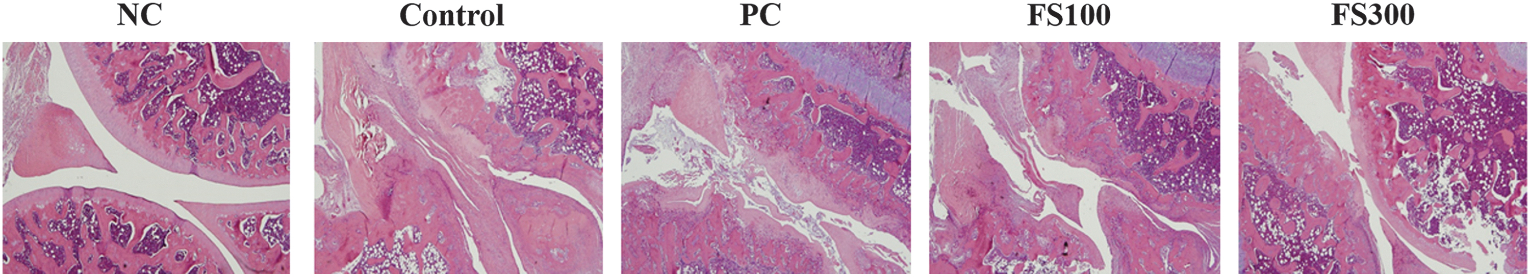
Representative histological changes in articular cartilage from MIA-induced osteoarthritic rats. NC: AIN93M diet; Control: AIN93M diet+MIA injection group; PC: AIN93M diet+MIA-injected group supplemented with MSM (300 mg/kg b.w.); FS100: AIN93M+MIA-injected group supplemented with a mixture containing fermented Achyranthes japonica Nakai (100 mg/kg b.w.); FS300: AIN93M+MIA-injected group supplemented with a mixture containing fermented A. japonica Nakai (300 mg/kg b.w.). b.w., body weight; MIA, monosodium iodoacetate; MSM, methylsulfonylmethane; NC, normal control; PC, positive control.
Analysis of architectural and mineralization parameters
The morphological change, bone mineral density (BMD), bone volume/total tissue volume (BV/TV), trabecular number, and trabecular separation from the articular cartilage of rats were measured by micro-CT. The joints of the normal control group possessed a smooth and shiny articular surface, and induction with MIA caused an irregular articular cartilage surface. BMD, BV/TV, and the trabecular number significantly decreased, and the trabecular separation levels significantly increased in the MIA-induced rats (control). The groups that received FS as a dietary supplement, however, experienced significantly suppressed changes in these architectural and mineralization parameters (P < .05) (Fig. 2).
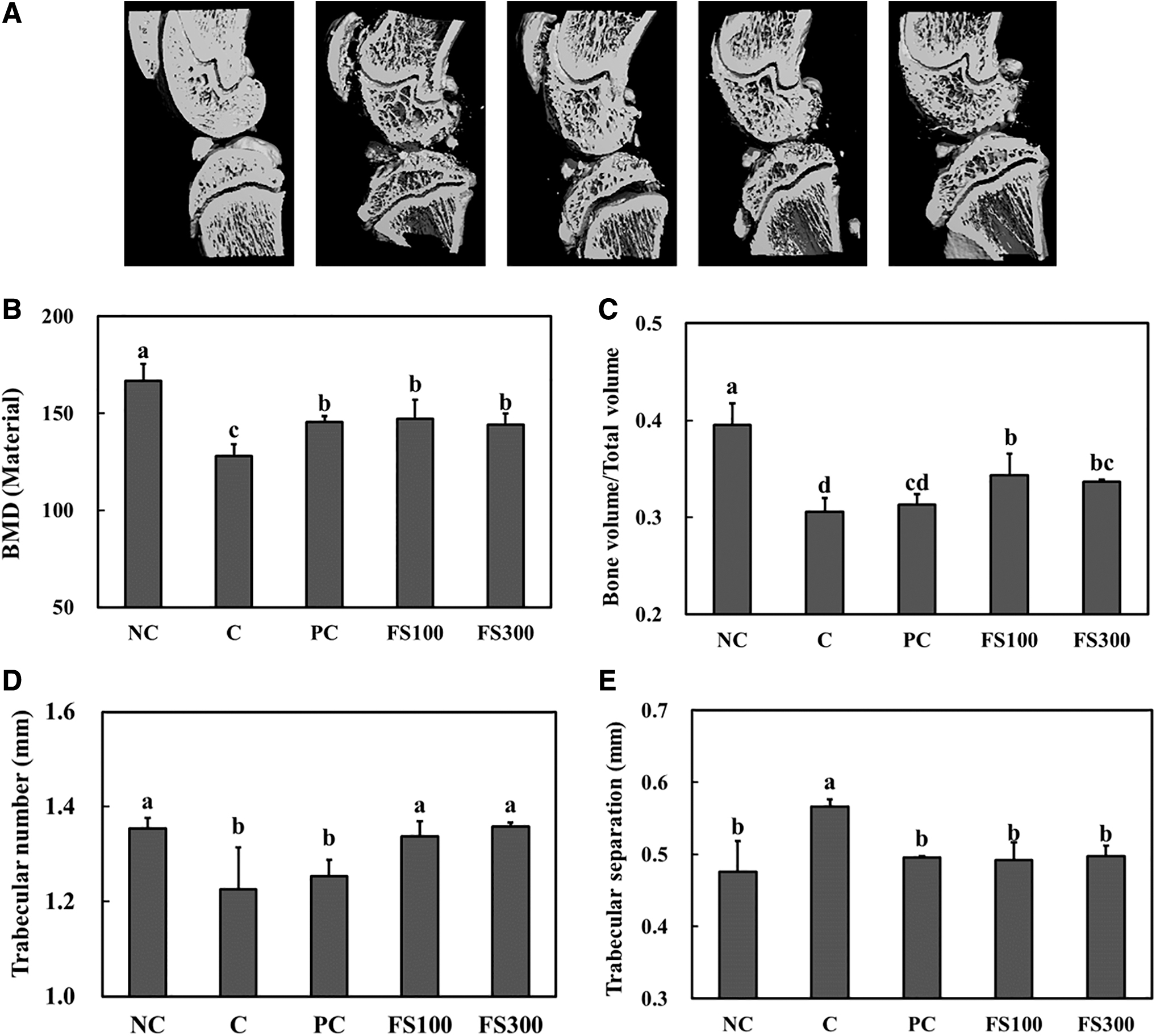
Architectural changes (
Measurement of the levels of PGE2 and proinflammatory cytokines in the serum
Proinflammatory cytokines play a role in the degradation of articular cartilage by stimulating proteases and the synthesis of PGE2. 12 To confirm the protective activity of FS during osteoarthritis development by MIA induction in the articular cartilage, we measured the level of PGE2 and proinflammatory cytokines, including IL-1β, TNF-α, and IL-6, in the serum. When compared with the normal control group, MIA induction caused a significant increase in the levels of PGE2, IL-1β, TNF-α, and IL-6. Dietary supplementation with FS, however, significantly reduced the levels of PGE2, IL-1β, TNF-α, and IL-6 compared with those of the control group (P < .05) (Fig. 3).
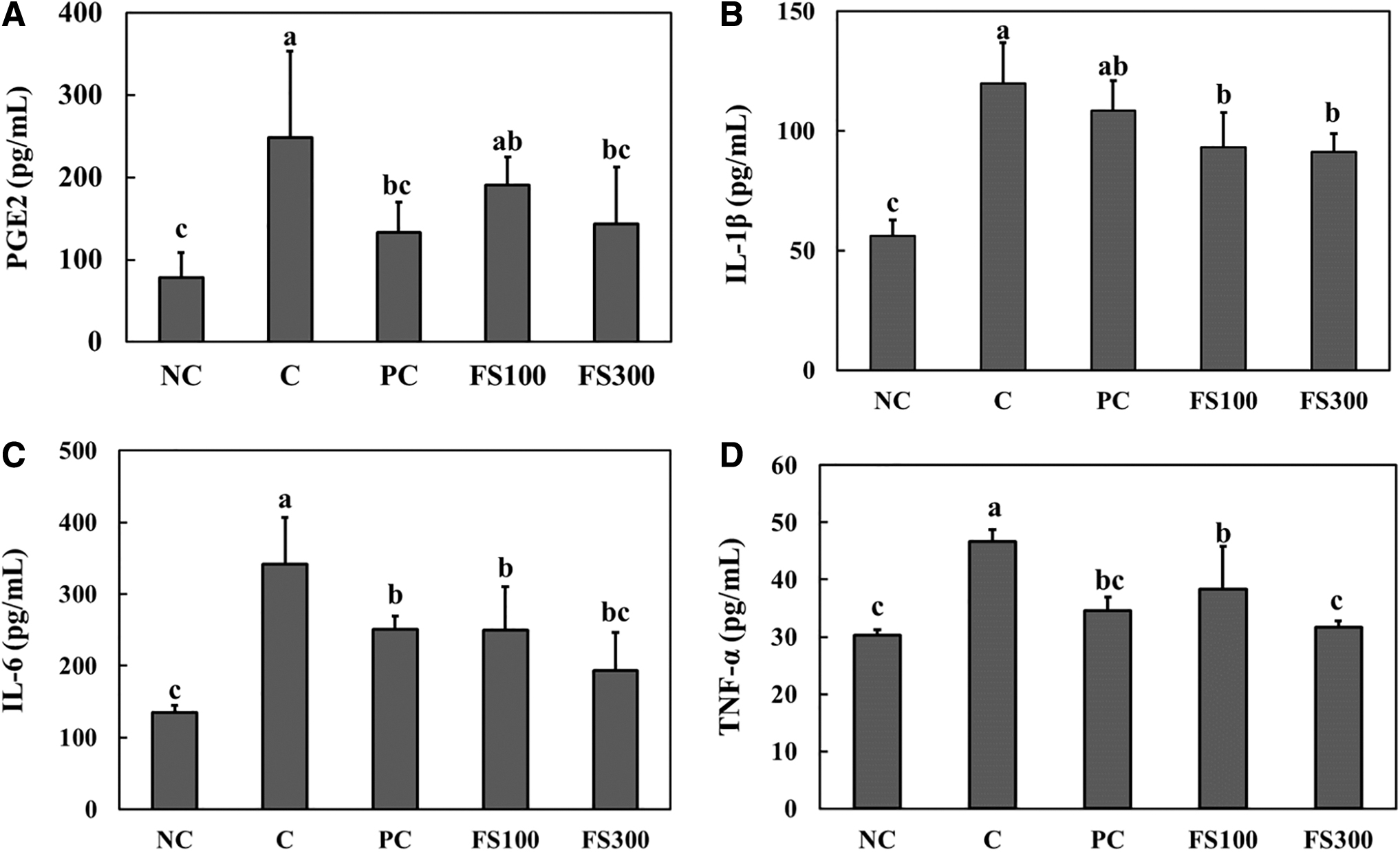
Inhibitory effects of fermented A. japonica Nakai on serum PGE2
The mRNA expression in articular cartilage
We investigated the mRNA expression of the anabolic and catabolic factors in the articular cartilage to assess the effect of FS on osteoarthritis. The mRNA expression of collagen type I and type II increased significantly in the groups supplemented with FS compared with that of the control group; however, aggrecan mRNA expression was not observed. The catabolic factors MMP-3 and MMP-7 decreased significantly in the groups supplemented with FS 300 compared with levels observed in the control group. In addition, dietary supplementation with FS significantly reduced mRNA expression of nuclear factor kappa B (NF-κB), IL-1β, TNF-α, and cyclooxygenase-2 (COX-2) compared with expression levels observed in the control group. Consequently, we observed that dietary supplementation with FS suppressed the development of inflammation and stimulated collagen synthesis by MIA induction (P < .05) (Fig. 4).
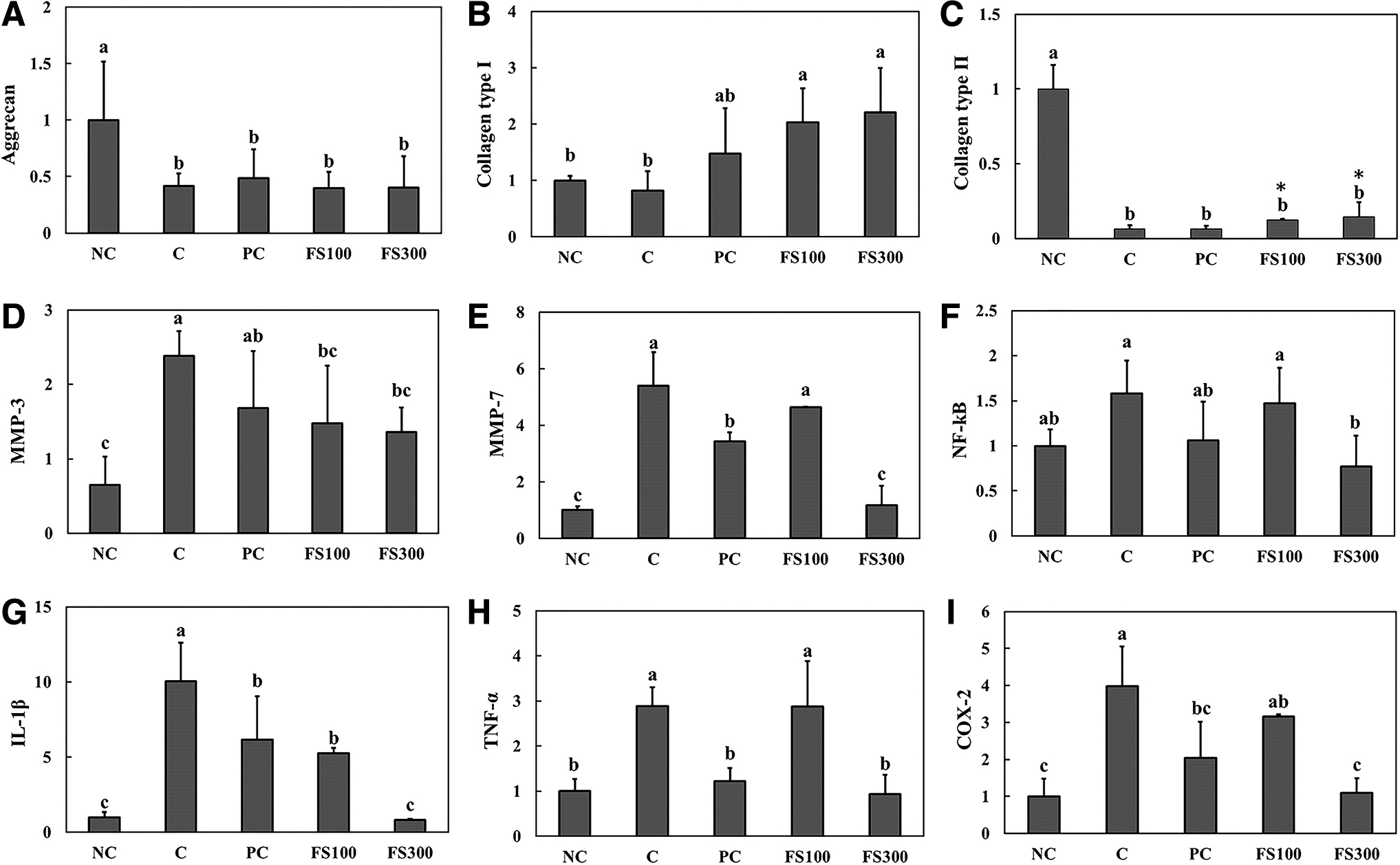
mRNA expression of aggrecan
Discussion
Traditionally, nonsteroidal anti-inflammatory analgesics (NSAIDs) have been used for the treatment of osteoarthritis to reduce pain and inflammation. NSAIDs inhibit the activity of COX-1 and/or COX-2, which are important mediators of inflammation during the development of osteoarthritis. 13 NSAIDs, however, cause gastrointestinal side effects such as stomach pain, ulcers, and heartburn. 13,14 Thus, herbal medicinal products are used in the treatment of osteoarthritis due to the low risk of side effects and toxicity; however, they are not yet one of the recommended treatments due to a lack of scientific evidence. 15 Therefore, we demonstrated the efficacy of a mixture containing fermented A. japonica Nakai (FS) that is used as an alternative medicine treatment for osteoarthritis.
To study the effect of new compounds for the treatment of osteoarthritis, it is necessary to use animal models that can reflect the actual state of joint pathology. Thus, an MIA intra-articular injection-induced osteoarthritic rat model was used. 16 MIA intra-articular injection inhibits glyceraldehyde-3-phosphate dehydrogenase that induces the development of osteoarthritis by chondrocyte death in articular cartilage. 17 We observed that MIA induced the formation of an irregular articular cartilage surface and caused matrix degradation both in histological and architectural analyses. We examined if dietary supplementation of FS affects the inhibition of osteoarthritis development in animals induced by MIA intra-articular injection. Our results indicated that dietary supplementation with FS effectively suppressed pathological changes in the articular cartilage when compared with observations of a group injected with MIA (control group). We also measured the amount of inflammatory factors to determine if the inhibitory effect of FS on osteoarthritis development was caused by an anti-inflammatory action.
According to numerous reports, osteoarthritis is associated with increased inflammatory mediators that interfere with the catabolic and the anabolic process of articular cartilage. 17,18 Chondrocytes produce proinflammatory cytokines that stimulate the synthesis and release of inflammatory mediators. 18 These inflammatory mediators lead to degradation of the cartilage, thus acting as instigators. 18,19 We found that MIA intra-articular injection caused an increased production of PGE2, IL-1β, TNF-α, and IL-6. This suggested that inflammatory reactions were involved in the development of osteoarthritis. When MIA-injected rats were administered with FS, the levels of PGE2, IL-1β, TNF-α, and IL-6 significantly decreased compared with those of the group injected with MIA (control group). Kothavade et al. reported that the saponin-rich fraction from Achyranthes aspera decreased the plasma level of TNF-α and IL-6 in adjuvant-induced arthritic rats. 20 These findings further support the idea that A. japonica Nakai possesses anti-inflammatory properties.
Extracellular matrix components such as aggrecan and collagen are key factors for the formation of the articular cartilage structure and normal function of joints. 21 Degradation of extracellular matrix is mediated by MMPs, a family of proteinases that are related to the pathological conditions of osteoarthritis. MMPs are expressed at a low level in normal articular cartilage; however, this expression is greatly increased under osteoarthritic conditions. 22 We found that the mRNA expression of aggrecan and collagen type II was decreased in the articular cartilage of MIA-injected rats. Dietary supplementation with FS induced an increase in the mRNA of collagen type I and type II and a decrease in the mRNA of MMP-3 and MMP-7. It did not, however, affect the mRNA expression of aggrecan. We need to confirm the protein level of aggrecan in MIA-injected rats to investigate the molecular function of dietary supplementation with FS.
In addition, FS dietary supplementation also decreased the mRNA expression of inflammatory factors. In a study performed by Lee et al., fermented A. japonica Nakai showed anti-inflammatory activity in lipopolysaccharides-treated RAW 264.7 cells preventing destruction of collagens and proteoglycan in chondrocytes. 23 These results suggest that FS may reduce the inflammatory response and improve clinical symptoms of osteoarthritis.
Based on these previous findings, we investigated the effect of FS on MIA injection-induced osteoarthritis development in rats, and we found that this substance exerted an inhibitory effect on inflammation and articular cartilage degradation. Therefore, we postulate that FS can be used for the development of new treatments for osteoarthritis.
Footnotes
Author Disclosure Statement
No competing financial interests exist.
Funding Information
This work was supported by the Korea Institute of Planning and Evaluation for Technology in Food, Agriculture, Forestry and Fisheries (IPET) through the High Value-Added Food Technology Development Program, funded by the Ministry of Agriculture, Food and Rural Affairs (MAFRA) (grant no.: 117054-3).