
Abstract
Acrocomia aculeata fruits are rich in monounsaturated fatty acid, β-carotene, tocopherol, and other antioxidant compounds. The aim of our study was to investigate and compare the protective effects of A. aculeata pulp oil and microencapsulated pulp oil on brain oxidative damage induced by chronic restraint stress (CRS) in rats (cortex, hippocampus, and striatum). Thirty-six Wistar rats were divided into six treatment groups: C, P, and M groups received 1 μL/g of body weight of distilled water, pulp oil, and pulp oil microcapsules by daily gavage, respectively. The SC, SP, and SM groups received 1 μL/g of body weight of distilled water, pulp oil, and pulp oil microcapsules by daily gavage, respectively, and were then subjected to uninterrupted 6 h of CRS. After 21 days of testing, the rats were euthanized and the brain tissue of the groups was removed for evaluation for oxidative damage markers and antioxidant enzymes. Endpoints of oxidative stress (OS) markers (lipid peroxidation, protein carbonylation, and reduced glutathione [GSH]) and antioxidant enzymes (superoxide dismutase and catalase) were evaluated. By imposing chronic stress on rats, pulp oil and microcapsules of pulp oil induced positive antioxidant responses, mainly by increasing the GSH content, increasing the ability of neural tissues to deal with inherent OS, thus protecting against neurodegenerative diseases. The administration of A. aculeata pulp oil and microencapsulated pulp oil made the reversal of the oxidant parameters, which may protect the brain tissue of rats altered by CRS. The Clinical Trial Registration number: n° 1.008/2018 CEUA/UFMS.
Introduction
Deprivation of spontaneous movements is a simple and painless animal stress inducing model already established. 1 –3 It consists of physically confining the animal preventing its locomotion and turning, 4 resulting in a great energy demand after several hours of restriction with behavioral and biochemical changes caused by oxidative damage in brain tissues. 5
Chronic stress can cause neurotoxicity, inducing exacerbated production of reactive oxygen species (ROS), cytotoxicity, and cell death. 6 In this neurotoxic environment formed by oxidative stress (OS), glucocorticoid increases are responsible for the formation of ROS and the decrease of antioxidants present in the central nervous system (CNS). 7
The intake of natural antioxidants is essential to counteract the adverse effects caused to the organism during exposure to pro-oxidant conditions, serving as therapeutic tools. 8 Many vegetable oils deserved special attention due to their potential beneficial effect on health, especially in neurological disorders, because they have several bioactive compounds and phytochemicals with neuroprotective capacity. 1,9 Among these, Acrocomia aculeata stands out for being among the plants with the highest oil production in the world. 10
A. aculeata (Jacq.) Lodd (“Bocaiuva” or “Macauba”) is a typical palm of the Brazilian Cerrado and Pantanal biomes, and its fruits are widely used in local cuisine. Many bioactive chemical compounds of A. aculeata have been already described. Its pulp is rich in monounsaturated fatty acids, 11 β-carotenes, and α-tocopherol, 12 all of them recognized antioxidants. 9
A. aculeata pulp oil has already been shown to have beneficial effects against diabetes in a rat model, 13 obesity, and metabolic syndrome in C57 BL/6 strain mice, as well as protection against lipoperoxidation in rat synaptosomes. 14 Also, Lescano et al. 15 demonstrated that the microencapsulation of A. aculeata pulp oil by complex coacervation was efficient in preserving the bioavailability of the oil and its antioxidant activity also showing an anti-inflammatory effect and diuretic action in Wistar rats.
Brain tissue is subject to OS due to its high polyunsaturated fatty acids (PUFA) content, 16 and considering the difficulty of medications to overcome the critical blood–brain barrier (BBB), several strategies are being addressed for the targeted delivery of molecules to the brain. 17 Considering also that several of the bioactive compounds are susceptible to degradation and, consequently, to the loss of properties and biological activities, the microencapsulation technique may favor the preservation of these compounds, 18 promoting not only viability but also protection of functionality with a targeted release of bioactive components. The microencapsulation system can minimize the harmful effects of the deterioration of sensitive components such as carotenoids in adverse environmental conditions such as heat, light, and oxygen. 19
Therefore, the aim of this study was to characterize the physicochemical properties as well as to investigate and compare the neuroprotective effects of A. aculeata pulp oil and microencapsulated pulp oil on brain oxidative damage induced by chronic restraint stress (CRS) in young Wistar rats.
Materials and Methods
A. aculeata pulp oil
A. aculeata fruits were collected in January of 2019 in Campo Grande, Mato Grosso do Sul (20°27′54.39″S 54°38′43.732″O), under the permission of the National Management System of Genetic Heritage and Associated Traditional Knowledge (SISGEN; no. A0F9FD6).
The fruit pulp was dehydrated at 50°C in a tray dryer with airflow of 0.5 m/s for ∼18 h and the pulp oil was obtained by direct extraction in the Soxhlet apparatus (about 12 h at 50°C, 2 cycles/min), using petroleum ether as solvent. 20
Centesimal composition of A. aculeata pulp
The centesimal composition of the homogenized pulp was evaluated according to the official analytical methods 21 for following parameters: moisture, ash, lipids, protein, and fiber. Moisture and ash contents were determined by the gravimetric method in a chamber at 105°C and by incineration at 550°C, respectively; lipid content was measured by the Bligh–Dyer method using a cold extraction method with chloroform and methanol; and protein content was estimated using the Kjeldahl method. The carbohydrate levels were calculated using the following formula: 100 – (% protein + % lipid + % total dietary fiber + % ash) in dry basis. Total fiber was determined according to the gravimetric enzymatic method in dry matter. In the digestion process: the samples were heated at 100°C with α-amylase and incubated at 60°C with protease and amyloglucosidase enzymes. The total energy value was estimated using the conversion factors of 4 kcal/g for protein or carbohydrate content and 9 kcal/g for the lipid content. Ascorbic acid content was determined according to Association of Official Analytical Chemists (AOAC) 22 by titration with 2,6-dichlorophenolindophenol reagent. All determinations were carried out in triplicate.
Physicochemical characterization of the A. aculeata pulp oil
Official standard methods (American Oil Chemists' Society [AOCS]) 23 were used to determine the physicochemical characterization of the pulp oil, including density (Cc 10a-25), iodine (Cd 1c-85), refraction (Cc 7-25), peroxide (Cd 8b-90), saponification (Cd 3-25), and acid index (Cd 3d-63). Tocopherols and carotenes were quantified following the method by Silva et al. 24
Microcapsules of the A. aculeata pulp oil
The microcapsules of A. aculeata pulp oil (5 g of pulp oil/100 g of microcapsules) were produced by the complex coacervation technique. 25
Animals
Thirty-six inbred male Wistar rats 3 months old weighing 205–250 g were obtained from the Central Bioterium, Mato Grosso do Sul Federal University, Campo Grande. Rats were housed in cages with free access to standard diet and water ad libitum. After acclimatization period, the rats were randomly assigned in six groups. The study protocol was approved by the Institutional Animal Use Ethics Committee (no. 1.008/2018), complying with the Principles of Laboratory Animal Care (National Institutes of Health, Bethesda, MD, USA).
Experimental protocol
The six groups (n = 6) received the treatments daily by gavage (1 μL/g of body weight) as shown in Table 1: (1) control group (C group): distilled water; (2) P group: A. aculeata pulp oil; (3) M group: microcapsules of A. aculeata pulp oil; (4) SC group: distilled water with CRS; (5) SP group: A. aculeata pulp oil with CRS; and (6) SM group: microcapsules of A. aculeata pulp oil with CRS (Table 1). During the daily restriction period, the rats remained deprived of food and water. At the end of the experimental period (21 days), the rats were killed by overdose of chemical anesthetics and their brains dissected in three distinct regions: cortex, striatum, and hippocampus (kept frozen at −190°C until analysis).
Experimental Groups and Protocols
Thirty minutes after the gavage procedure.
C, control; CRS, chronic restraint stress; M, microcapsules; P, pulp oil; SC, control with CRS; SM, microcapsules with CRS; SP, pulp oil with CRS.
CRS protocol
The groups submitted to CRS (SC, SP, and SM) were stressed daily (from 9:00 am to 3:00 pm) for 21 consecutive days in an individual box with the following dimensions: 8 cm (high) × 7 cm (wide) × 14 cm (long). 5 To prevent the development of adaptation strategies, different daily stressing stimuli were applied for 2 h during the restraint period. 26 They included the inclination of the box by 45° and the inversion of the light–dark cycle. Groups that were not submitted to CRS remained in originals cages (30 × 30 × 20 cm).
Body weight and food intakes
Body weight, weight gain, and food consumption were estimated weekly throughout the experimental period.
Open-field test
The open-field test 27 was used to assess the locomotor/exploratory activity and anxiety-like behavior. The test was performed 1 day before the end of the experiment, immediately after the restraint period, once with each rat, using an open-field apparatus (AccuScan Instruments, Inc., Columbus, OH, USA). Each rat was placed in the center of the cage (30 cm high; 25 cm long; 25 cm wide) and open-field activity was measured for 30 min with infrared sensors that recorded simultaneously horizontal and vertical walking and total time in motion. After each test, the cage was cleaned with a solution of 10% alcohol.
Sample preparation and biochemical determinations
Harvesting and preparing brain tissues was carried out according to Parisotto et al. 28 Results were corrected by the protein content in samples. 29
Lipid peroxidation assay
Lipid peroxidation in tissue homogenates was determined by detection of substances that react with thiobarbituric acid (TBARS). 30 The values were expressed in nmol MDA/g protein.
Protein carbonyls assay
Oxidative damage in proteins was measured by protein carbonyl (PC) contents, by the method of Levine et al. 31 The PC concentrations were expressed in mol/mg protein.
Reduced glutathione assay (nonprotein thiols)
Reduced glutathione (GSH) content was measured by the method of Beutler et al., 32 expressed in μmol/mg protein, by using ɛ = 14.1/mmol/cm.
Antioxidant enzymes
Superoxide dismutase (SOD) activity was determined according to Misra et al. 33 and modified by Boveris et al., 34 expressed in USOD/mg protein. The catalase (CAT) activity was determined according to Aebi 35 and expressed in mmol H2O2/min/mg protein.
Statistical analysis
Data are summarized as mean ± standard deviation. All data were analyzed by two-way analysis of variance followed by Bonferroni corrections when all groups were compared, and Tukey post hoc test when two individual groups were compared, using a minimum level of significance of P < .05. GraphPad Prism 8.0 software was used to create the graphs.
Results
Physicochemical characterization of the A. aculeata pulp
The physicochemical characterization of the A. aculeata pulp and pulp oil are shown in Table 2. The main components observed in the pulp were lipids, fiber, and proteins (22.5%, 12.7%, and 8.7%, respectively).
Physicochemical Characterization of the Acrocomia aculeata Pulp and Pulp Oil
Values expressed on a dry basis. Values represent means ± SD.
KOH, potassium hydroxide; SD, standard deviation.
Body weight and food intakes
The data of the body weight, body weight gain, and food intake are shown in Table 3. There was a significant reduction (P < .05) in the weight gain of the animals submitted to stress when compared with the control or the respective nonstressed group. The stressed control showed ∼22.2% reduction in weight gain compared with the control group, as well as the SP and SM groups showed an ∼15.0% reduction concerning the P and M groups, respectively. The SP group decreased by about 12.0% of body weight gain compared with the SC group (P < .05), unlike the SM group, which showed no significant difference with SC. Although the M group did not show a significant difference in weight gain with C group, P group showed a significant reduction in this parameter (P < .05). During the experiment, the food intake of the control group was higher than that of the other groups (P < .05), showing similar results between themselves and with the SC group.
Body Weight, Body Weight Gain, and Food Intake of Rats Submitted to Chronic Restraint Stress and Control Groups During the 21-Day Trial Period
Values represent means ± SD (n = 6). Two-way ANOVA and Bonferroni post-test. Different alphabetical superscripts (a, b) are significantly different (P < .05).
ANOVA, analysis of variance.
Open-field test
The SC group showed less locomotor activity when compared with C group (Fig. 1), suggesting a decrease in their exploratory capacity (53.0% below the control, P < .01). Stressed rats treated with oil (SP and SM) showed recovery of their locomotor activity concerning the SC group (P < .01), without a significant difference with C group.
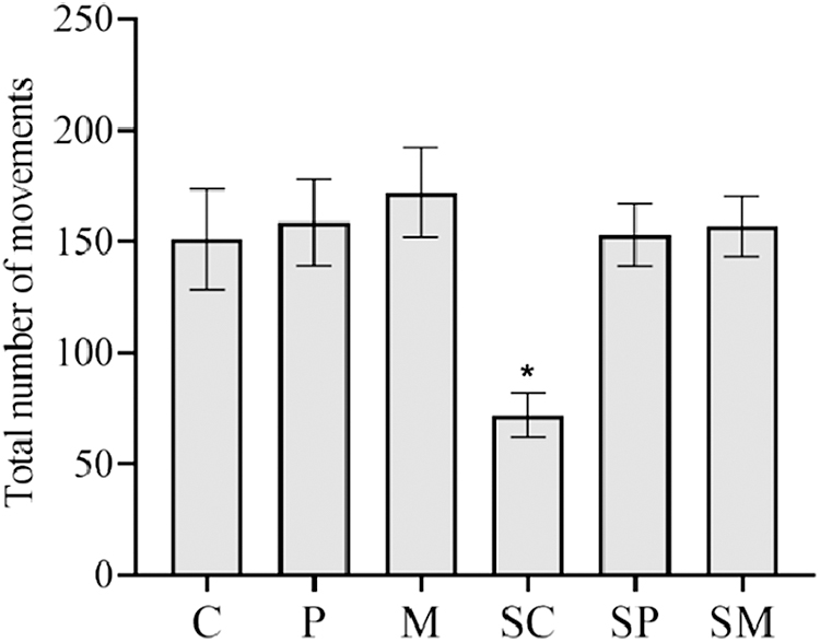
Total number of movements in the open-field test of rats submitted to CRS and control groups. Values represent means ± SD (n = 6). One-way ANOVA followed by Tukey post hoc test; *P < .01 versus control. ANOVA, analysis of variance; C, control; CRS, chronic restraint stress; M, microencapsulated pulp oil; P, pulp oil; SC, control and CRS; SD, standard deviation; SM, microencapsulated pulp oil and CRS; SP, pulp oil and CRS.
Lipid peroxidation assay
The TBARS content (Fig. 2) of the SC group was significantly higher (P < .05) than that of C group and the groups of stressed treatments in the three brain regions analyzed, except in the striatum SP group, which did not show any difference compared with the SC group, but showed a significant difference compared with the P group (P < .05). Among the stressed groups that received different treatments, there was a difference between SP and SM in the regions of the cortex and striatum. The hippocampus was the only brain region that showed no significant difference between the treatment groups, unlike the cortex and striatum, whose values in the SM group were significantly higher (P < .05) than M group.
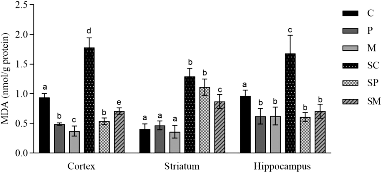
Effect of Acrocomia aculeata oils on TBARS contents in cortex, striatum, and hippocampus of rat's brain submitted to CRS and control groups. Values represent means ± SD (n = 6). Two-way ANOVA and Bonferroni post-test. Values with different superscript letters (a, b, c, d, e) indicate significantly different means at P < .05. MDA, malondialdehyde; TBARS, thiobarbituric acid.
Protein carbonyls
Similar to the concentrations of TBARS, which increased in the SC group in the three brain regions analyzed compared with the control C group, the concentrations of PC revealed parallel results (Fig. 3). The pulp oil administration prevented alterations in SP groups in the three brain regions with significant differences when compared with the SC group. However, the SM group was effective in preventing changes in PC only in the striated and hippocampal tissues, with significant differences in comparison with the SC group (P < .05 and P < .01, respectively). We found that only in the hippocampus region there was a significant decrease among the treatments that were stressed (CRS) and the respective groups that were not stressed, comparing M with SM groups and P with SP groups.
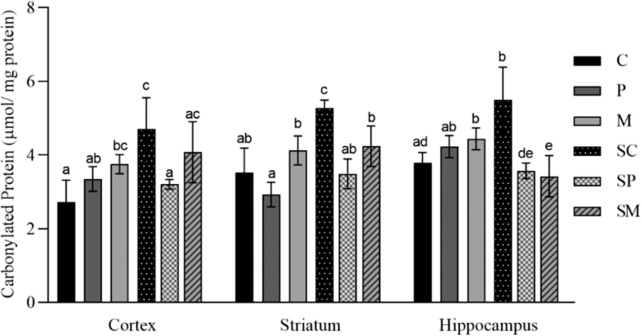
Effect of Acrocomia aculeata oils on PC contents in cortex, striatum, and hippocampus of rat's brain submitted to CRS and control groups. Values represent means ± SD (n = 6). Two-way ANOVA and Bonferroni post-test. Values with different superscript letters (a, b, c, d, e) indicate significantly different means at P < .05. PC, protein carbonyl.
GSH assay (nonprotein thiols) and antioxidant enzymes
Table 4 shows the results found for GSH, SOD, and CAT in the cortex, striatum, and hippocampus of the rat brain. The GSH content of the SC group was significantly lower (P < .01) than that of C group in the striatum and hippocampus regions. In the SP and SM groups, the difference was significantly higher (P < .01) compared with the SC group in the three evaluated regions. The GSH contents of the SP groups in the cortex and hippocampus were significantly higher (P < .05) than those values found in P groups. Similarly, SM levels were significantly higher in the three regions when compared with the corresponding M groups (P < .01). Besides, SM presented significantly higher results (P < .01) than those shown in the SP groups for all the three regions evaluated.
Effect of Acrocomia aculeata Oils on Reduced Glutathione (μmol/mg Protein) Contents, Superoxide Dismutase (U/mg Protein), and Catalase (mmol/min/mg Protein) Activities in the Cortex, Striatum, and Hippocampus of Rats' Brain Submitted to Chronic Restraint Stress and Control Groups
Each measurement was done in triplicate. Values represent means ± SD (n = 6).
Two-way ANOVA and Bonferroni post-test versus SC.
Two-way ANOVA and Bonferroni post-test versus P.
Two-way ANOVA and Bonferroni post-test versus M.
Two-way ANOVA and Bonferroni post-test versus SP.
P < .01; # P < .05.
CAT, catalase; GSH, reduced glutathione; SOD, superoxide dismutase.
The SOD activity of the SC groups was significantly lower (P < .01) than that presented in C, SP, and SM groups in the cortex and hippocampus regions. In the striatum, the only significant change occurred in the SP group, which showed lower values than the P group (P < .01). The SM groups in the cortex and hippocampus regions showed significantly higher values compared with the SC group (P < .01), whereas only the SM group of the hippocampus showed significantly higher activity than the M group (P < .01).
The CAT activity in the SC group was significantly lower than that of C groups for the cortex and striatal tissues (P < .01) and hippocampus (P < .05). The SP groups in the cortex and hippocampus showed significantly higher activities when compared with the SC group (P < .05 and P < .01, respectively), in contrast to SM, whose difference was significantly higher than SC in the three regions evaluated (P < .01 for cortex and striatum and P < .05 for the hippocampus).
Discussion
Chronic stress is associated with energy imbalance and reduced body weight 2,36 and food intake 37 in rodents. This affirmation was also confirmed in this study, where CRS was responsible for the lower body weight gain in rats, whereas the oils reversed this situation. The natural and protective antioxidant effect of A. aculeata pulp oil may be related to its rich content in monounsaturated fatty acids, 11 which, in turn, stabilize redox status and maintain body weight. 3 We observed a reduction in food intake in response to stress when comparing stressed groups to the respective nonstressed groups.
We also found that the P group showed a reduction in body weight gain of ∼18.0% compared with the control C group. Jacobowski et al. 14 demonstrated that daily intake of 2 μg/g of A. aculeata pulp oil for 8 weeks by obese and nonobese C57Bl6C mice caused a significant reduction in body weight of the groups when compared with controls. Oleic acid has an anorectic effect, 38 signals the CNS as “nutritional abundance” that limits the entry of nutrients into the circulation and, therefore, reduces food intake. Probably the oil content, rich in oleic acid, regulated body weight homeostasis and lipid metabolism. 39 However, this behavior was not observed in the M group, which showed no difference with the control group.
Comparing the composition of the pulp of A. aculeata found in this study with that described by Lescano et al., 40 there is no significant difference in the contents of moisture, lipids, and carbohydrates. In contrast, ash and protein contents were ∼53% higher in our study, whereas there was no significant difference in the total calculated energy value. The ascorbic acid content (31.2 mg/100 g) was approximately twice found by Lescano et al. 40 compared with the pulp of A. aculeata (15.41 mg/100 g). The variations in physicochemical values in fruits of the same species can be attributed to climatic conditions, soil type, fruit collection period, and other climatic factors. 40 CRS induced a deficit in locomotor activity and increased anxiety-like behavior, including decreased total movements in the open-field test. Suppression of exploratory behavior in response to stress appears to be mediated in the hippocampus, 41 which revealed an increase in the contents of the two marker of oxidative damage here examined, TBARS and PC, as well as decreased activity of the antioxidant enzymes in the SC group. The intervention of pulp oil and microcapsules of pulp oil restored this anxiety behavior.
We showed that CRS-induced markers for OS in the cortex, striatum, and hippocampus, and that A. aculeata pulp oil protected against these CRS-induced deleterious effects. In this regard, the brain is susceptible to OS due to its high demand for oxygen and high concentration of PUFA, which are extremely susceptible to lipid peroxidation, thereby may result in the accumulation of lipid hydroperoxides. 10 The BBB has a critical role in CNS homeostasis, which selectively excludes most blood-borne substances from entering the brain. 42
Most of antioxidants and neuroprotective agents have relatively low permeability at the BBB; however, ascorbic acid seems to improve other compounds to cross such barrier 43 by glucose transporters. 44 Also, α-tocopherol is actively taken up by the brain and is directly involved in protection of nervous cell membranes, whereas the concentrations of α-tocopherol in cerebrospinal fluid correlated significantly with its concentration in serum. 45,46 The pulp oil showed to be also rich in α-tocopherol and ascorbic acid, both with synergic antioxidant properties 16,47 (Table 2).
The relatively high content of several nutritional antioxidants present in A. aculeata pulp oil is similar to those found in other seeds and fruits already studied. The olive oil (Olea europaea) showed values of α-tocopherol (11.2 mg/100 g) similar to that of A. aculeata pulp oil (12.6 mg/100 g), 48 whereas the ascorbic acid content of A. aculeata pulp oil (31.2 mg/100 g) is similar to that exhibited by Eugenia dysenterica (29.7 mg/100 g), another economically important fruit of Cerrado biome. 49
The important presence of both antioxidants in A. aculeata pulp oil makes us believe in its antioxidant property and potential use in humans. In this regard, attenuation of systemic OS markers was detected in the blood of obese nurses submitted to a hypocaloric diet combined with the intake of soybean or olive oils, both also rich in α-tocopherol contents, after 2 months of supplementation. 50 Ainsah et al. 51 demonstrated that α-tocopherol supplementation (90 mg/kg) for 8 weeks in rats submitted to restraint stress significantly decreased the locomotor changes observed in controls in an open-field test. They suggested that α-tocopherol was effective in reducing stress, probably due, among other antioxidant benefits, to the release of endorphins, thus reducing the effects of stress on the locomotor activity of rats.
We have previously demonstrated that A. aculeata pulp oil is also rich in β-carotene, a ROS scavenger of singlet oxygen ( 1 O2), protecting cells from the toxic action of oxidizing agents, and preventing further degeneration in an OS scenario. 14 Carotenoids also appear to alter the condition of cognitive decline according to the results obtained in the open-field test. The underlying mechanism is unclear but possibly related to antioxidant activity 52 by scavenging ROS. 53
A higher level of protein carbonylation was demonstrated to be linked to various pathologies of the CNS. 54,55 The antioxidant protection effect of the oil was clearly shown in the different brain structures of the young rats treated with oil and its microcapsules, especially in the hippocampus, in which there was a decrease in the PC content in this region of these animals (Fig. 3).
Vitamin E can control decreasing ROS by modulating the enzyme SOD by reducing the expression of this enzyme. Also, α-tocopherol can inhibit kinase C protein in several cell types, inhibiting the production of O2 •− in monocytes and macrophages, by the nicotinamide adenine dinucleotide phosphate oxidase system. 3 –56
After treatment with the pulp oil or microcapsules of pulp oil, CAT activity decreased in all analyzed brain fractions, possibly due to a compensation of the oil supplementation and/or the synergistic effect of α-tocopherol, ascorbic acid, and β-carotene. 16
GSH has a fundamental role as a general antioxidant, including the detoxification of •OH, in addition to supporting GPx (glutathione peroxidase) and GST (glutathione S-transferase) activities. 16 Furthermore, GSH also participates in ascorbic acid and α-tocopherol regeneration, among other essential antioxidant functions. 16 The increased activity of SOD and CAT together with decreased in the GSH content are probably a consequence of the oxidative damage verified through the high lipoperoxidation and protein carbonylation found in the different brain tissue examined in this study.
These findings are in accordance with previous similar studies that showed the efficacy of other antioxidants in attenuating the oxidative insult promoted by system. 3,5,14,56
In conclusion, CRS-induced oxidative damage in brain in male Wistar rats increased lipid peroxidation, depleted GSH concentrations, and modulated enzymatic antioxidants. The intake of pulp oil of A. aculeata was able to ameliorate the cerebral oxidative damage, probably due to the presence of powerful nutritional antioxidants found in pulp oil. The microcapsules of pulp oil demonstrated a better antioxidant and neuroprotective effect compared with nonencapsulated pulp oil, demonstrating that encapsulation protects bioactive compounds that could act physiologically in the brain regions altered by CRS. A. aculeata pulp oil and especially microencapsulated pulp oil attenuated behavior alterations and several OS parameters in cortex, striatum, and hippocampus caused by CRS-associated oxidative damage.
Footnotes
Author Disclosure Statement
No competing financial interests exist.
Funding Information
This study was carried out with support from the Federal University of Mato Grosso do Sul—UFMS/MEC—Brazil. It also had the support of the following funding agencies: National Council for Scientific and Technological Development/MCTIC—Brazil (CNPq), Financing Code 430694/2016; Support Foundation for the Development of Education, Science and Technology of the State of Mato Grosso do Sul (FUNDECT), Financing Code: T.O. 009/2015 and T.O. 047/2018.