
Abstract
Selenium has an anti-inflammatory, antioxidant, and possibly antitumoral action. Thus, we hypothesized that this element could be an ally in cancer treatment. We evaluated the effect of chelated selenium treatment of BALB/c mice with Erhlich Tumor on tumor size, histology, and biochemical parameters of the liver. A total of 96 male mice were treated for 7, 15, and 30 days with different doses of chelated selenium. During the 7 days of treatment, livers presented mild hydropic degeneration; after 15 days, the livers presented mild hydropic degeneration, inflammatory infiltrate, and steatosis, which was intensified in the animals treated for 30 days. Biochemical analysis showed an increase of the alanine transaminase enzyme in those animals, indicating hepatotoxicity. At the beginning of treatment, selenium was able to inhibit tumor growth. After 30 days of treatment, however, hepatotoxicity could be seen.
INTRODUCTION
Breast cancer is one of the most common cancer among women. Its incidence and mortality rise from the age of 40 years. In women younger than 40 years, 10 deaths occur per 100,000 women. In women older than 60 years, the mortality is 10 times higher. 1 For experimental breast cancer studies, the Erhlich transplantable tumor model (TET, from Transplant of Erhlich Tumor) is used. TET originates from murine mammary adenocarcinoma and is covered by a pseudocapsule that allows monitoring of the evolution of the tumor. 2,3
The importance of diet and nutrition in cancer prevention is widely recognized and was based on a series of clinical and laboratory evidence. 4 Food is naturally complex and provides numerous bioactive substances that can act individually and/or synergistically to influence processes such as cell differentiation and apoptosis, as well as hormonal regulation of cellular functions. 5
The two most studied nutrients used as antioxidants in cancer prevention and treatment are selenium and vitamin E. The effect of organic selenium and vitamin E on the growth of the Ehrlich tumor in the solid form in BALB/c mice has already been evaluated. 6 Selenium supplementation did not reduce tumor growth and no histological change was observed. However, selenium generated an increase in the percentage of natural killer cells in the spleen of animals without the tumor. These cells have cytotoxic potential that is important for the action response against tumor cells. Other studies have shown results that demonstrate the antineoplastic action of selenium since its supplementation in many cases was able to reduce or inhibit tumor growth both in humans and in murine models. 7,8
The ideal daily supplement for adults is 100–300 μg/day of selenium. 9 The toxic dose of this element was established at 400 μg/day. Values above this can cause selenosis, 9 characterized by nausea, diarrhea, fatigue, vomiting, hair loss, nail changes, changes in mental status, nervous system abnormalities with peripheral neuropathy, and, eventually, cirrhosis. Therefore, up to 400 μg/day of selenium supplementation is considered to be a safe dose. In male Swiss mice, the lethal dose 50 (LD50) was established at 133 mg/kg. 10
In light of the above, we hypothesized that selenium could be an ally in cancer treatment. Thus, we proposed to evaluate the action of subclinical doses of selenium treatment in BALB/c mice with transplanted Erhlich tumor to elucidate the possible antitumor action of selenium and its toxicity in the liver, lung, kidneys, quadriceps, and spleen.
MATERIALS AND METHODS
Animals
A total of 96 male BALB/c mice (Mus musculus) with 4–8 weeks of age (adults) were used and obtained from the Centro Universitário Saúde ABC/Faculdade de Medicina do ABC (CUSABC/FMABC) Vivarium. The animals were kept in groups of four per polypropylene box with a stainless-steel lid filled with autoclaved wood shavings. The animals were kept under controlled ventilation (20 air changes/h), with temperatures between 22°C and 28°C and relative humidity of 60–80% and received filtered water and a ready-to-use ration (free of selenium) indicated for the feeding of mice and laboratory rats Nuvilab CR-1 (Nuvital Nutrientes, Premix, Brazil) ad libitum. Diet composition can be found in Table 1.
Composition of Animal's Diet
BHT, butylated hydroxytoluene; IU, international unit.
This work was approved by the Animal Use Ethics Committee of FMABC (protocol no. 04/2017).
Tumor cell inoculation in animals
Tumor cells were extracted from the ascitic fluid of mice with transplantable Ehrlich tumor kept at CUSABC. Tumor cells were counted in a Newbauer chamber (Grid Optik, Brazil), and the concentration adjusted to 2 × 10⁵ cells per 0.2 mL of sterile saline. Forty-eight mice of the different groups were inoculated subcutaneously with 0.2 mL of the TET cell suspension in the dorsal-lateral region.
Preparation of selenium
With the knowledge that mice weigh an average of 30 g, we obtained 1.43, 2.86, and 4.29 μg/(kg·day) as final concentrations of selenium to be used in the animals. These doses were based on previous results obtained by the group, 11,12 adapted for mice. Pure selenium was in the chelated form with glycine 1.078% of crude mass (Pharmanostra, Brazil); 0.9276 g of glycine was weighed, with a selenium mass of 0.01 g. This mass was diluted to a concentration equal to 0.001 g/mL. This solution was diluted 10 × , resulting in a concentration equal to 0.0001 g/mL; this new solution was diluted until it reached a final concentration of 5.0 μg/mL. From this last solution, volumes of 0.1, 0.2, and 0.3 mL were administered to the animals, corresponding to the concentrations mentioned above.
Treatment of animals
The animals were divided into 8 groups, of which 4 groups were composed of animals with the tumor, and the other 4, animals without the tumor. Each group was further divided into three subgroups (7, 15, and 30 days of treatment) composed by 4 animals/subgroup, who received daily the different doses of selenium (gavage) orally, as described in Table 2.
Distribution of Groups of BALB/c Mice Without Tumor and With Ehrlich Tumor Supplemented with Selenium in Different Concentrations
Each group composed 12 animals, subdivided into 3 subgroups (n = 4) according to treatment.
Mice with and without Erhlich tumor were treated by gavage on consecutive days according to the treatment time (7, 15, and 30 days), always performed at the same time (13:00–14:00).
Euthanasia of animals
At the end of the treatment times (7, 15, and 30 days), the animals were euthanized with an overdose of the anesthetic sodium thiopental, injected intraperitoneally (100 mg/kg). Then, the abdominal cavity was opened to puncture the caudal vena cava and obtain a blood sample (1 mL) that was placed in a bottle with pediatric clot retractor.
Dosage of liver function enzymes
The serum dosages of liver transaminases (alanine transaminase—ALT and aspartate transaminase—AST) were determined with the aid of a Siemens® Advia 1200 Chemistry Analyzer (Siemens Healthcare, Deerfield, IL, USA). The results were evaluated following the Good Practices in Clinical Analyses. 13
Assessment of tumor growth
After euthanizing the animals of the different groups, the tumors were removed from the dorsal region and weighed individually on an analytical scale.
Histological analysis of the tumor, muscles, liver, kidney, and spleen of the animals
Histological fragments of the liver, kidney, spleen, quadriceps muscle, and tumor were analyzed. These were fixed in a 10% formaldehyde solution for 24 h and then dehydrated in an increasing gradient of ethanol, diaphanized in xylol, and included in paraffin. Serial slices 7-μm thick were collected on glass slides using a LEICA—RM 2145 manual microtome and stained with hematoxylin-eosin for histological evaluations.
The histological sections were dewaxed in xylol, hydrated in a decreasing gradient of ethanol (100%, 95%, and 70%), stained for 2 min in Harris Hematoxylin, washed for 3 min in distilled water, and stained with aqueous Eosin for 4 min. After staining, the slides were washed in distilled water, dehydrated in an increasing gradient of ethanol (70%, 95%, and 100%), and mounted with Entellan®. The sections were then analyzed under light microscopy and photographed using a Nikon Eclipse E200® photo microscope.
Statistical analyses
Data were expressed as medians and 95% confidence intervals. We used the Dunnett's test for multiple comparisons. The program used was GraphPad Prism version 7.0. (GraphPad Software, San Diego, CA, USA). A significance level of 5% (P < .05) was considered.
The study was conducted in accordance with the Basic and Clinical Pharmacology and Toxicology policy for experimental and clinical studies. 14
RESULTS
The influence of selenium treatment on the growth of the Ehrlich tumor in the animals was initially evaluated by comparing the weight of the tumors in the control group in relation to the weight of the tumors of the supplemented animals. The animals that were on treatment for 7 days showed a significant decrease in the weight of the tumors compared to the control in the three tested concentrations of selenium. The animals treated with selenium for 15 days did not show significant differences in the weights of the tumors in relation to the control in the three evaluated concentrations. In the group treated for 30 days with selenium, we noticed a significant increase in the weight of the tumors compared to the control in the different tested concentrations (Table 3; Fig. 1).

Photomicrograph of transplantable Ehrlich tumor stained with HE obtained from animal treated with saline solution for 7
Median and Confidence Interval of Weight (g) of Solid Ehrlich Tumors of Animals Supplemented with Different Concentrations of Selenium in the Periods of 7, 15, and 30 Days
Control = 0.2 mL saline, T0.1 = animals with Ehrlich tumor treated with selenium at 1.43 μg/kg, T0.2 = animals with Ehrlich tumor treated with selenium at 2.86 μg/kg, and T0.3 = animals with Ehrlich tumor treated with selenium at 4.29 μg/kg.
Dunnett's test (P < .05). n = 4/group.
CI, confidence interval.
After 7 days of selenium treatment, the tumor was observed to have a thick capsule that possibly inhibited its growth. After 15 days of treatment, the thickness of the capsule decreased, which favors the growth of the tumor mass; after 30 days, this capsule is represented by a thin layer, no longer inhibiting the growth of the tumor; these data are corroborated with the analysis of tumor weights (Fig. 1).
Histological analysis of the liver, kidney, spleen, and quadriceps muscle showed that only the liver tissue presented morphological changes with selenium treatment (Figs. 2 –4).

Photomicrograph of livers stained with HE of animals treated for 7 days with saline solution of liver in the tumor group
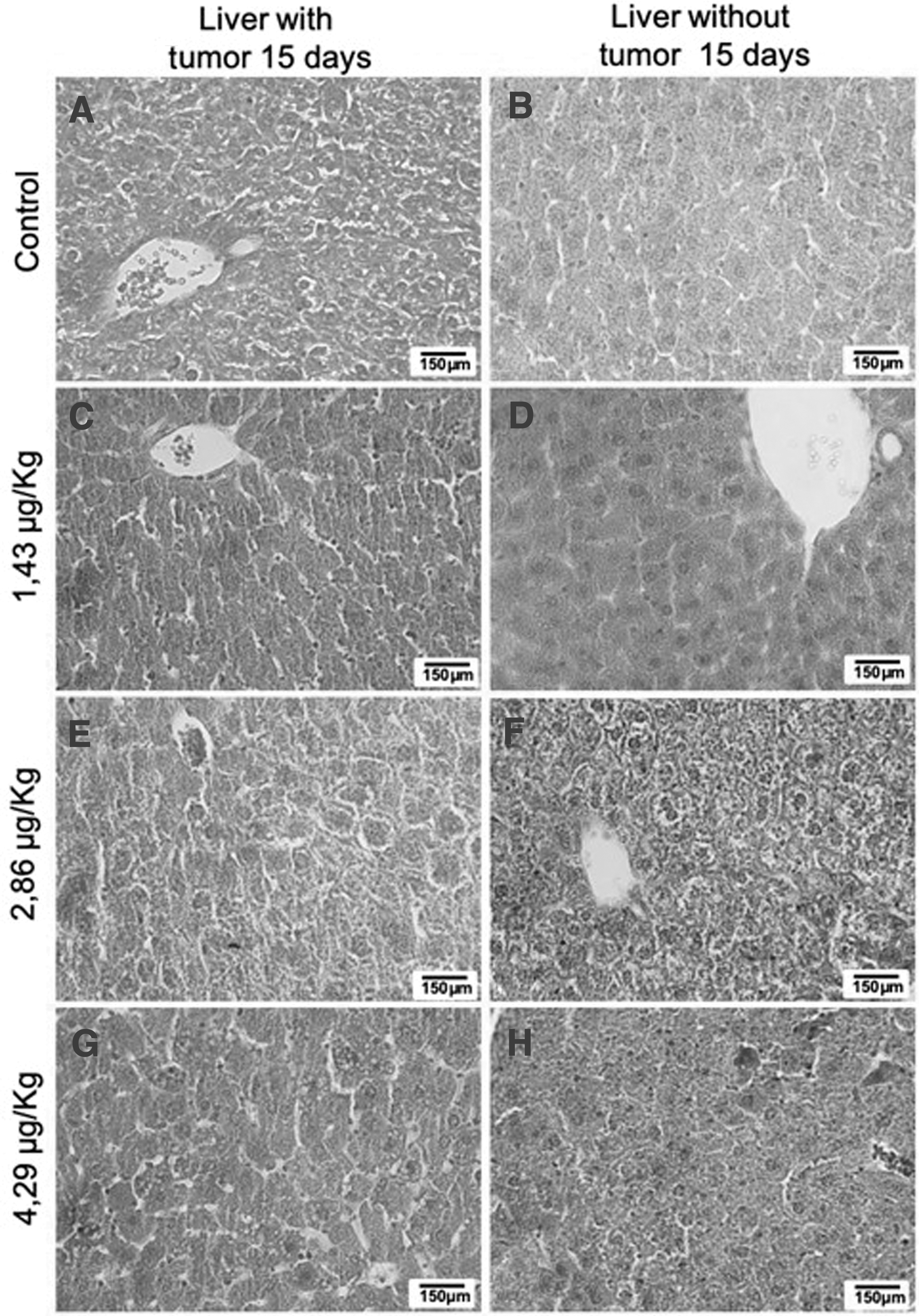
Photomicrograph of livers stained with HE from animals treated for 15 days with saline solution of liver in the tumor group
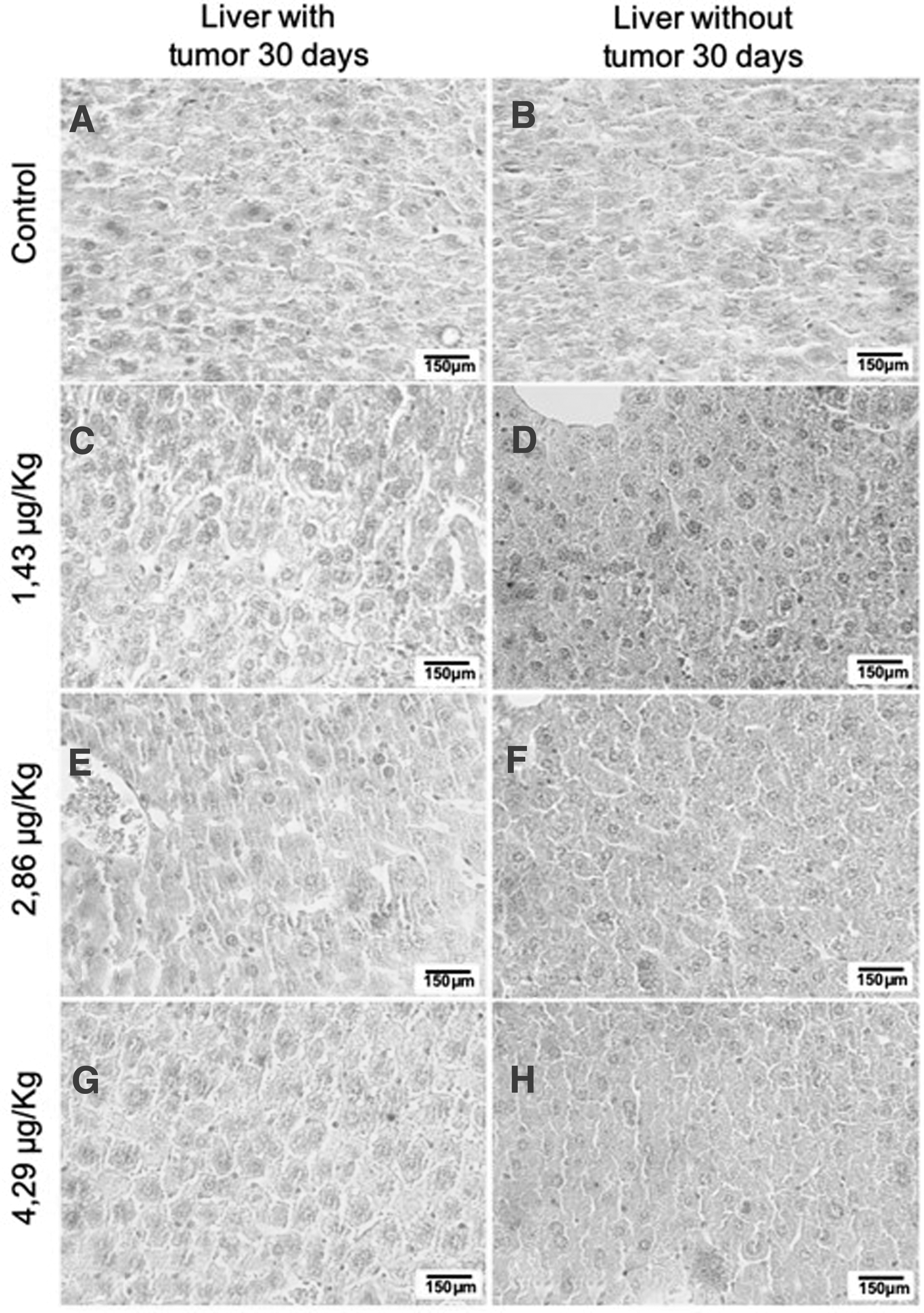
Photomicrograph of livers stained with HE of animals treated for 30 days with saline solution of liver in the tumor group
Livers of animals with tumor showed mild edematous degeneration compared to the control; livers of animals without tumor showed no change. After 15 days, the liver of animals with TET treated with 1.43 μg/kg of selenium showed possible inflammatory infiltrate, edematous degeneration, and congestion in relation to the control. The liver of animals with TET treated with 2.86 μg/kg of selenium also showed hydropic degeneration and congestion, while the liver of animals with TET treated with 4.29 μg/kg of selenium showed, in addition to hydropic degeneration, the presence of inflammatory infiltrate and steatosis in relation to the control. The liver of animals without tumor, in turn, showed hydropic degeneration after treatment with all doses; animals treated with 4.29 μg/kg of selenium also had inflammatory infiltrate in the liver.
After 30 days, livers of the animals with TET and treated with 2.86 μg/kg of selenium presented inflammatory infiltrate, edematous degeneration, and congestion in relation to the control. Livers of animals treated with 4.29 μg/kg of selenium with and without TET showed hydropic degeneration in relation to the control and the presence of inflammatory infiltrate. These data are corroborated by the biochemical analysis of liver function, according to which animals with tumors treated with selenium for 30 days showed a significant increase in the ALT enzyme in relation to the control, indicating liver changes (Table 4). Animals without a tumor showed significant changes in the concentration of 4.29 μg/kg of selenium compared to the control (Table 5).
Serum Aspartate Aminotransferase and Alanine Aminotransferase Measurements in International Unit of Animals with Erhlich Tumor and Supplemented Orally with Different Selenium Concentrations During the Periods of 7, 15, and 30 Days
Control = 0.2 mL saline, T0.1 = animals with Ehrlich tumor treated with selenium at 1.43 μg/kg, T0.2 = animals with Ehrlich tumor treated with selenium at 2.86 μg/kg, and T0.3 = animals with Ehrlich tumor treated with selenium at 4.29 μg/kg.
Dunnett's test (P < .05). n = 4/treatment time groups.
ALT, alanine aminotransferase; AST, aspartate aminotransferase.
Serum Aspartate Aminotransferase and Alanine Aminotransferase Measurements in International Unit of Animals Without Erhlich Tumor and Supplemented Orally With Different Concentrations of Selenium During the Periods of 7, 15, and 30 Days
Control = 0.2 mL saline, S0.1 = animals without Ehrlich tumor treated with selenium at 1.43 μg/kg, S0.2 = animals without Ehrlich tumor treated with selenium at 2.86 μg/kg, and S0.3 = animals without Ehrlich tumor treated with selenium at 4.29 μg/kg.
Dunnett's test (P < .05). n = 4/treatment time groups.
DISCUSSION
Selenium has an anti-inflammatory, antioxidant, and possibly antitumoral action. In this work, the evolution of the Erhlich tumor in the solid form according to the weight of the tumor mass of each animal treated with selenium was evaluated. Our results demonstrate that selenium does not inhibit tumor growth after 7 days of treatment. Since the doses given here have shown hepatotoxicity, selenium would not be a good ally in the treatment of cancer.
Studies demonstrated that selenium functioned as a substance capable of preventing breast carcinogenesis. 15 However, selenium supplementation of 5 ppm/kg for 30 days in female BALB/c mice with solid Erhlich tumor inoculated into the footpad did not inhibit or reduce tumor growth. In turn, castrated female mice with solid Ehrlich tumor and under the effect of hypothyroidism showed a reduction in tumor growth through thyroid hypofunction. 16
In this work, final concentrations of selenium 1.43, 2.86, and 4.29 μg/(kg·day) were used in animals with an average weight of 30 g. Selenium treatment with 15 μg/kg of body weight per day does not cause damage to health. 15 However, caution should be exercised with the consumption of this mineral, as there are doses that are potentially toxic and its therapeutic window is relatively small. The maximum safe dose can reach 600 μg for 84 days in humans. Therefore, it would be prudent to restrict the adult diet to 400–450 μg/day, with a safe dose of 300 μg/day. 17
Metabolism of selenium occurs in the liver, especially in hepatocytes. To evaluate the liver functions in animals with and without TET treated with selenium, serum enzymes AST and ALT were measured. When these values are increased, they indicate lesions in the hepatocyte membrane, and the increase in AST is indicative of serious injury. 18 In addition to the measurement of liver enzymes, histological analyses of the liver of these animals were performed. Although some works demonstrate the hepatoprotective role of selenium, 11,19 our results show that dietary selenium treatment does not lead to a decrease in the evolution of TET, but contributes to the hepatic alterations observed in animals with the tumor. Morphological results demonstrate that the treatment time is more significant than the tested dose, that is, the longer the treatment time with selenium in animals with TET, the more morphological changes were observed.
The hydropic degeneration observed represents an accumulation of water in the intracellular environment, which indicates imbalance in liver function. 18 These changes are observed after 7 days and during treatment; other changes complement hepatotoxicity, such as congestion and the presence of an inflammatory infiltrate. Serum biochemical measurements showed an increase in the AST enzyme in all administered doses and tended to increase according to the time of treatment. Urbankova et al. 20 also demonstrated hepatic alterations caused by selenium. In this case, the changes were dose dependent, not time dependent as our work shows: rats treated for 28 days with different doses of selenium (0.5, 1.5, 3.0, and 5.0 mg Se/kg) had morphological changes in the liver, but unchanged enzyme rates.
A study carried out with male Swiss mice strain shows that the LD50 indicating acute intoxication after 72 h of administration of the selenium micronutrient was 133 mg/kg. 10 It is possible that our results may be different because it is strain specific.
Therefore, selenium is known to have some therapeutic functions, but an excess of the mineral can cause hepatotoxicity and trigger symptoms of intoxication, such as diarrhea, nausea, fatigue, hair loss, vomiting, nail changes, nervous system abnormalities with peripheral neuropathy, changes in mental status, and eventually cirrhosis. A low concentration can favor the development of diseases, such as Keshan disease, cancer, and cardiovascular changes. 21 An effective clinical evaluation method is the serum selenium measurement, both for deficiency and toxicity. 22 Our results suggest the importance of individual good health when ingesting selenium amounts since its therapeutic window is relatively small.
Our data demonstrate that up to the 7th day of treatment, selenium was effective in inhibiting tumor growth without interfering with liver function at all doses. However, in longer treatment periods (15 and 30 days), there is a significant increase in tumor mass, liver enzymes ALT and AST, and histomorphological changes, such as hydropic degeneration and presence of inflammatory infiltrate and congestion, indicating hepatotoxicity in the treatment doses.
Footnotes
AUTHORS' CONTRIBUTIONS
JFRS and BB: literature search, experiments; GP, AHA and JV: animals care; GLV: statistical analysis; TP, FLAF and BCAA: Conceptualization; all authors participated in writing original draft and editing.
AUTHOR DISCLOSURE STATEMENT
No competing financial interests exist.
FUNDING INFORMATION
No funding was received for this article.