
Abstract
Acorn (Quercus acutissima CARR.) has been used in traditional food and medicinal ethnopharmacology in Asia, and it has shown multifarious functions such as antidementia, antiobesity, and antiasthma functions. However, there is limited scientific evidence about the efficacy of acorn for ameliorating skin problems. Treatment with ethanol-extracted acorns (EeA's) ablated the expression of inducible nitric oxide synthase (iNOS), cyclooxygenase 2 (COX2), monocyte chemoattractant protein-1 (MCP-1), and interleukin (IL)-8 stimulated by tumor necrosis factor (TNF)-α in human adult low calcium high temperature (HaCaT) cells under sublethal dosages. In addition, treatment with EeA dose dependently inhibited the ex vivo hyper keratin formation induced by TNF-α in HaCaT cells in conjunction with the blockade of cytokeratin-1 (CK-1) and cytokeratin-5 (CK-5) expression. Moreover, EeA treatment stimulated the expression of hyaluronic acid (HA) expression in human fibroblasts in a dose-dependent manner. Linoleamide was identified as the functional component of EeA using preparative high-performance liquid chromatography and ultra high performance liquid chromatography-mass spectrometry-mass spectrometry analysis, and the anti-inflammatory features and enhanced HA expression were verified. Collectively, these results suggest the efficacy of EeA supplementation in improving skin problems via anti-inflammation and upregulating HA production.
INTRODUCTION
Atopic dermatitis (AD) is a complex inflammatory skin disease characterized by various clinical phenotypes originated from multifunctional genetic and environmental factors, including infections, ultraviolet, and chemical allergens. It affects ∼20% of children and 5% of adults. 1 AD is associated with food allergy, bronchial asthma, and allergic rhinitis, and it is commonly accompanied by a number of other immune disorders. 2 The morbidities of AD include scratching the affected skin, which impairs the structure of skin barriers and aggravates the subsequent inflammatory response, which coexists with erythema, lichenification, sores, and itchiness involving the actions of cytokines and chemokines. 3
Keratinocytes play a critical function in the pathogenesis of chronic inflammatory skin disease including AD. 4 Activated keratinocytes initiate the secretion of T helper cells-2 (Th-2)-related chemokines such as CCL5, CCL17, CCL22, IL-8, and MCP-1, 5 which is characterized by small proteins secreted by various cells, including keratinocytes and immune cells, and the level of chemokines is correlated to the severity of AD. 6 Chemokines have been reported to contribute to the pathogenesis of AD. 7 Chemokines and their receptors play a critical function in the selective recruitment of various immune cells such as leukocytes, and some chemokine receptors are differentially expressed on Th-1 and Th-2 cells. 8 Moreover, chemokine and Th-2-related immunity have been reported to be associated with dysregulation of stratum corneum development, which is one of the epidermis layers composed of keratinocytes through the repeated cell division in the basal and spinous layers. 9
Therefore, the inhibition of chemokine might be a promising therapeutic target for AD treatment. 10 In addition, the released cytokines contribute to ILC2 and Th-2 cell activation and hypersensitivity in response to environmental stimuli, that is, microbial antigens and allergens in AD skin. Cytokine-mediated crosstalk between T cells and keratinocytes also plays a key role in AD features. 11,12 Nevertheless, the understanding of the relative contributions of chemokine, cytokine, keratinocytes, and hyper-stratum corneum remains limited in AD.
Acorn (Q. acutissima CARR.) is distributed in subtropical regions of Asia, and it has been used in traditional food and folk medicine for the inhibition of colitis, labor pains, laryngopharyngitis disease, and stomatitis. 13 Foods that contain acorn have been reported to be those that serve as a source of tannins, gallic acid, and gallotannin. 14 Although extracts from acorns have reportedly shown antidementia, antiobesity, antioxidant, and antiasthma functions, 15,16 the sedation effects and the underlying mechanism involving hyper-stratum corneum and hyaluronic acid (HA) production remain unclear.
In the present study, we investigated the dual ameliorative effects of ethanol-extracted acorn (EeA) through the blockade of chemokine expressions in keratinocytes in conjunction with the enhanced expression of HA expression in human fibroblasts. Our results suggest that extracted acorn might be a safe, natural reagent for anti-
MATERIALS AND METHODS
Cell culture
Human keratinocytes human adult low calcium high temperature (HaCaT) were obtained from Thermo Fisher Scientific (MA, USA). Human dermal fibroblasts Detroit-551 and HDF were purchased from the Korean Cell Line Bank (KCLB, Seoul, Korea). The HaCaT cells were grown in a keratinocyte growth media bullet kit (Lonza Biologics, Hopkinton, MA, USA), 1 × 10⁵ U/L penicillin, and 100 mg/L streptomycin (Invitrogen, Carlsbad, CA, USA) at 37°C in a humidified atmosphere containing 5% CO2. Detroit-551 and HDF were maintained as described in a previous study. 17
Reagents
Antibodies for phosphor-JNK (Thr183/Tyr185, #4668), JNK (#9252), phosphor-p38 (Thr180/Tyr182, #4511), p38 (#8690), cytokeratin-1 (CK-1) (#2655), cytokeratin-5 (CK-5) (#25807), TNF-α (#3707), and GAPDH (#5174) were purchased from Cell Signaling Technology (Beverly, MA, USA). Antibodies against iNOS (sc-7271), COX2 (sc-376862), IL-8 (sc-8427), and MCP-1 (sc-52701) were purchased from Santa Cruz Biotechnology (Santa Cruz, CA, USA). Tannic acid and linoleamide were purchased from Sigma-Aldrich (St. Louis, USA). Recombinant TNF-α was obtained from R&D Systems (Minneapolis, MN, USA).
Preparation of EeA
Acorns (Q. acutissima CARR.) were obtained from Hongchun farm, agricultural producer corporation (Gwangwon-do, Korea). One kilogram of unshelled acorn was washed and then air-dried. The dried acorn was bored with a gimlet before being extracted with 40% ethanol at room temperature for 15 days. The EeA was centrifuged at 277 g for 10 min, and the supernatants were evaporated. The powdered extract was stored at −70°C until use.
Cell viability
To evaluate cell viability, WST-1 reagent (Nalgene, Rochester, NY) was used as described previously. 18 Varying volumes of EeA were treated in HaCaT, Detroit-551, and HDF cells for 72 h. Ten microliters of WST-1 solution was added to each well and incubated for 10 min; absorbance was then measured at 450 nm (Bio-Rad Laboratories, Richmond, CA).
Ex vivo organotypic keratin formation
For formation of keratin, an SPL Insert™ Hanging six-well plate was used (SPL Life Science, Pocheon, Korea). To begin, collagen matrices (1.5 mL, 1 mg/mL collagen) were polymerized and stabilized for 24 h in a mixture of DMEM (Gibco-BRL, Gaithersburg, MD, USA): Ham's F12 media (Hyclon, Logan, UT, USA) at a ratio of 3:1 using Pure-colR Bovine Collagen type I (Thermo Fisher Scientific). HaCaT cells (1 × 10⁷ cells/mL) were incubated on collagen matrices for 2 weeks at 37°C in a humidified atmosphere containing 5% CO2 according to a previously described method. 19 At the end of the incubation period, the matrices were fixed with 3.7% paraformaldehyde and images were captured, and they were then prepared for further western blot analysis.
Western blot analysis
To evaluate the alteration of intracellular signaling molecules against EeA treatment in HaCaT cells, western blot analysis was performed as described previously. 20
HA enzyme-linked immunosorbent assay
Enzyme-linked immunosorbent assay (ELISA) for studying the efficacy of enhancing cellular HA production was performed using Human Hyaluronic Acid (HA) ELISA KIT (R&D Systems) as per the manufacturer's direction. The production of HA was measured at 450 nm (Bio-Rad Laboratories).
Preparative high-performance liquid chromatography
Preparative high-performance liquid chromatography (prep-HPLC) was used to isolate a, b, or c fractions in EeA with high purity. The prep-HPLC (Sigma-Aldrich) system consisted of a system controller (CBM-20A), photodiode array detector (SPD-M20A), pump (LC-20AP), and autosampler (SIL-10AP). Qualitative analysis was performed in a step gradient mode using a mixture of acetonitrile and water (2:8, 4:6, 7:3, and 2:8) for different time periods at a flow-rate of 1 mL/min. The total run time was 30 min. Detection was performed by monitoring the absorbance signals at 270 nm. Chromatographic analysis was performed by comparing the retention time of each peak with that listed in the reference HPLC data. According to this peak, the a, b, or c fraction was collected by using the supplier's manual.
Analysis of functional components in EeA by high HPLC and ultra high performance liquid chromatography-mass spectrometry-mass spectrometry
An HPLC system (Shimadzu Scientific Instruments, Columbia, MD, USA) equipped with a photo diode array detector and a Luna C18 (2) column (5 μm particle size, 4.6 mm × 250 nm; Phenomenex, Torrance, CA, USA) was used as reported previously. 21 The prepared EeA samples were analyzed using Q-Exactive Orbitrap hybrid mass spectrometer (Thermo Fisher Scientific) along with an Ultimate 3000 UPLC system (Thermo Fisher Scientific). Fifty centimeters × 75 μm ID packed with 2 μm C18 resin analytical columns were used to analyze the components in EeA. The flow rate of the mobile phase solution through the column was 0.15 mL/min for equilibrium.
In a whole gradient elution, 0.1% formic acid (FA; Sigma-Aldrich) in water was used as mobile phase A and 80% acetonitrile (Sigma-Aldrich) was used as mobile phase B to further separation. Gradient steps were performed with a linear gradient of 12.5% B in 20 min, 12.5–25.0% B in 5 min, 25.0–37.5% B in 5 min, 37.5–80.0% B in 6 min, and 2.5% B until it finished for 60 min.
Statistical analysis
Data are presented as mean ± standard deviation. The levels of significance for comparisons between two independent samples were determined using the Student's t-test. Groups were compared by one-way analysis of variance with Tukey's post hoc test applied to significant main effects (SPSS 12.0 K for Windows; SPSS, Inc., Chicago, IL, USA).
RESULTS
Effects of EeA treatment on human keratinocytes and fibroblasts
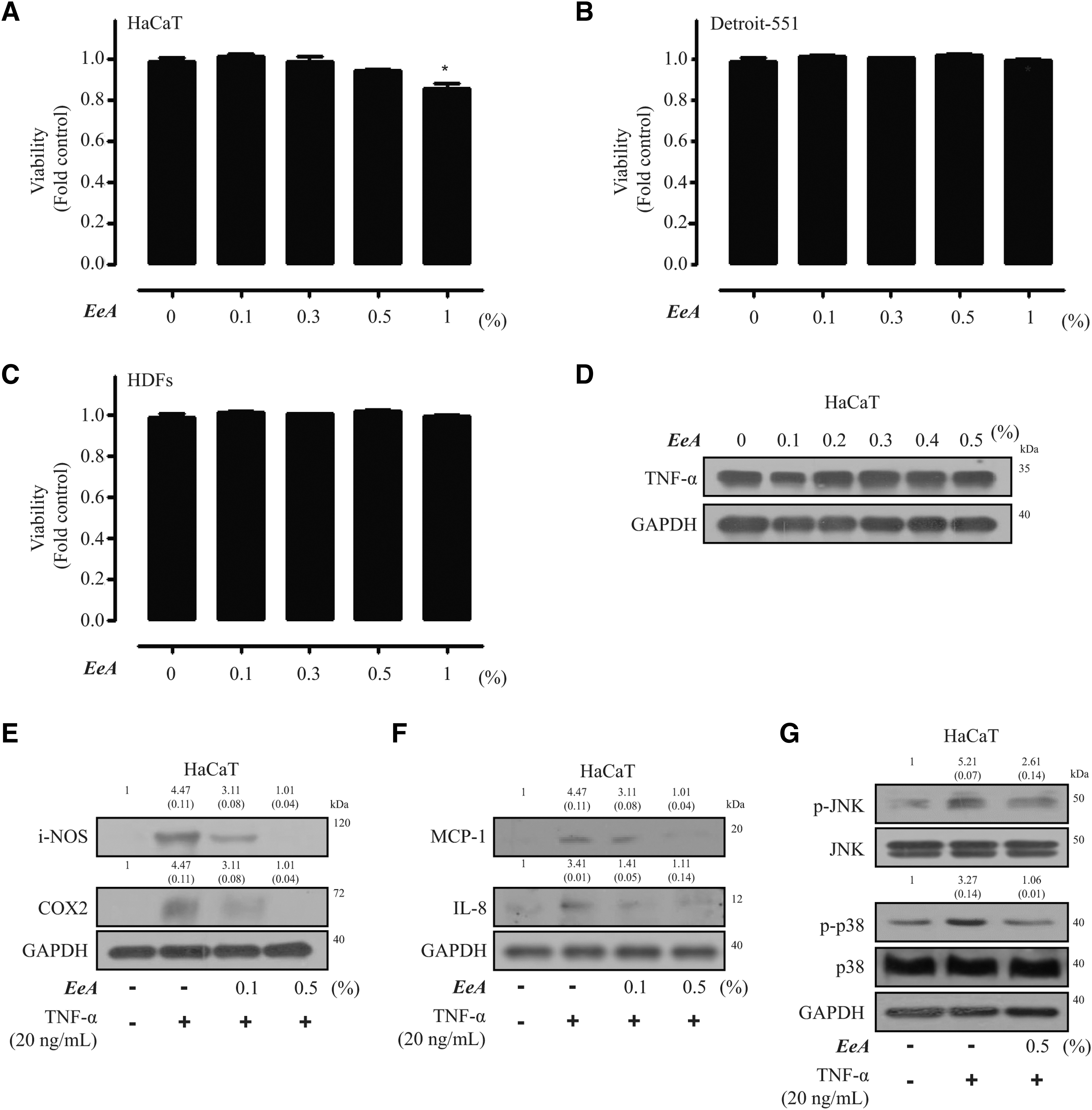
The cell viabilities of HaCaT and human skin fibroblasts (Detroit-551 and HDF) were examined following varying doses of EeA treatments to evaluate in vitro basal toxicity. EeA exposure weakly decreased the viability (∼0.05-fold at 0.5% or 0.14-fold at 1%) of HaCaT cells at the treated dose in a dose-dependent manner (Fig. 1A). Further, EeA treatment had no effects on viability in Detroit-551 and HDF cells (Fig. 1B, C). To perform further research, we decided that 0.5% of total volume was an appropriate nontoxic dosage of EeA. To demonstrate the decrease of the representative inflammatory-inducer of TNF-α expression, 22 we treated human keratinocytes with sublethal dosages of EeA (0.1%, 0.2%, 0.3%, 0.4%, and 0.5%). Treatment with EeA had no effect on basal TNF-α expression in HaCaT cells (Fig. 1D).
Effects of EeA treatment on human keratinocytes and fibroblasts.
To explain the expressions of iNOS and COX2, which have been shown to play critical functions in inflammatory features, 23,24 we investigated the expression alteration stimulated by exogenous TNF-α under nontoxic levels of EeA exposure (0.1% and 0.5%) in human keratinocytes using western blot analysis. Treatment with different dosages of EeA inhibited the expressions of iNOS and COX2 activated by exogenous TNF-α (20 ng/mL) treatment in HaCaT cells (Fig. 1E). To elucidate the expression of chemokines MCP-1 and IL-8 activated by functional cytokine treatment, we investigated the expression transduction regulated by nontoxic levels of EeA exposure (0.1% and 0.5%) in human keratinocytes using western blot analysis. Treatment with different dosages of EeA inhibited the expressions of MCP-1 and IL-8 induced by functional cytokine TNF-α (20 ng/mL) treatment in HaCaT cells (Fig. 1F).
To delineate intracellular signaling by EeA treatment in human keratinocyte, we examined the signal transduction pathways specifically regulated by EeA treatment in HaCaT cells compared to control cells. Exposure to an EeA treatment effectively suppressed the phosphorylation of JNK and p38 stimulated by functional TNF-α treatment (Fig. 1G), but not in AKT and ERK1/2 (Data not shown), which were involved in inflammatory features in HaCaT cells, as described previously. 25
Effects of EeA treatment on keratin formation in human keratinocytes and HA expression in human fibroblasts
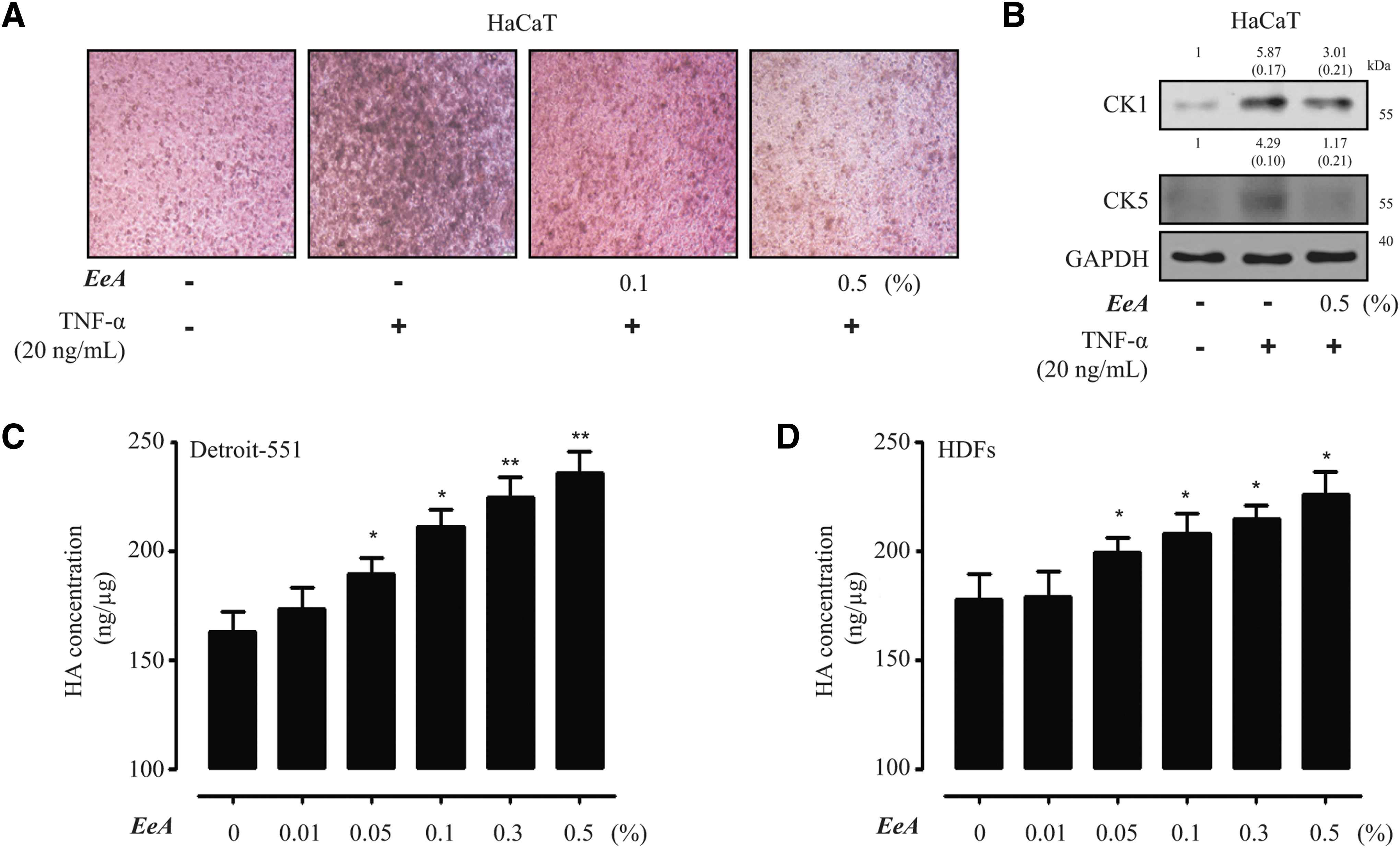
To evaluate the ameliorative effect of EeA treatment on hyperkeratin formation of keratinocyte, we established the ex vivo keratin formation using HaCaT cells. Treatment with TNF-α significantly activated the hyper formation of keratin compared to that of control HaCaT cell, and different dosages of EeA treatment restored the hyper keratin formation induced by TNF-α treatment (Fig. 2A). Dysregulated keratinocyte differentiation has been reported to involve various functional molecules, including CK-1 or CK-5. 26,27 Western blot analysis validated these results using EeA-treated HaCaT cells against TNF-α treatment, which induced the expression of CK-1 and CK-5, and different dosages of EeA treatment significantly inhibited the enhanced CK-1 and CK-5 expressions stimulated by TNF-α treatment in HaCaT cells (Fig. 2B). We also verified the effect of EeA on HA production in human fibroblast cells, which has previously been reported to be associated with inflammatory features. 28 Treatment with various dosages of EeA significantly stimulated the production of HA compared to that of control cells in a dose-dependent manner in Detroit-551 and HDFs cells (Fig. 2C and 3D).
Effects of EeA treatment on keratin formation in human keratinocytes and HA expression in human fibroblasts.
Identification of functional components in EeA.
Identification of functional components of EeA
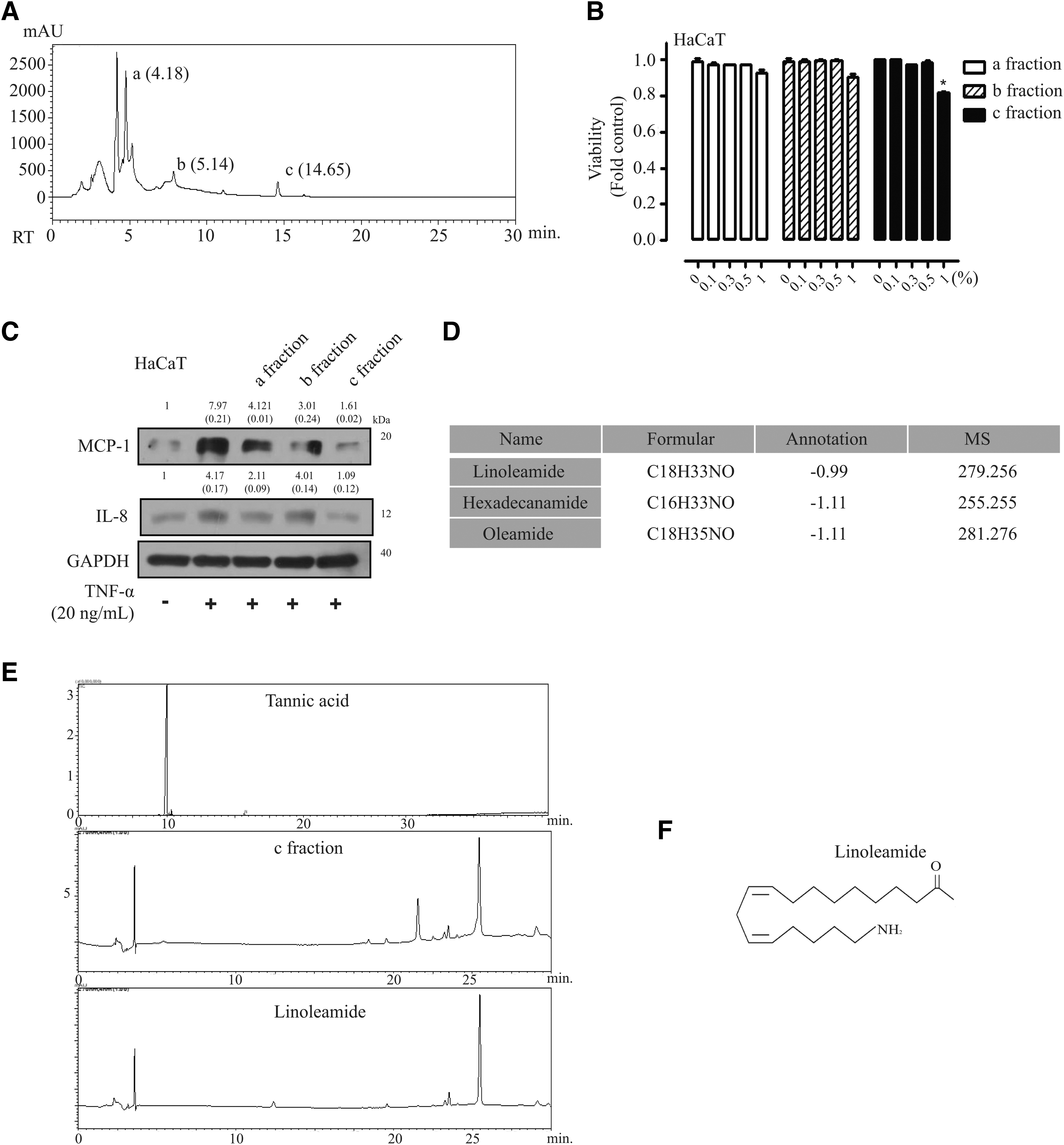
To identify the functional components of EeA, we analyzed the compositions of EeA using Preparative HPLC, HPLC, and ultra high performance liquid chromatography-mass spectrometry-mass spectrometry (UPLC-MS/MS). The Preparative HPLC chromatograms of the three peaks (a, b, and c) exhibited good resolution, which were separated as the a, b, and c peaks of EeA (Fig. 3A). Then, we investigated the basal toxicity of a, b, or c fraction on HaCaT cells viability. As shown in Figure 3B, the dose-dependent treatment of a, b, or c fraction produced a weak or nonexistent decrease in the viability of HaCaT cells, excluding the dosage of 100 μg/mL. To demonstrate the effect of a, b, or c fraction on MCP-1 and IL-8 expressions, we used the a, b, or c fraction at a 50 μg/mL dosage to treat HaCaT cells.
Treatment of c fraction significantly decreased the enhanced MCP-1 and IL-8 expression levels activated by TNF-α treatment compared to a or b fraction in HaCaT cells (Fig. 3C). To identify the isolated c fraction from EeA, UPLC-MS/MS analysis potentially suggested the c fraction to be oleamide, hexadecanamide, or linoleamide (Fig. 3D). To clarify the correspondence between c fraction in EeA, we performed HPLC analysis using c fraction, three candidates, and tannic acid, which has been identified as the major component inducing antiasthma effects in acorn extract. 16 In the HPLC chromatogram of c fraction, the peak of linoleamide was consistent compared to that of tannic acid (Fig. 3E, F). In addition, the peak of oleamide or hexadecanamide was quite different from that of c fraction (Data not shown).
Effects of linoleamide treatment on keratin formation in human keratinocyte and HA expression in human fibroblasts
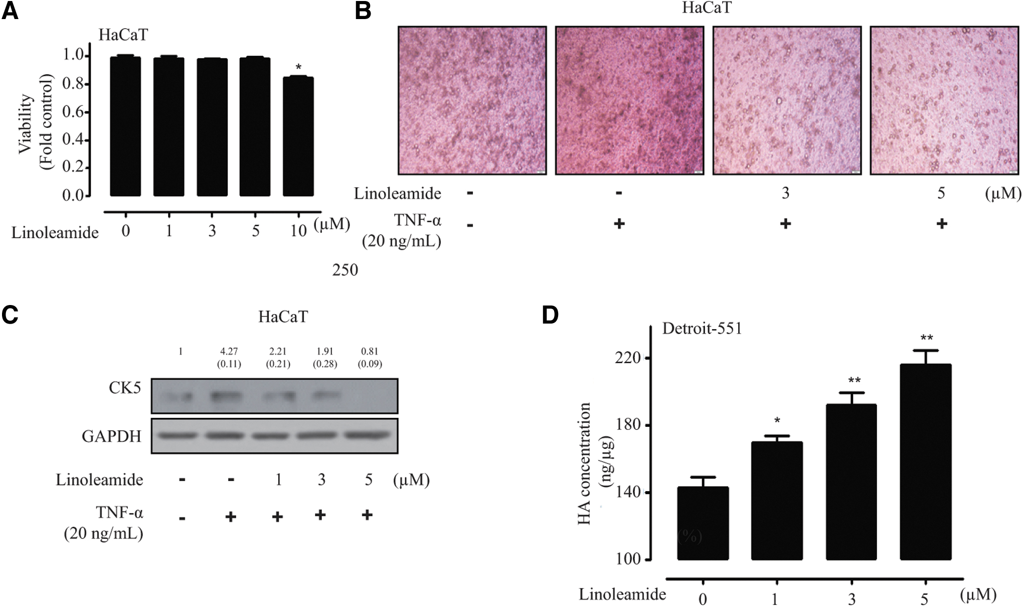
To verify the functional sedative effect of linoleamide treatment on the hyperkeratin formation of keratinocytes, we determined the sublethal dosage of linoleamide in HaCaT cells (Fig. 4A). Then, we conducted the keratin formation assay using the exogenous TNF-α and noncytotoxic dosage of linoleamide in HaCaT cells. Exogenous TNF-α stimulated the hyperkeratin formation of keratinocyte, and different dosages of linoleamide treatment restored the hyperkeratin formation that was activated by TNF-α treatment (Fig. 4B). In addition, linoleamide treatment significantly inhibited cytokeratin-5 (CK-5) expressions stimulated by TNF-α treatment in HaCaT cells in a dose-dependent manner (Fig. 4C). We also verified the sublethal dosage of linoleamide to human fibroblasts (Data not shown). Then, we validated the effect of linoleamide on the production of HA in Detroit-551 cells under treatment with sublethal dosages (Fig. 4D).
Effects of linoleamide treatment on keratin formation in human keratinocyte and HA expression in human fibroblasts.
Collectively, linoleamide containing EeA shows anti-inflammatory features in human keratinocyte and enhances the expression of HA in human fibroblasts (Fig. 5).
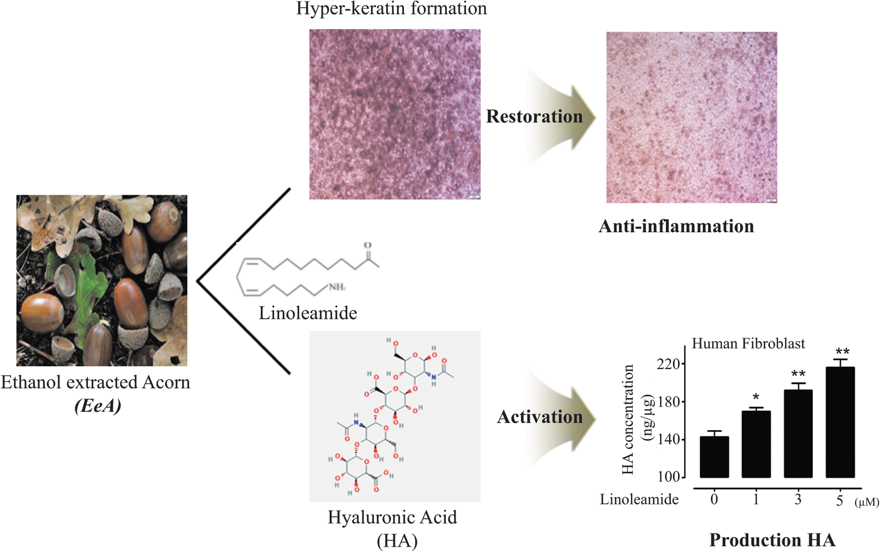
Graphical abstract of EeA's dual sedative effects via anti-inflammation and upregulating HA production.
DISCUSSION
In the present study, we verified that EeA has an anti-inflammatory effect in human keratinocyte and enhances the expression of HA in human fibroblasts. We also verified that EeA contains functional linoleamide. To our knowledge, this is the first scientific evidence showing that EeA has the dual effects of anti-inflammatory and HA induction features for the judicious use as an ameliorative intervention against skin disorders, including
Acorn-processed foods are consumed as local foods that have been shown to be beneficial for treating colitis, stomatitis, labor pain, obesity, and laryngopharyngitis disease in Korea. 15,16 Although derivatives from acorns have been reported to have multiple functions in antidementia, antiobesity, antioxidant, or antiasthma treatments, the anti-inflammatory effects involved in the action of regulating chemokine function remained ambiguous. In the present study, we clearly validated the scientific evidence of EeA that is responsible for the anti-inflammatory effects involved in regulating chemokine expressions even under sublethal dosage treatment in human keratinocytes.
Cytokines are small (<30 kDa) key signaling molecules regulating various cellular functions through the frequent overlapping and functional redundancies in the human immune system. 29 Differences in the regulation of cytokines might be expected in genetically diverse patients due to the natural fate of cytokines; however, targeting cytokines has been reported to be accompanied with an increased susceptibility to various infections such as mycobacteria, salmonellae, and fungal pathogens. 30 Further, ablating cytokine-correlated histamine using antihistamine reagent has been reported to have an adverse effect profile despite the recognition of the clinical usefulness of these drugs due to their similar potency in competing for histamine-receptor sites, which has resulted in a variety of side effects such as dopaminergic, serotonergic, and cholinergic responses. 31
Since the first discovery of the biological activity of chemokines in the late 1980s, these unveiled proteins have been a critical target for drug discovery based on the similarity with the G protein-binding site, which has motivated the exploration of new antichemokines. 32 Consistent with a previous report, we verified the anti-inflammatory effect of EeA in downregulating the representative functional chemokines, MCP-1 and IL-8 expressions activated by TNF-α treatment, even under sublethal dosages, to human keratinocyte. Notably, EeA had no effect on basal TNF-α expression even at a maximal dosage, thus implying that treatment with EeA can safely induce an anti-inflammatory effect by regulating functions of chemokine without that of cytokine in conjunction with regulating p38 and JNK pathway, as described previously. 33
Previous reports have shown that the amount of chemokine in the stratum corneum is correlated with the severity of inflammation in
Moreover, EeA treatment stimulated the production of HA in human fibroblast in a dose-dependent manner, and this production has been reported to have a correlation with anti-inflammation features, 28,35 suggesting that EeA might be a safe and effective reagent through dual-effects concerning the decrease in inflammatory features and the increase in HA production.
Acorn and the various components it contains, such as tannins, gallic acid, and gallotannin, have been reported to have antidementia, antiobesity, antioxidant, and antiasthma effects. 14 –16,21 In the present work, we further identified the functional component of acorn using preparative HPLC and UPLC-MS/MS analysis. Throughout the analysis using combined preparative HPLC and UPLC-MS/MS in EeA, the linoleamide was clearly identified as the critical component in EeA showing the regulation of chemokine expression in human keratinocytes and the induction of HA production in human fibroblasts. Notwithstanding the wide search in previous reports for an associated anti-inflammatory effect of linoleamide in EeA, there is no scientific evidence that disputes the anti-inflammatory effects of linoleamide.
Therefore, this is the first report of an evidently functional linoleamide from EeA as a dual intervention with anti-inflammatory and healing effects that facilitates HA production in human skin, implying that utilizing linoleamide or EeA might be an effective strategy against skin problems, including
Collectively, EeA has beneficial effects on human skin conditions through the selective reduction of the inflammatory response in human keratinocyte in conjunction with the induction of HA production in human fibroblast. Altogether, the present results indicate that EeA might be a safe and authentic reagent against human skin problems such as
Footnotes
AUTHORS' CONTRIBUTIONS
J.L. and S.J. conceived and supervised the experiment. J.L. designed and performed the experiments. J.L., S.J., S.H., and J.K. analyzed the data. J.L. and J.K. obtained the funding. J.L. and S.J. wrote and proofread the manuscript.
AUTHOR DISCLOSURE STATEMENT
S.-E.J. is the founder and CEO of Chanchanhee, Inc., and J.L., S.-I.H., and J.-H.K. have no competing interests.
FUNDING INFORMATION
This research was supported by the Basic Science Research Program through the National Research Foundation of Korea (NRF) funded by the Ministry of Education (2016R1A6A1A03012862 and 2021R1I1A1A01041462).