
Abstract
Given the importance of discovering plant species from the Brazilian Cerrado biome with anticancer potential, this study evaluated the antitumor activity of two extracts of Campomanesi adamantium fruits in in vitro and in vivo models of melanoma lung metastasis. Pulp and peel extracts (DEGPU and DEGPE, respectively) were extracted from fresh fruit using dichloromethane as a solvent. As cytotoxicity parameter, concentration values that inhibited 50% cell growth (GI50), total growth inhibition (TGI), and selectivity index (SI) were established. The melanoma lung metastasis model was obtained by injecting 5 × 10⁵/50 μL B16-F10 cells via the tail vein of mice, which received treatment on the 15th day. Metastatic lungs were collected for fluorescence analysis with the IR-780 marker and also macro- and microscopic assessment. In vitro analyses showed that DEGPU was active in K562 (GI50 32.99; TGI 47.93) and U-251 (GI50 32.10; TGI 249.92), whereas DEGPE showed better cytotoxicity results for all tumor cell lines, but was more efficient in K562 (GI50 27.42; TGI 40.20) and U-251 (GI50 4.89; TGI 12.77). Both showed a cytocidal effect on B16F10 at the highest concentration tested, with approximately 25% (DEGPU) and 88% (DEGPE) of cell death. In vivo analyzes showed that both extracts showed significant activity in metastatic lung. Fluorescence images showed differences in intensity between groups owing to greater tumor involvement. Macro- and microscopic images showed that treatments with extracts limited tumor growth and prevented proliferation. The extracts tested have promising activity, thus requiring further research on their active compounds.
INTRODUCTION
Cancer is considered the second leading cause of death worldwide, justifying the concern of health services and countries to develop more studies aimed at discovering active ingredients with activity against cancer. In this regard, considering the importance of using natural resources for this purpose, Brazil stands out with significant visibility for possessing the second largest biome in South America, concentrating a vast biodiversity available for study in approximately 2 million km2 of Cerrado. 1 – 3
Campomanesia adamantium (Cambess.) O. Berg, locally known as “guavira,” is a fruit found abundantly in the Brazilian Cerrado, being widely and popularly used as a medicine and in the food industry. 4 As a result, given its virtually omnipresence in the culture of the population of the State of Mato Grosso do Sul, including in music, art, and food, this fruit is considered a local symbol according to a State Law established in 2017.
Guavira is used as a folk medicine for poor digestion, anemia treatment, as an anti-inflammatory, for cholesterol control, diabetes, rheumatism, and menopause, among other uses. In 2018, Lima e Silva et al. 5 described the great in vitro antiproliferative potential of C. adamantium fruit extract on cancer cells, which could be considered a potential anticancer agent for treating this disease. From this perspective, the present research describes the analysis of two fruit extracts evaluated for their in vitro antiproliferative potential and proposes a new B16-F10 pulmonary metastasis animal model.
MATERIALS AND METHODS
Campomanesia adamantium fruits
The fruits were acquired in the District of Taunay, Municipality of Aquidauana, Mato Grosso do Sul, Brazil (20°15′12.4″ S 56°04′04.3″ W). Harvest was carried out in November 2020. The fruits were stored at a temperature of −8°C until processing. At the time, exsiccate No. 53328 was prepared and deposited at the CGMS-UFMS Herbarium.
Preparation of fruit extracts
The fruits were separated into peel and pulp (no seeds), which were dried in a convection oven at 37°C until dehydration (after about six days). Then, the peel and pulp portions separately were exhaustively macerated in dichloromethane for 15 days at room temperature, with the solvent being changed every five days. Subsequently, the extracts were filtered and concentrated in a rotary evaporator for complete elimination of the solvent and stored at 4°C. The samples obtained, named DEGPU (dichloromethane extract of guavira pulp) and DEGPE (dichloromethane extract of guavira peel), were stored in an amber flask at 4°C. 5
Cell lines
The cell lines used in the study were: chronic myeloid leukemia (K562 ATCC® CCL-243TM), two human colorectal adenocarcinomas (HT-29 ATCC® HTB-38TM and CACO-2), murine fibroblast (NIH-3T3 ATCC® CRL-1658TM), murine macrophage (J774 ATCC® TIB-67TM), human glioma (U-251), and murine melanoma (B16-F10). The experiment was carried out in the Cell Culture Laboratory of the Federal University of Mato Grosso do Sul (FACFAN-UFMS) in Campo Grande, MS, Brazil.
The NIH-3T3, B16-F10, and J774 cell lines were grown in Dulbecco’s modified Eagle’s medium (DMEM) produced by Sigma-Aldrich (MO, USA), whereas the other lines were grown in RPMI-1640 (Roswell Park Memorial Institute Medium), also produced by Sigma-Aldrich, both containing 10% fetal bovine serum (Gibco) (Thermo Fisher) and 1% streptomycin/penicillin (100 μg/mL and 100UI/mL, respectively) (Sigma-Aldrich).
In vitro antiproliferative activity—GI50, TGI, and SI
The cells were subsequently distributed in 96-well plates (100 μL/well) and exposed for 48 h to increasing DEGPE and DEGPU concentrations (0.25, 2.5, 25, and 250 μg/mL) from a 0.1% stock solution in dimethyl sulfoxide (DMSO) (Sigma-Aldrich), with each concentration being added to three wells (triplicate).
Cell proliferation was determined using colorimetric methods with sulforhodamine B (SRB) (Sigma-Aldrich), for HT-29, CACO-2, NIH-3T3, J774, U-251, B16-F10; and MTT [3-(4,5-Dimethylthiazol-2-yl)-2,5-Diphenyltetrazolium Bromide] (Sigma-Aldrich) for K562. Doxorubicin was used as a positive control at the concentrations of 0.025, 0.25, 2.5, and 25 μg/mL. 6,7
Using a concentration–response curve for the cell lines, the GI50 (concentration causing 50% cell growth inhibition) and total growth inhibition (TGI) was determined by nonlinear regression analysis (sigmoidal fitting) using the software Origin 6.0. 6 The selectivity index (SI) was calculated based on the GI50 results of nontumor cell line NIH-3T3 divided by the GI50 obtained for the other tumor cell lines. A higher SI result is considered more effective and safer. 5
IR-780 iodide, Sigma-Aldrich
IR-780 iodide was prepared from a stock solution (1% in DMSO) by diluting 0.9% sterile saline solution to a final concentration of 0.45 mg/kg, stored at 4°C.
In-Vivo Xtreme™, Bruker
An In-Vivo Xtreme BI 4MP device (Bruker) was employed for fluorescence analysis, operating with the following settings: foreground at fluorescence modality, illumination source multiwavelength, with excitation filter in 760 and emission filter in 830, camera control with standard exposure type, high speed mode, 2.0 sec of exposure time, Bin 2 × 2 pixels, FOV 19 cm, fStop 1.1, focal plane 0 mm, and rainbow histogram. Background at x-ray modality, kVp 45, 0.1 sec of exposure time, Bin 1 × 1 pixel, fStop 2.8, focal plane 0 mm, and gray scale histogram. To obtain images, the animals were previously sedated by anesthetic inhalation with isoflurane.
Lung metastasis animal model
The experiments were carried out after approval by the Ethics Committee on Animal Use (CEUA) of the Federal University of Mato Grosso do Sul, under protocol no. 1140/2020, as per directives of the National Council for Animal Experimentation Control (CONCEA). Mus musculus BALB/c males weighing 25–35 g and 4–6 weeks old were acquired from the Central Animal Facility, a sector under the authority of the Pro-rectorate of Research and Graduate Studies of UFMS (PROPP-UFMS).
The animals were weighed on the first day of the experiment, and the lung metastasis model was obtained by a single administration of B16-F10 cells (5x10⁵/50 μL) IV via the lateral tail vein using isoflurane to restrain the animals.
In vivo antiproliferative activity
On the 15th day, the animals were weighed and groups were randomly distributed (n = 6), totaling five groups named Control, Olive, DOX, DEGPU, and DEGPE. The control animals did not receive any type of treatment. The Olive group received 100 μL of pure olive oil intraperitoneally. Animals in the DOX group received 200 μL of the chemotherapy drug doxorubicin solubilized in phosphate buffered saline at a final concentration of 5 mg/kg, administered intraperitoneally. For use in animal treatments, both extracts were solubilized in pure olive oil at a final concentration of 92.5 mg/kg/100 μL, which was defined by previous experiments at the laboratory. DEGPU and DGEPE were also administered intraperitoneally. Animal feed consumption was evaluated in the last three days of experiment (from 28th to 30th), when animals began to show behavioral differences between groups.
On the 30th day, fluorescence images were acquired using an In-Vivo Xtreme equipment. All animals received 200 μL of IR-780 fluorescence marker at 0.45 mg/kg intraperitoneally, 24 h before the images were taken. The animals were weighed and euthanized by immersion in isoflurane followed by exsanguination, after which the ex vivo images of metastatic lungs were obtained.
Lung macroscopic analysis
Metastatic nodules present on the pleural surface of the lungs were counted using an Olympus optical stereomicroscope. Results were presented as group mean and standard deviation.
Lung histological analysis
The left lung lobe stained with hematoxylin and eosin (HE) was used for quantitative analysis of metastasis using a Leica DM 2000 Led microscope and the software LAS V4.12. Parenchyma involvement, location of nodules, and possible tissue injuries were analyzed. Calculation of the percentage of implantation was observed based on total area of metastasis by total area of tissue × 100. 8
Statistical analysis
The data obtained from the surface count of lung nodules, animal weight, and feed consumption were analyzed using one-way ANOVA, followed by Tukey’s post-test (P < .05) using the software Bioestat 5.0.
RESULTS
Antiproliferative activity in vitro—GI50, TGI, and SI
Table 1 presents data obtained in antiproliferative activity studies of extracts in seven different cell lines, including tumor and nontumor. The lowest GI50 results (<30) 9 are considered indicative of promising samples, as they indicate greater activity of the extract on the target cell lineage.
Antiproliferative Activity of Guavira Fruit Pulp (DEGPU) and Peel (DEGPE) Extracts against Six Cell Lines
GI50: µg/mL dose that inhibits 50% cell growth; SI: selectivity index calculated by the ratio of NIH-3T3 GI50 to each tumor’s GI50; TGI: µg/mL dose that completely inhibits cell growth.
No-tumor cell line; data in bold demonstrate the most promising results.
Thus, the pulp extract (DEGPU) proved to be more effective against tumor cell lines K562 (leukemia) and U-251 (glioma), with a GI50 of 32.99 and 32.10 μg/mL and a TGI of 47.93 and 249.92 μg/mL, respectively. The peel extract (DEGPE) also showed better results in same strains (K562 and U-251), with a GI50 of 27.42 and 4.89 μg/mL and a TGI of 40.20 and 12.77 μg/mL, respectively.
Results demonstrate that the peel extract has greater effects against the HT-29, B16-F10, and CACO-2 strains compared to the pulp extract owing to lower GI50 results obtained. None of the extracts showed cytotoxic effect against nontumor cell line NIH-3T3. The pulp extract also did not inhibit the growth of nontumor cell line J774 (macrophage); thus, at tested concentrations, DEGPU did not have a toxic effect on this cell, whereas for other cell lines this extract did not show a cytostatic effect.
SI was determined based on the GI50 results obtained previously by calculating the ratio of nontumor cell line NIH-3T3 (fibroblast) to GI50 results of other tumor cell lines. The higher the SI value, the more selective the sample is; thus, both the pulp and peel extracts were efficient against the K562 and U-251 strains. The best result obtained with the peel extract against the U-251 strain, with an SI of 51.26.
The results referring to the antiproliferative activities of both extracts against all strains tested are shown in Figure 1. The peel extract results were more effective than the pulp extract.
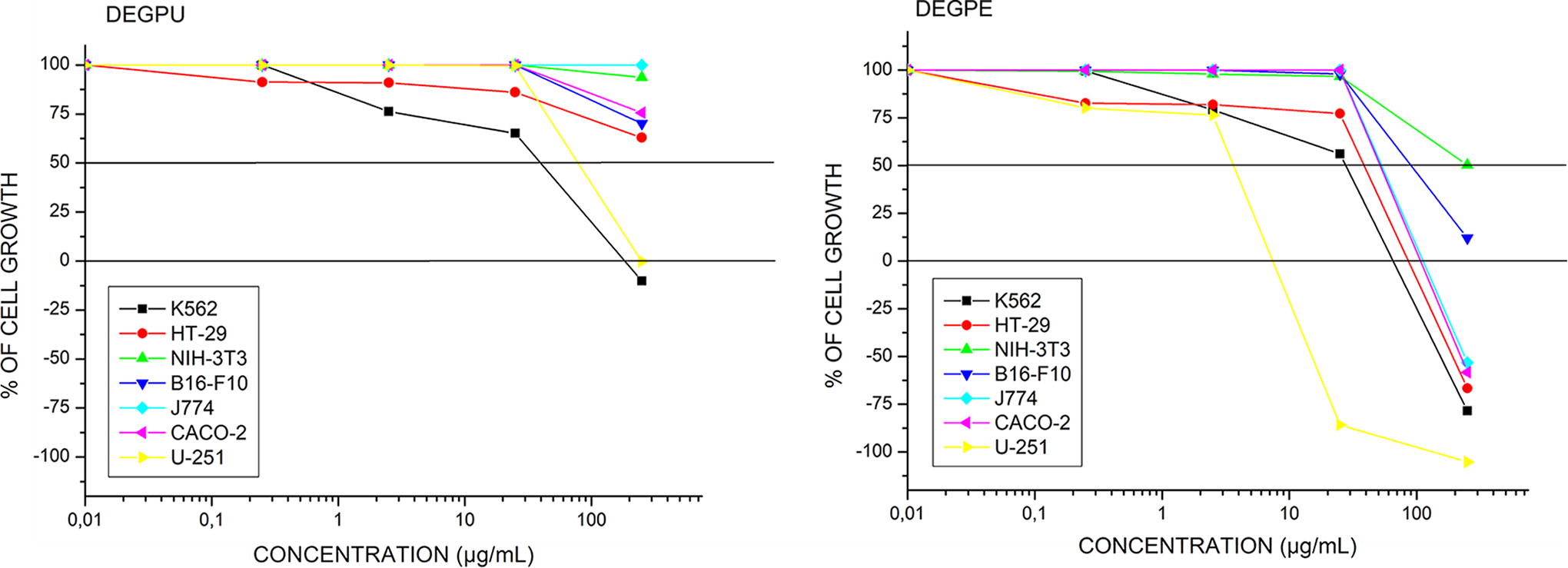
Representation of the antiproliferative activity of DEGPU and DEGPE against strains K562 (leukemia), HT-29 (colorectal adenocarcinoma), NIH-3T3 (fibroblast), B16-F10 (melanoma), J774 (macrophage), CACO-2 (colorectal adenocarcinoma), and U-251 (glioma). The result was expressed as the percentage of cell growth by the concentration of extracts (0.25, 2.5, 25, 250 µg/mL) (log10 scale). Origin 6.0 Software.
Figure 2 depicts the antiproliferative activity of the two strains that showed best results, K562 and U-251, treated with doxorubicin (standard sample) after 48 h. In both cases, the peel extract showed more satisfactory results than doxorubicin, a reference chemotherapeutic drug widely used to treat various types of cancer.
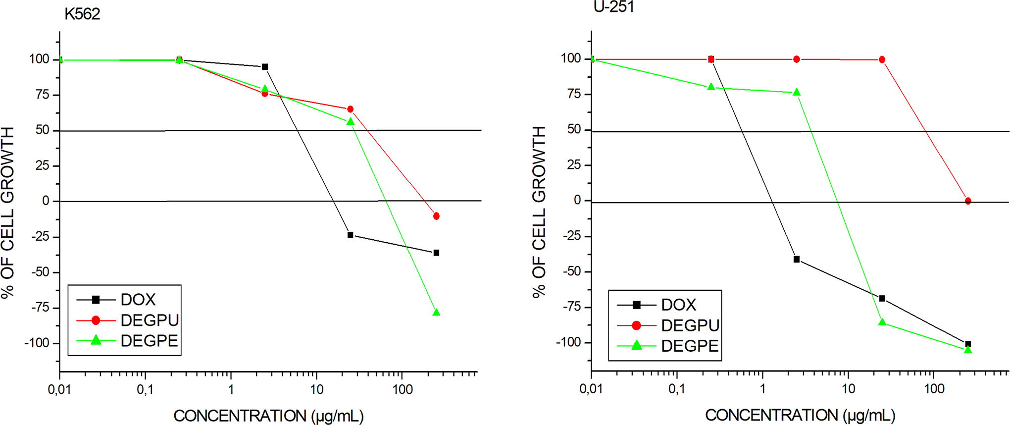
Representation of antiproliferative activity to K562 (leukemia) and U-251 (glioma) treated with doxorubicin (DOX—black square), DEGPU (red circle), and DEGPE (green triangle). The result was expressed as the percentage of cell growth by the concentration of the treatments (0.25, 2.5, 25, 250 µg/mL) (log10 scale). Origin 6.0 Software.
With regard to the B16-F10 cell line, the percentage of cell viability was also low in both treatments compared to control (untreated cells). Therefore, at the highest dose tested, there was total cell growth inhibition and cell death, demonstrating that both treatments were cytocidal for these cells (Fig. 3).
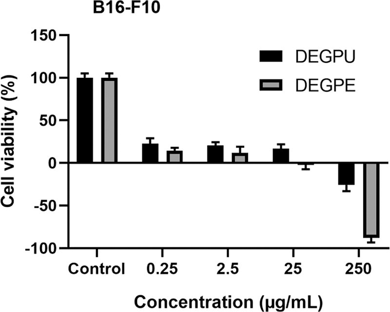
Cell viability (%) with standard deviation of the B16-F10 line treated with DEGPU and DEGPE at 0.25, 2.5, 25, and 250 µg/mL. GraphPad Prism 8.0.1 Software.
In vivo antiproliferative activity
The animals were divided into the Control, Olive, DOX, DEGPU, and DEGPE groups, and were evaluated for feed consumption and weight. All animals were weighed on the 1st, 15th, and 30th days of the experiment. However, there were no statistically significant differences between them, indicating that the treatments with extracts are not toxic to animals and, therefore, do not interfere with weight gain. With regard to feed consumption, the animals in the control group showed a reduction in feed consumption in the last three days of the experiment, with only a statistically significant difference between the Control and Olive groups (P < .02) (Fig. 4).
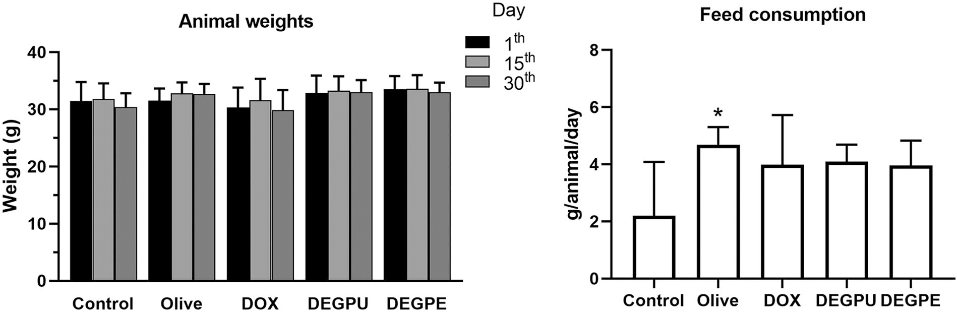
Graph of animal weight values with standard deviation on days 1th, 15th, and 30th; and graph of feed consumption in the last three days of experiment with standard deviation. *P < .05. GraphPad Prism 8.0.1 Software.
The images obtained by In-Vivo Xtreme showed that the lungs had their surface compromised by nodules, characterized by reduced fluorescence (Fig. 5a–c). As melanin absorbs a wide range of the light spectrum, 10 it is possible to evaluate these images in that way because less fluorescence is emitted when there is greater tissue involvement.
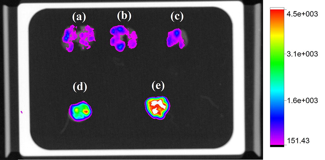
Fluorescence image of lungs and their respective fluorescence intensity scale acquired by In-Vivo Xtreme equipment. Introducing groups: Control
Lung macroscopic analysis
The lung metastasis model was confirmed by macroscopic examination, where the presence of tumor nodules on the entire pleural surface of the lungs was evident (Fig. 6).
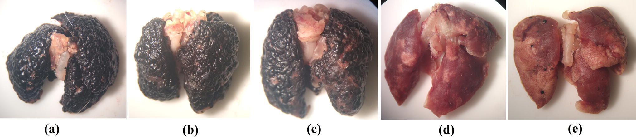
Macroscopic image of lungs, affected by numerous nodules of malignant tumor origin on pleural surface. Image acquired with an Olympus optical stereomicroscope. Introducing groups: Control
The Control, Olive, and DOX groups showed no statistical difference regarding the surface count of nodules (Fig. 7). The DEGPU and DEGPE groups showed a reduction of 97.5% and 94.38%, respectively, compared to the Control group (P < .001). Both groups also showed a significant difference between the Olive (P < .01) and DOX (P < .05) groups.
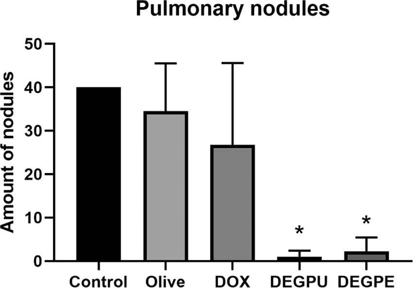
Number of pulmonary superficial nodules with standard deviation. *P < .05. GraphPad Prism 8.0.1 Software.
Lung histological analysis
The control group had a high percentage of implantation (>95%) in its parenchyma (Fig. 8a). In turn, the Olive group had 90% (Fig. 8b), followed by group DOX with 70% implantation (Fig. 8c). Treated groups DEGPU and DEGPE had the lowest percentages, with 5% and <5%, respectively (Fig. 8d–e). Histological analysis did not indicate inflammatory or necrotic processes in the lung parenchyma.
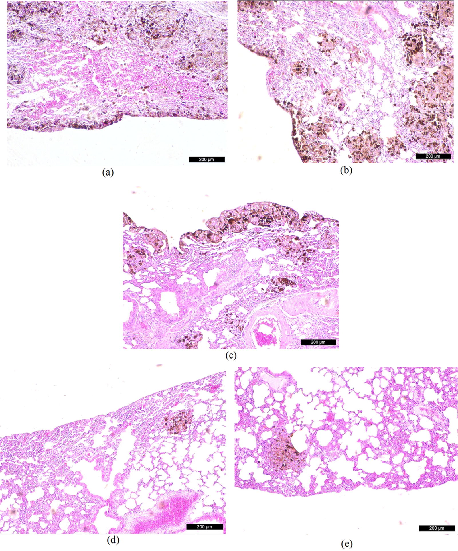
Histological images of lung parenchyma. Blackened areas showing presence of malignant metastatic nodules. Pleural surface showing nodules in Control
DISCUSSION
Melanoma skin cancer is considered one of the most serious types of disease owing to the high possibility of generating metastasis. It has a favorable prognosis if detected in an early phase. However, when in metastasis, patient survival in five years is only 31.9%. 11,12 For this reason, studies on drugs that can delimit or even inhibit the progression of this disease are of great interest.
The analyses showed that both pulp and peel extracts have antitumoral activity. Both extracts showed marked antiproliferative activity against neoplastic leukemia and glioma strains, and, at the highest concentration tested, the peel extract showed cytotoxic activity against most strains. Despite these results, the pulp extract did not show toxicity against nontumor strain J774, whose cell proliferation was greater than the control, demonstrating its selectivity against neoplastic strains. The in vivo test also showed that both extracts showed activity against melanoma, reducing metastatic cell implantation and limiting its growth.
Several compounds were found in research carried out with whole fruits of C. adamantium, 13 including limonene and two types of terpenes, which have antitumor activity described in the scientific literature. 14,15
According to Lima e Silva et al., 5 the chemical profile of two dichloromethane extracts, pulp with seed and peel of C. adamantium fruit, showed the same substances. In their study, three flavones, two chalcones, and two champanones were identified, and the antiproliferative activity test showed that substance 4′,6′dihydroxy-3′,5′-dimethyl-2′-methoxy-chalcone (dimethylchalcone) had a GI50 of 7.11 (±1.75) when tested in isolation.
Because of the presence of these compounds, numerous studies have attributed biological activities to leaves and fruits of this plant, e.g., anti-inflammatory, 16,17 antimutagenic, 18 antioxidant, 19 –22 antimicrobial, 23,24 antiproliferative, 25 and antidiarrheal action. 26
Chalcones have been the subject of studies since the 1800s and occur naturally in several plants, many of which are described in literature as having medicinal activities, including antitumor activity. 27 Species with potential anticancer activity include Bidens spp., 28 Morus alba, 29 Dorstenia sp., 30 Desmodium renifolium (Linn.), 31 Psoralea corylifolia L. (Buguchi), 32 Sophora flavescens Ait., 33 Glycyrrhiza glabra, 34 Angelica keiskei, 35 and Dalbergia velutina. 36
Considering the anticancer activity found in our research, as well as the presence of chalcones in both extracts, we can infer that the possible mechanism of action is associated with the fact that chalcones are a precursor molecule of flavonoids, with the ability to donate hydrogen atoms and thus have an antioxidant effect.
Other studies demonstrate that a possible mechanism of action of chalcones is through the generation of cellular oxidative stress owing to formation of superoxides, which are reactive oxygen species that promote cell apoptosis. 37 The ability to inhibit angiogenesis and cell growth by inhibiting kinases responsible for epidermal (EGFR) and endothelial (VEGFR-2) growth factor receptors, 38 the activation of cell apoptosis by regulating caspases, and the cell cycle regulation by inhibiting cyclins and tubulins and inhibiting kinase, responsible for the signal transduction pathway, are other forms of tumor inhibition described in the literature. 39
CONCLUSION
The results of this research prove that C. adamantium extracts are effective against tumor cell lines, showing a high SI for these cells. Corroborating in vitro analyses, the activity shown in lung metastasis models confirms the ability to limit growth and prevent tumor implantation in metastasized lungs without posing toxicity to animals, justifying the need to deepen research of their isolated active ingredients for the elaboration of new drugs with antitumor effect.
Footnotes
ACKNOWLEDGMENTS
We thank the Graduate Program in Health and Development in the Central-West Region and the Federal University of Mato Grosso do Sul-UFMS for their support. The authors thank the Coordination for the Improvement of Higher Education Personnel (Coordenação de Aperfeiçoamento de Pessoal de Nível Superior-CAPES).
AUTHORS’ CONTRIBUTIONS
All authors contributed to the study conception and design. E.A.S.: Material preparation, data collection, analysis, and the first draft of the article. All authors commented on previous versions of the article. All authors read and approved the final article.
AUTHORS DISCLOSURE STATEMENT
The authors have no relevant financial interests to disclose.
FUNDING INFORMATION
This work was supported by Coordenação de Aperfeiçoamento de Pessoal de Nível Superior—Brasil (CAPES)—Finance Code 001.