
Abstract
Pearl millet (PM) (Pennisetum glaucum L.) contains a wide variety of bioactive compounds, such as polyphenols, mostly flavonoids and phenolic acids. In the present study, we investigated the effects of PM activity against hydrogen peroxide (H2O2)-induced behavior impairment and oxidative damage in rats. The rats were divided into four groups based on the treatments they received over 30 days: Control, H2O2, PM + H2O2, and PM. The phytochemical screening, total polyphenols content (TFC), and total flavonoid content (TFC) were determined using colorimetric analysis. All animals were subjected to behavioral test (elevated plus maze test). Thereafter, oxidative stress response (malondialdehyde [MDA], H2O2, and Thiol groups [–SH]) contents and antioxidant enzymes superoxide dismutase (SOD), catalase (CAT) were estimated in brain, liver, and kidney tissues. We evaluated the levels of liver enzymes, such as alanine aminotransferase (ALAT) and aspartate aminotransferase (ASAT). Our investigation showed that PM is rich in total phenolic content and TFC and exhibited an important in vitro antioxidant activity. In vivo, we first found that H2O2-treated rat exhibited anxiogenic behavior in the elevated plus maze test and the genesis of oxidative stress in the brain, liver, and kidney was measured by an increase of MDA and antioxidant enzyme activity depletion, such as SOD and CAT. Moreover, H2O2 increased levels of liver enzymes (ALAT and ASAT). Pearl Mille administration improved emotional behavior impairments and significantly reversed H2O2-induced biochemical alterations. Thus, we suggest that the Pearl Mille may have an anxiolytic-like effect and prevent biochemical disorders associated from the oxidative stress (H2O2), confirming its potential therapeutic capability.
INTRODUCTION
Oxidative stress has been associated with many diseases such as damage to the nervous system, for example, neurodegenerative diseases and neuropsychiatric diseases, such as schizophrenia and major depressive disorder. 1,2 Many studies have shown oxidative stress can alter neurotransmission, neuronal function, and overall brain activity. 1,2
Hydrogen peroxide (H2O2) as an oxidizing agent-induced cell death through apoptosis and necrosis in vitro 3 and reduced to repair skin, 4 because of its genotoxicity and DNA damage. 5 In vivo ingestion of H2O2 in water caused toxicity via corrosive damage, oxygen gas formation, and lipid peroxidation, 6,7 in addition H2O2 oxidizing caused high anxiety-related behavior associated with hyperactivity in mice. 8
There is a growing interest in uses of natural antioxidants as a protective strategy against the brain problem. Poaceae species were found to be rich in mainly flavonoid derivatives that significantly contribute to their neuroprotective properties. 9 Pearl millet (PM) is one of the most resilient cereal crop that belongs to the family of Poaceae and grows essentially in marginal environments in Africa and Asia. Millet is a grain that is utilized as a traditional food for many populations in various countries worldwide, particularly in arid and semiarid areas. 10,11 PM has a great adaptation to harsh conditions like drought and it cultivation in Tunisia is very considerable especially in the region of Kairouan, Cap Bon, and Medenine. 12
PM is a good source of well-balanced food, which has shown anti-inflammatory effects, 13 hypoglyceamic, 14 and improves adipogenesis by lowering the bad cholesterol Low-density lipoprotein (LDL decrease). 13 –15 Furthermore, it is a good food for protection against cardiovascular diseases by lowering blood pressure 15 and can be consumed for treatment of severe constipation, stomach ulcer, and obesity because of high fiber contain. 16 PM exhibits a phytochemical profile, comprising polyphenols, flavonoids, tannins, and phytosterols, which collectively contribute to its diverse biological effects. The grain demonstrates robust antioxidant activity, stemming from its rich polyphenolic content, capable of mitigating oxidative stress. 17 Additionally, certain bioactive compounds in PM, such as flavonoids, confer anti-inflammatory properties, suggesting potential applications in addressing inflammatory conditions. The grain’s antimicrobial effects, attributed to specific phytochemicals, imply a role in combatting microbial infections. 18
PM grain is highly recommended in Folklore for increasing toughness and stiffness of bone, 19,20 because of high contain of calcium and antioxidant compounds. Indeed, the studies on therapeutic effects of Millet are limited, moreover, some studies have shown antioxidant potential due to the wealth of phenolic, flavonoids, phytates, protein, vitamins, and minerals. 12,21
The rat model involving the induction of oxidative stress and cognitive impairment, followed by treatment with PM, and the subsequent study of its effects on the brain, kidneys, liver, along with various biochemical and behavioral analyses, presents a comprehensive approach to understanding the potential health benefits of PM. The cognitive impairment and oxidative stress aspects of the model align with features observed in neurodegenerative diseases like Alzheimer’s, while investigations into kidney and liver functions relate to conditions affecting these organs in humans. The study’s exploration of biochemical markers reflects its relevance to metabolic disorders and inflammatory conditions. Furthermore, the behavioral analyses provide insights into the potential impact of PM on health, including oxidative stress, inflammation, and metabolic disorders. 13,20 Recognizing the limitations of animal models, these findings conduce for further research, highlighting the potential translational significance of PM in promoting health across multiple physiological domains in humans. However, subsequent clinical studies in human populations are essential to validate and extend these promising preclinical observations. The experimental protocol involves using H2O2 to induce oxidative stress and anxiety-related effects in rats 22,23 with the choice of PM as an intervention aimed at exploring its potential therapeutic benefits due to its antioxidant properties and historical significance as a staple food in various cultures.
We hypothesized that PM treatment may inhibit the development of H2O2-induced oxidative stress. However, neuroprotective and antioxidant properties of PM together with brain cortex, cerebellum, and hippocampus function have not yet been reported. Therefore, keeping the above facts in view the purpose of the present study was conducted to examine the hepatoprotective and neuroprotective activity of PM during 30 days prevent H2O2-induced anxiety and oxidative damage in brain tissues and liver of rats.
MATERIAL AND METHODS
Animals
Male Wistar rats Rattus norvegicus (weighing 200–250 g at the beginning of the experiment) were provided by SIPHAT (Ben Arous Tunisia) and were treated in accordance to the Tunisian code of practice for the care and use of animals for scientific purposes using the protocols monitored by the faculty Ethics Committee of the Faculty of Sciences of Bizerte, Tunisia.
All animals were housed during 2 weeks of acclimatization in cages two per cage in 12:12 light dark cycle environment and were maintained at a constant temperature (23 ± 2°C) the water and standard rodent show were provided ad libitum. All measures were taken to minimize the number of used animals and their suffering. The experiment was performed over a period of 30 days.
Plant preparation procedure
PM was provided from the region of Mateur in Bizerte/Tunisia. The seeds of Millet were dried to the sun for 48 h and powdered in an electric blender (Moulinex Ovatio 2, Fr). The powdered mixture of millet 66% was mixed with 33% of normal feed (composition of normal feed [corn, soya, oil, certified mineral compound]), water was added, and then feed was exposed to the sun. 24
Phytochemical screening
Plant Extraction
Two grams of powder were extracted with 20 mL of 80% methanol in mortar. The mixture was heated at 60°C for 1 h in marine bath and cooled to the room temperature. The mixture was filtered with Whatman filter paper n°1 and the filtrates were lyophilized then stored at −20°C during analysis. 12
Total phenolic content
Quantification of polyphenols was carried out with the reagent of Folin–Ciocalteu. The extract was added to 125 μL of Folin–Ciocalteu reagent. After 2 min, 1250 μL of Na2CO3 (7 g/100 mL) was supplied to promote an alkaline medium and to start the oxidation-reduction reaction. After adjusting with distilled water to a final volume of 3 mL, these extract solutions were kept in the dark for 90 min at room temperature. The absorbance of each extract solution was read as described at 760 nm. Gallic acid was used as a standard to estimate the total amount of polyphenols. 25
Total flavonoid content
The flavonoid amount was carried out by a method based on the formation of complex between phenolic compounds and aluminum trichloride (AlCl3) from the stock solution of millet grains extracts we added 0.125 mL from the solution to 75 μL of NaNO2 solution (5%) and 0.15 mL of AlCl3 (10%). All was mixed thoroughly for 6 min. The mixture that was obtained was then added to 0.5 mL of 1 M NaOH solution. The assay was carried out by ultraviolet-visible spectrophotometry at 510 nm and total flavonoid content was expressed as mg quercetin equivalent (QE) per gram of dry weight (mg QE/g DW) through its calibration curve. 26
DPPH antioxidant capacity
The antioxidant capacity of the extract was realized with the help of DPPH.
27
In brief, various concentrations of extract (0.5, 1, 2, 3, 4, and 5 mg/mL) were added to 1 mL of 0.1 mM methanol solution of DPPH and incubated at 27°C for 30 min. The optical density of the sample was measured at 517 nm. DPPH radical scavenging (RSA), expressed as a percentage, was estimated using the following formula:
Throlox was used as a reference molecule in the same concentration as the test extract. All the analyses were executed in duplicate. The efficacy concentration 50 (EC50) value was determined as the concentration (in mg/mL) of the compound required to scavenge 50% of The 2,2-Diphenyl-1-picrylhydrazyl (DPPH) radical.
Animal treatment
A total of 24 rats were divided into four groups (six per group) and assigned to the following dietary groups. Group 1: Control: control rats received water (1 mL/kg) per os and fed with normal-based diet. Group 2: H2O2: rats treated by H2O2 1.5% (1 mL/kg) per os and fed with normal-based diet. Group 3: H2O2+PM: rats treated by H2O2 1.5% (1 mL/kg) per os and fed with 66% of PM. Group 4: PM: rats received 1 ml/kg water per os and fed with 66% PM.
During 4 weeks of experimentation, the body weight of the rats, and food and water intake were measured daily. At the end of the last week of treatment, animal behavioral tests were assessed.
Emotional behavior
Animals were tested in the elevated plus maze on the first day following the 30 consecutive days of PM. The behavioral test was performed during the light part of the light/dark cycle between 8:00 A
Elevated plus maze
The maze was made of clear painted wood and elevated at a height of 50 cm. The arms were 50 cm of length and 10 cm of width. Closed arms were surrounded by a 50 cm wall while open arms had 0.5 cm edges. 28 The animals were allowed to explore the maze for 5 min. The percent of time spent on the open arms (%OAT [the ratio of latencies on open arms was calculated relatively to total time spent in open and closed arms ×100]) was used as a measure of anxiety. The percent of entries into the open arms (%OAE [the ratio of entries made onto open arm was calculated relatively to total number of entries in open and closed arms ×100]). The maze was cleaned with a 10% alcohol solution following each animal trial.
Biochemical assessment in brain, liver, and kidney tissues
After the behavioral assessment, the animals underwent an overnight fasting period during which they were provided only with water. On the 32nd day of the experiment, the animals were weighed, anesthetized, and then euthanized by decapitation. The liver, kidney, and brain were rapidly excised (the hippocampus, prefrontal cortex, and cerebellum were excised from brain) all tissues were homogenized in Tris-buffered saline, 50 mM, pH 7.4 with ultra-turrax homogenizer. After homogenization samples were centrifugated at 10,000 g for 10 min at 4°C. Than the supernatant was used later for the biochemical determination.
Oxidative stress assessment
For oxidative stress evaluation, levels of malondialdehyde (MDA) were determined using the thiobarbituric acid (TBA) method. Tissue aliquots were combined with a butylated hydroxytoluene (BHT)–trichloroacetic acid (TCA) solution (1% BHT w/v dissolved in 20% TCA w/v), followed by centrifugation at 1000 g for 5 min at 4°C. The resulting supernatant was then mixed with a solution containing 0.5N hydrochloric acid and 120 nM TBA. Subsequently, the mixture was heated at 80°C for 10 min and after cooling, the absorbance was measured at 532 nm. 29
The H2O2 level was performed according to Dingeon et al. 30 briefly, 100 µL of aliquots from each tissue mixed with 900 µL of glucose reagent (100 mM tris buffer pH7, 0.3 mM phenol, 10,000 U/L glucose oxidase, 1000 U/L peroxidase, and 2.6 mM amino 4-antipyrine), the reaction mixture was incubated at 37°C for 15 min. the absorbance was measured at 505 nm.
To evaluate the antioxidant system, the activity of superoxide dismutase (SOD) was determined using the method described by Misra and Fridovich. 31 The enzyme extract was mixed with a solution comprising 10 µL bovine catalase (CAT, 0.4 U/mL), 20 µL epinephrine (5 mg/mL), and 62.5 mM sodium carbonate/bicarbonate buffer (pH 10.2). Subsequently, the absorbance was measured at 480 nm. CAT activity was assessed following the procedure outlined by Sinha. 32 The method involved measuring the initial rate of H2O2 disappearance at 240 nm. Tissue aliquots were prepared with 33 mM H2O2 in 50 mM phosphate buffer (pH 7). The Thiol groups (–SH) level was measured conforming to Sedlak and Lindsay. 33 Samples from various tissues were mixed with 800 μL of 0.25M phosphate buffer (pH 8.2) and 100 μL of 20 mM ethylenediaminetetraacetic acid. The initial optical density at 412 nm (A1) was recorded, and subsequently, 100 μL of 10 mM 5-5″-Dithio-bis(2-nitrobenzoic acid) was added. After incubating the reaction mixture at 37°C for 15 min, a new value (A2) was measured. Thiol group concentration was calculated as the difference between A2 and A1.
Protein determination
Total proteins were measured by the Bradford method, 34 in brain structures, liver, and kidneys.
Biochemical indexes assay in blood
The blood was collected and centrifugated at 9000 g for 10 min at 4°C than the supernatant was served for the different assays of alanine aminotransferase (ALAT), aspartate aminotransaminase (ASAT), calcium, glucose, triglycerides, and cholesterol using commercial kits (BioMagreb, Tunisia).
Statistical analysis
Statistical analysis of the obtained data was performed by Statview ® software using analysis of variance for comparison between groups, followed by a Fisher’s protected least significant difference post-hoc test for multiple comparisons between all groups. Values for P < .05 were considered statistically significant. The data are shown as mean ± standard error of the mean.
RESULTS
Total polyphenol and flavonoid content of pearl millet
The result of colorimetric analysis of chemical constituents of PM indicates that the mean polyphenols and flavonoid contents were equivalent to 22.047 ± 0.33 mg GAE/g DM, 15.46 ± 0.55 mg QE/g DM.
In vitro DPPH antioxidant capacity
Several concentrations ranging from 0 to 5 mg/mL of the PM were tested for their antioxidant activities in different in vitro model. The EC50 value calculated from the graph demonstrated that the RSA of PM (EC50= 1.91 ± 0.01 mg/mL) and Throlox (EC50= 0.27 ± 0.01 mg/mL).
Elevated plus maze
As seen in Figure 1, H2O2-treated rats showed a significant decrease (P < .05) in %OAE when compared to three rats groups. The percent of time spent on the open arms (%OAT) was significantly decreased (P < .001) by H2O2 compared to PM and control rats. The associated treatments (PM + H2O2) induced increase in the %OAT compared to H2O2-treated rats (Fig. 2). We find statistically (P < .05) increased fecal boli in H2O2-treated rats compared to all groups (Table 1). Thus, H2O2-treated rat exhibited anxiogenic behavior in the elevated plus maze. PM treatment exhibited anxiolytic-like effects in H2O2-treated rats.
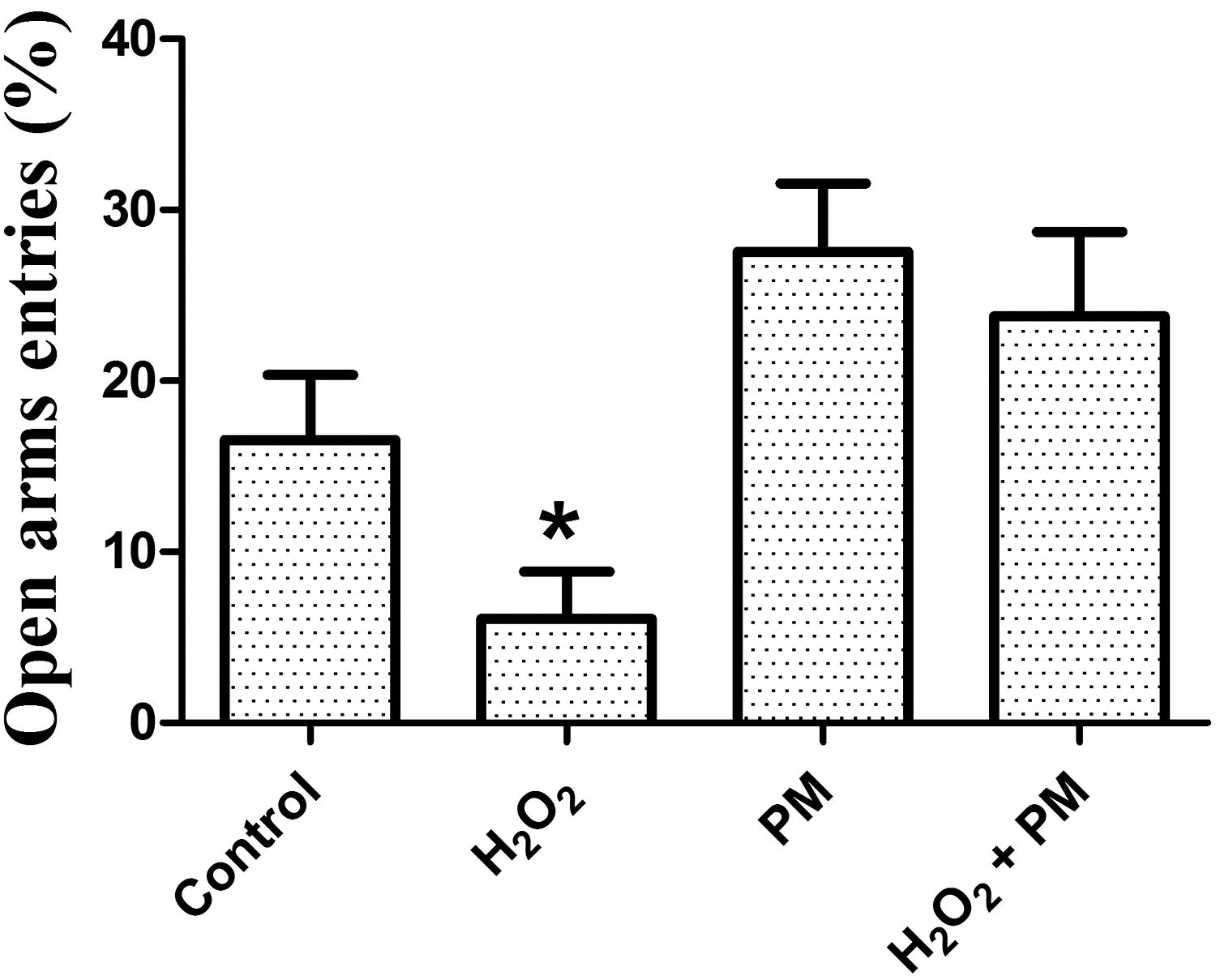
Percentage of open arms entries in the elevated plus maze in control (n = 6), H2O2 (n = 11), PM (n = 6), and H2O2 + PM (n = 6) rats. Data are given as mean ± S.E.M. *P < .05 H2O2 versus (Control; PM; H2O2 + PM). H2O2, hydrogen peroxide; PM, Pearl millet; S.E.M., standard error of the mean.
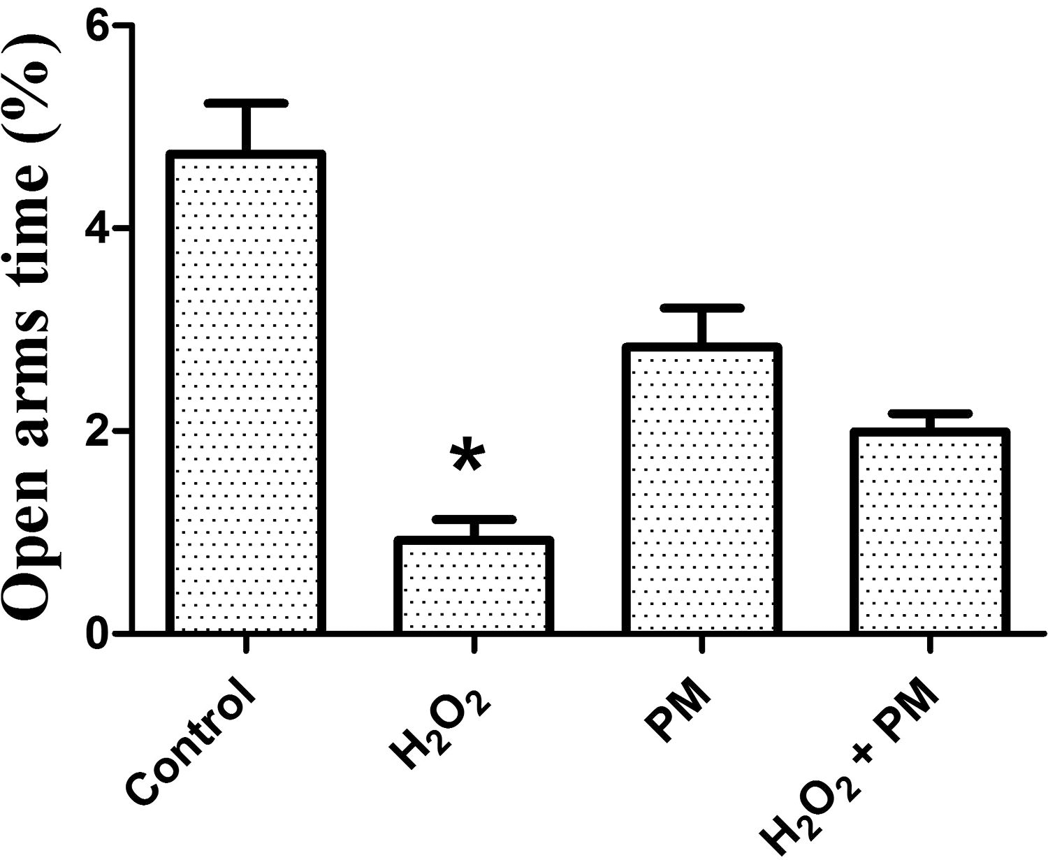
Percent time in open arms of the elevated plus maze in control (n = 6), H2O2 (n = 11), PM (n = 6), and H2O2 + PM (n = 6) rats. Data are given as mean ± S.E.M. *P < .05 H2O2 versus (Control; PM; H2O2 + PM).
Behaviors of Control (n = 6), H2O2 (n = 11), PM (n = 6) and H2O2 + PM (n = 6) Rats in the Elevated Plus Maze
Data are given as mean ± S.E.M.
P < .05 Control versus H2O2.
P < .05 H2O2 versus (Control; PM; H2O2 + PM).
P < .001 H2O2 versus (Control; PM; H2O2 + PM).
H2O2, hydrogen peroxide; PM, Pearl millet.
Oxidative stress
Oxidative stress parameters in brain
As shown in Table 2, H2O2 treatment-induced oxidative stress response by significantly increasing (P < .05) MDA level as compared to control and both PM and PM+ H2O2 treated groups in brain cells (hippocampus, prefrontal cortex, and cerebellum).
Oxidative Stress in Rat Brain in Control (n = 6), H2O2 (n = 6), PM (n = 6), and H2O2 + PM (n = 6) Rats
Data are presented as mean ± S.E.M.
P < .05 H2O2 versus (Control; PM; H2O2 + PM).
CAT, catalase; MDA, malondialdehyde; SOD, superoxide dismutase.
H2O2 levels were affected in the brain structures. H2O2 treatment induced significantly increasing H2O2 levels in brain cells (P < .05). PM treatment significantly reversed H2O2-induced increase H2O2 levels in the brain cells.H2O2 treatment-induced decrease thiol groups of protein (SH) content in hippocampus in comparison with the control, PM and PM+ H2O2 groups.
SOD and CAT activities are widely employed to indicate antioxidant responses. Results obtained here showed that administration of H2O2 significantly decreased (P < .05) the CAT and SOD activities in brain structures of the treated groups compared to control, PM, and PM+ H2O2 treated rats. However, administration of PM in H2O2-treated rats increased the activity of antioxidant enzyme in the brain cells of rat compared with the control groups (P < .05).
Oxidative stress parameters in liver and kidney
As shown in Table 3, H2O2 treatment induced oxidative stress response in liver and kidney tissues. We observed a significant (P < .05) decreasing of CAT and SOD activities and increasing of MDA and H2O2 levels. However, PM treatment in H2O2-treated rats repairing the increase of MDA and H2O2 levels (P < .05) but increased the activity of antioxidant enzyme in the liver and kidney tissues of rat (P < .05).
Oxidative Stress in Rat Liver and Kidney in Control (n = 6), H2O2 (n = 6), PM (n = 6), and H2O2 + PM (n = 6) Rats
Data are presented as mean ± S.E.M.
P < .05 H2O2 versus control and H2O2 + PM.
P < .001 H2O2 versus (Control; PM; H2O2 + PM).
***P < .05 H2O2 versus (Control; PM; H2O2 + PM).
Serum biochemical assays
As shown in Table 4, in the H2O2 rats, a statistically significant increase (P < .05) ALAT and ASAT levels compared to control, PM, and PM+ H2O2 treated rats, while no changes in the glucose, calcium, and triglycerides levels in comparison to those in control PM and PM+ H2O2 treated rats. Administration of PM in H2O2 rats caused decreases in ALAT and ASAT levels as compared with the H2O2 rats.
Biochemical Indices in the Blood Serum in Control (n = 6), H2O2 (n = 6), PM (n = 6), and H2O2 + PM (n = 6) Rats
Data are presented as mean ± S.E.M.
P < .001 H2O2 versus (Control; PM; H2O2 + PM).
P < .05 H2O2 versus PM.
ALAT, alanine aminotransferase; ASAT, aspartate aminotransferase.
DISCUSSION
In the present study, we evaluated the neuroprotective and hepatoprotective activity of PM administered at 66% of normal feed for 30 days in rat which prevent H2O2-induced anxiety, and oxidative damage in brain tissues, liver, and kidney of rats.
The elevated plus maze is the commonly employed behavioral paradigm to explore anxiety-like behaviors in rodents. 35 Animals that spend an increased time in the closed arms is considered to exhibit a anxiogenic behavior than animals that spends a longer time in open arms of the elevated plus maze. 36 Our behavioral data showed that H2O2 induced decrease in the percentage of time spent into the open arms and the percentage of open arms entries in the open arms of elevated plus maze, which evidently signified the anxiety-like behavior by H2O2. These findings are consistent with previous studies reporting provide that H2O2 caused high-anxiety-related behavior associated with hyperactivity in mice in the elevated plus maze and open Field tests. 8 Boualam et al. 37 showed that H2O2 exposure significantly decreased the locomotion activity of rat evaluated by the open field and rotarod tests and decreased in the spatial working memory and the spatial reference memory of rat in the Y-maze test. Also, H2O2 has dramatic effects on fly movement and daily locomotor rhythms of Drosophila. 38
H2O2 is a reactive oxygen species (ROS) that contributes to oxidative stress directly as a molecular oxidant and indirectly through free radical generation. 39 Hovatta et al. 40 showed that a relation between oxidative stress and trait anxiety. Masood et al. 41 showed that oxidative stress-related anxiety behavior can be reversed in mice upon inhibition of nicotinamide adenine dinucleotide phosphate oxidase or phosphodiesterase-2, enzyme that is indirectly implicated in oxidative stress mechanisms. The brain is highly vulnerable to oxidative stress because of contains plentiful polyunsaturated fatty acids the brain tissue is highly susceptible lipid peroxidation, due to its high O2 consumption, its modest antioxidant defenses. 42,43 Endogenous antioxidant enzymes such as CAT, SOD, and glutathione peroxidase (GSH-Px) have an important part to scavenge free radicals or prevent their formation. SOD catalyzes the superoxide anion to H2O2 and oxygen, and next as electron donor GSH-Px and/or CAT catalyze. The present results revealed that H2O2 induced oxidative stress in brain cells (hippocampus, prefrontal cortex, and cerebellum). This included a decrement of the activity levels of antioxidants such as CAT and SOD and an increase in the H2O2 level as well as lipid peroxidation biomarker. These results are in agreement with previous studies concluding a causative link between H2O2 and oxidative brain damage. 44,45 Therefore, in this study, we show that H2O2-induced oxidative damage in H2O2-treated rat affected the anxiety-related behavior. Oxidative stress has been also linked to cognitive decline in several medical conditions, including aging, traumatic brain injury, and Alzheimer’s disease. 46
In our study, the treatment with H2O2 and fed with 66% of PM during 30 days improved significantly the emotional behavior (elevated plus maze) in rats. Such anxiolytic effects of PM could be due to the antioxidant properties. Also, our results showed an improvement of oxidative markers in the brain cells of H2O2 + PM rats. PM reduces H2O2-induced elevated level of MDA and restores the antioxidant enzyme activity (SOD and CAT) in brain cells (hippocampus, prefrontal cortex, and cerebellum). These findings may be attributed to the high content of neuroprotective compounds in PM. The animals were fed with millet, which was incorporated into their diet at a concentration of 66%. Based on the findings, there was no observed cytotoxicity of PM; instead, it demonstrated positive effects by ameliorating oxidative stress parameters. This included a reduction in ROS levels and an increase in the activity of antioxidant enzymes.
In our study, the data demonstrated that the result of colorimetric analysis of chemical constituents of PM indicates the presence of polyphenols and flavonoids. Similar results showed that PM was the richest source of total polyphenols and total 3-Deoxyanthocyanidin Content. 47 The generous content of phenolic compounds in millet has made it a potent source of antioxidants. 48,49 Millet grains contained several natural occurring phenolic compounds which include phenolic acids, flavonoids, and tannins, in addition to xylo-oligosaccharides, insoluble fibers, and peptides. 50
It is well-intentioned that the antioxidant capacity of these constituents in natural products is due to their ability to provide electrons or hydrogen atom transfer to scavenge free radicals. 51 The antiradical DPPH test to confirm the potentiality of the bioactive components of PM to act as donors of hydrogen atoms. The results obtained showed that PM has a high antioxidant power with EC50 = 1.91 ± 0.01 mg/mL. Nutritionally, PM is superior to major cereals with reference to energy value, high-quality proteins, fat, and minerals such as calcium, iron, and zinc. Besides, it is also a rich source of dietary fiber and micronutrients. 52,53 In our study, the antioxidant properties of PM maybe are attributed to their polyphenols and flavonoids, which act as free RSA agents and protect against oxidative stress.
Our results depict that H2O2-exposed animals had decreased levels of antioxidant enzymes such as, SOD and CAT, while an increased level of lipid peroxidation, and H2O2 levels in liver and kidney. These results indicated the correlation between H2O2 administration with the oxidative stress in the adult rats. Hepatic biomarkers (ALAT and ASAT) were assayed as well in this work. H2O2 administration induces increase of ALAT and ASAT. These results are in agreement with the previous study of Boualam et al. 37 In the present study, PM attenuated the deleterious effects of H2O2 and improved all liver biomarkers. Butterworth (2013) shows that impaired liver function was predisposing factor for encephalopathy and neurodegenerative diseases. PM ability to regulate liver biomarkers may be effective attenuated the behavioral changes. Another study showed that millet grain can improve hepatic glucose hemostasis by alleviating obesity/type 2 diabetes mellitus-associated oxidative stress by its high content ofantioxidant-derived ingredients. 54,55 Alzahrani et al. 56 showed that powder and ethanolic extract of PM have an antiobesity, hypoglycemic, hypolipidemic, anti-inflammatory, and antisteatotic in high-fat diet fed rat.
CONCLUSIONS
In African and Asian regions, millet grains have been used efficiently to produce adult meals. PM (Pennisetum glaucum L.) contains a wide variety of bioactive compounds, such as polyphenols, mostly flavonoids and phenolic acids. The current study was conducted to examine the hepatoprotective and neuroprotective activity of PM in an in vivo model. PM furnished in vitro antioxidant activities, reduced the effects of H2O2-induced oxidative stress, and attenuated the associated behavioral changes. These data may provide important information for the future researches to elaborate the appropriate underlying mechanisms, the mediated molecular pathways, and the principal bioactive compounds of PM.
Footnotes
ACKNOWLEDGMENT
Financial support of the Tunisian Ministry of Higher Education and Scientific Research is gratefully acknowledged.
AUTHORS’ CONTRIBUTIONS
Mohamed Ammari and Soumaya Ghodbane designed and supervised the study, Latifa Hajri and Haifa othman conducted the experiments, Khémais ben Rhouma, Hafedh Abdelmelek and Mohsen Sakly performed the data analysis, Mohamed Ammari and Latifa Hajri participated in writing the manuscript. All authors contributed to the article and approved the submitted version of the manuscript.
AUTHOR DISCLOSURE STATEMENT
The authors report no declarations of interest. The authors alone are responsible for the content and writing of the article.
FUNDING INFORMATION
No funding was received for this article.