
Abstract
Our study aimed to investigate whether unripe pear extract (UP) could provide protection against UVB-induced damage to both mouse skin and keratinocytes. We observed that UVB exposure, a common contributor to skin photoaging, led to wrinkle formation, skin dryness, and inflammation in mice. Nevertheless, these effects were mitigated in the groups of UVB-irradiated mice treated with UP. Moreover, UP treatment at 400 μg/mL increased the antioxidant enzyme activities (sodium dodecyl sulfate, 2.22-fold higher; catalase, 2.91-fold higher; GPx, 1.96-fold higher) along with sphingomyelin (1.58-fold higher) and hyaluronic acid (1.31-fold higher) levels in UVB-irradiated keratinocytes. In the keratinocytes irradiated with UVB, UP 400 μg/mL resulted in reduced cytokine production (TNF-α, 33.2%; IL-1β, 45.3%; IL-6, 33.4%) and the expression of inflammatory pathway-related proteins. The findings indicate that UP has a direct protective effect on UVB-irradiated keratinocytes and is also able to shield against photoaging induced by UVB. Hence, it is suggested that UP could contribute to improved skin health by averting skin photoaging.
INTRODUCTION
Skin is a vital organ that acts as a defense to protect the body from water loss as well as various external factors. The skin is the most externally exposed organ of the body and is therefore vulnerable to adverse environmental factors. 1,2 Being the outermost layer, the epidermis, primarily made up of keratinocytes, is pivotal in strengthening the skin's structure. This cellular barrier defends against environmental harm and maintains skin integrity. 3 –5
Ultraviolet (UV) radiation is a form of electromagnetic radiation that causes skin photoaging. The keratinocytes in the epidermis can act as the initial line of defense against UV radiation, and particularly against UVB radiation, which causes the majority of skin photoaging through wrinkle formation, dryness, and thinning. 6 –11 It is well known that UVB radiation produces excessive reactive oxygen species (ROS) that cause oxidative stress in the epidermis. When ROS production overwhelms the cells' antioxidant capacity, oxidative stress occurs, which can contribute to the development of several diseases. ROS in the epidermis can mediate various inflammatory responses, including the production of proinflammatory molecules and the phosphorylation/activation of nuclear factor kappa B (NF-κB) p65. Proinflammatory molecules from the epidermis can stimulate matrix metalloproteinase (MMP) expression and its activation in the dermis, which is a key mechanism of wrinkle formation. 8 –11
In the present study, we used in vitro and in vivo models to investigate the effects of unripe pear extract (UP) on UVB-induced skin photoaging. Pears (Pyrus spp.) are mainly cultivated in Asia and Europe and are consumed worldwide, either fresh or in more processed forms such as juices. Pears have also been traditionally used to prevent respiratory diseases in Asia and have also been shown to act as an anti-inflammatory and antioxidative agent. 12 –14 Hong et al. 13 reported that the anti-inflammatory properties of arbutin from pears were found to inhibit the production of proinflammatory mediators, and it was identified for its skin-whitening capabilities. Sanchez et al. 14 demonstrated that the peel contains higher concentrations of chlorogenic acid, flavonols, and arbutin compared with the flesh, suggesting its potential use as an antioxidant agent. Therefore, we expected to find that pears could inhibit skin photoaging by suppressing oxidative stress and inflammation caused by UVB radiation.
We also investigated whether immature UP can contribute to skin health by examining skin hydration, antioxidant enzyme activities, inflammatory signaling, signaling pathways of MMP, and moisturizing-related factors in UVB-irradiated mice. In addition, we examined antioxidant enzyme, inflammatory signaling, and moisturizing-related factors in UVB-irradiated keratinocytes.
MATERIALS AND METHODS
Preparation of UP
Unripe pear (Pyrus spp.) fruits were obtained from Naju (Jeollanam-do, Korea) in May 2022. The whole unripe pears were washed and extracted with 100°C hot water for 8 hours, a method previously identified as yielding the most active extract in previous studies. 15 The extracts were then filtrated and concentrated in vacuo and the product was dried in a spray dryer with 20% dextrin to produce dried extracts (UPhenon®; UP).
Animals and UVB Irradiation
Male hairless mice (SKH-1) at 5 weeks of age were obtained from SaeRon Bio (Uiwang, Korea) and housed in a controlled environment (temperature: 23°C ± 2°C, humidity: 50% ± 5%, and lighting: 12-h light/12-h dark cycle).
The mice were allocated randomly into the following six groups (n = 8): normal control (non-UVB irradiation), control (UVB irradiation), L-ascorbic acid (positive control, UVB irradiation +
Pathological changes
Hematoxylin and eosin (H&E) staining was performed to assess morphological alterations in the dorsal skin. Initially, skin tissue samples were preserved in a formalin solution to maintain cellular integrity. Following this, the fixed tissues were embedded in paraffin wax to provide structural support for sectioning. Subsequently, fine sections of the skin tissue were sliced using a microtome and affixed to a glass slide. The tissue sections underwent deparaffinization with xylene, followed by rehydration through graded alcohol solutions. Finally, the sections were subjected to H&E staining.
Measurement of skin hydration
Skin moisture levels were assessed using the Howskin device (Seoul, Korea) following established protocols 16 in a controlled environment of 22–24°C and 55–60% humidity, as previously outlined.
Cell culture and treatments
HaCaT cells were cultured in line with previously described methods.
16
The cells were subjected to a UVB dose of 50 mJ/cm2 and treated with 100 μg/mL
Measurement of antioxidant enzymes
Skin tissue from mice and cells were collected, and enzymes were quantified using assay kits purchased from BioVision, Inc. (Milpitas, CA, USA) following the methods described in previous studies. 16
Quantitative real-time PCR
RLT buffer (lysis buffer; Qiagen, Valencia, CA, USA) with β-mercaptoethanol was used to isolate total RNA from the adipose tissue lysates. Next, reverse-transcript II reverse transcriptase (Invitrogen, Carlsbad, CA, USA) was used to generate cDNA from 1 μg of total RNA. Real-time PCR was then conducted using Universal SYBR Green PCR Master Mix as per the manufacturer's instructions (Bio-Rad, CA, USA), and amplification was performed with selective primer sets, as shown in Table 1.
Primer Sets Used for Real-Time PCR
Western blot analysis
Mouse skin tissues and HaCaT cells were lysed with a lysis reagent and then 100 μg of protein per lane was separated on a 10% sodium dodecyl sulfate gel. Following separation, the proteins were electrophoretically transferred onto membranes. Membrane blocking, incubation with primary antibodies and secondary antibody, visualization, and analysis were performed by our previous methods. 16
Measurements of the productions of cytokines
The cell production of cytokines was measured using the DuoSet ELISA Kit (R&D Systems, Minneapolis, MN, USA), according to methods described previously. 16
Measurement of sphingomyelin and hyaluronic acid
The quantities of sphingomyelin and hyaluronic acid were determined as per the guidelines provided by the manufacturer, utilizing the Sphingomyelin Assay Kit from Abcam (Cambridge, United Kingdom) and the Hyaluronic Acid ELISA Kit from BioVision, Inc.
Statistical analyses
The results are presented as means ± standard deviations (SD). Statistical analyses were performed using one-way Duncan's multiple range tests following one-way analysis of variance using SPSS software (SPSS PASW Statistic 23.0; SPSS Inc., Chicago, IL, USA). A significance level of P < .05 indicates statistical significance.
Results
Effect of UP on histopathological changes and antioxidant enzyme activities in UVB-irradiated hairless mice
The UVB-irradiated mice showed wrinkle formation, irregularly shaped skin (Fig. 1A), and decreased skin hydration (Fig. 1B) compared with the normal hairless mice (NC). The UVB-irradiated hairless mice also showed a significant decrease in antioxidant enzyme activity compared with the normal hairless mice (Fig. 1C–E). However, the dietary supplementation of UP attenuated some of the morphological and histopathological changes (Fig. 1A) in the UVB-irradiated hairless mice. The groups receiving UP dietary supplementation exhibited enhanced skin hydration and greater antioxidant enzyme activity in comparison to the control group (Fig. 1B–E) (P < .05). The findings indicated that UP protected the skin from UVB-induced skin dryness, wrinkle formation, and oxidative stress.
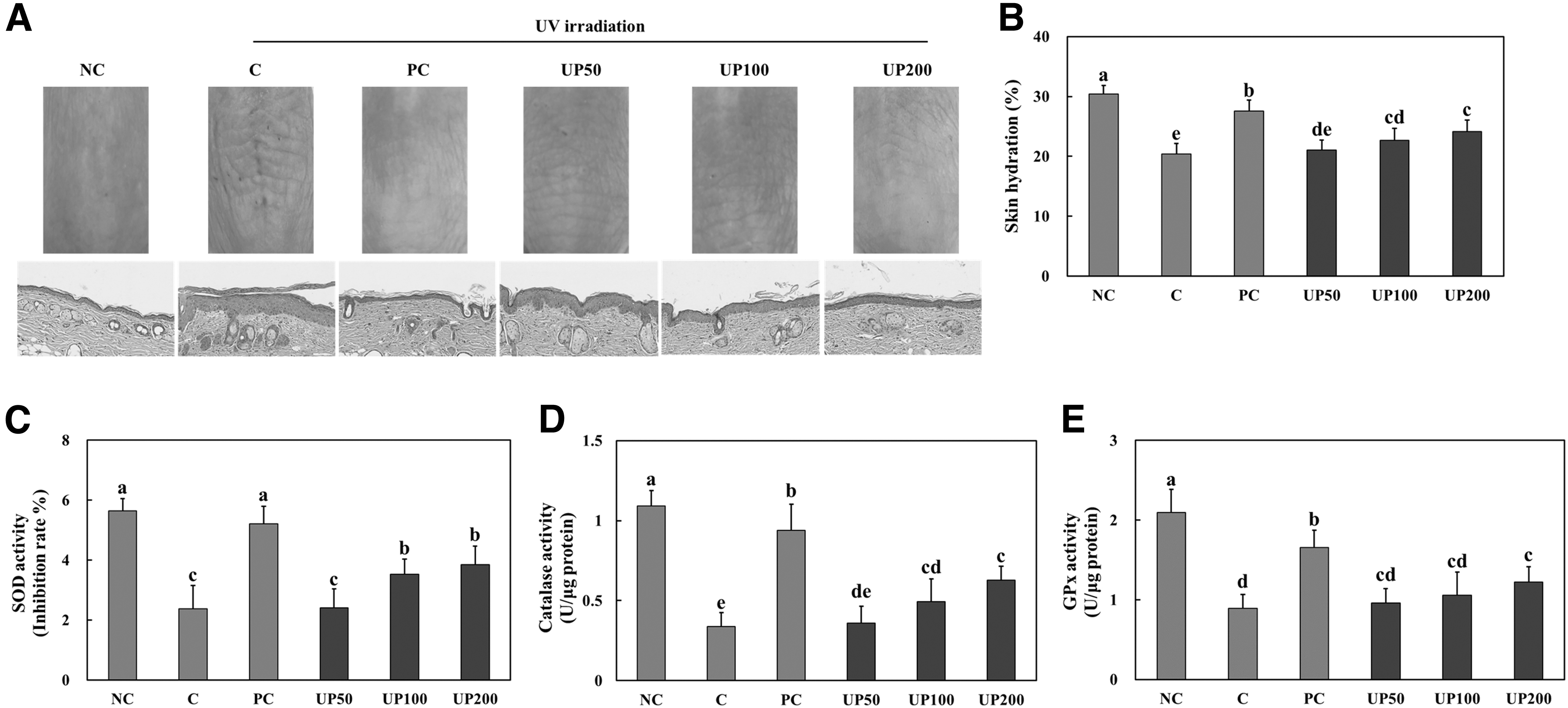
UP protected skin against UVB irradiation in hairless mice. Morphological and histopathological changes
Effect of UP on inflammation in UVB-irradiated hairless mice
The UVB-irradiated hairless mice showed a significant increase in mRNA expression levels of TNF-α, IL-1β, and IL-6 and protein expression of IkB-α phosphorylation, p65 phosphorylation, and cyclooxygenase-2 (COX-2) compared with the normal hairless mice (Fig. 2A–D). However, UVB irradiation also increased the levels of TNF-α and IL-1β, whereas IL-6 and protein expression of IkB-α phosphorylation, p65 phosphorylation, and COX-2 all decreased significantly in the PC and UP groups (Fig. 2A–D) (P < .05). These results suggest that a dietary supplementation of UP reduces the level of UVB irradiation-induced skin inflammation.
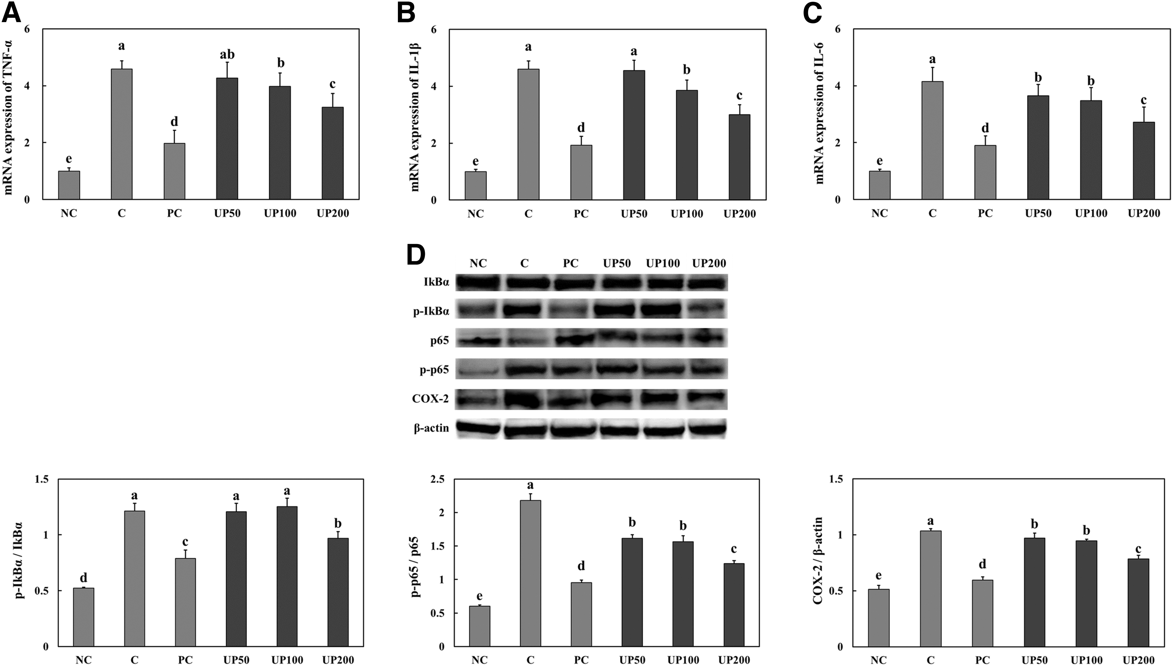
UP suppressed UVB irradiation-induced skin inflammation in hairless mice. mRNA expression of TNF-α
Effect of UP on wrinkle formation pathway in UVB-irradiated hairless mice
We confirmed the wrinkle formation pathway in the dorsal skin to investigate the molecular mechanism of UP effects. Figure 3 shows that wrinkle formation pathway-related protein activation increased, whereas TGF-β RI, procollagen C-endopeptidase enhancer protein, and collagen type I mRNA expression decreased in the skin of UVB-irradiated hairless mice compared with the normal hairless mice. However, wrinkle formation pathway-related protein activation decreased in the PC and UP groups compared with the control group. In addition, TGF-β RI, procollagen C-endopeptidase enhancer protein, and collagen type I mRNA expression increased significantly in the PC and UP groups compared with the control group (Fig. 3) (P < .05).
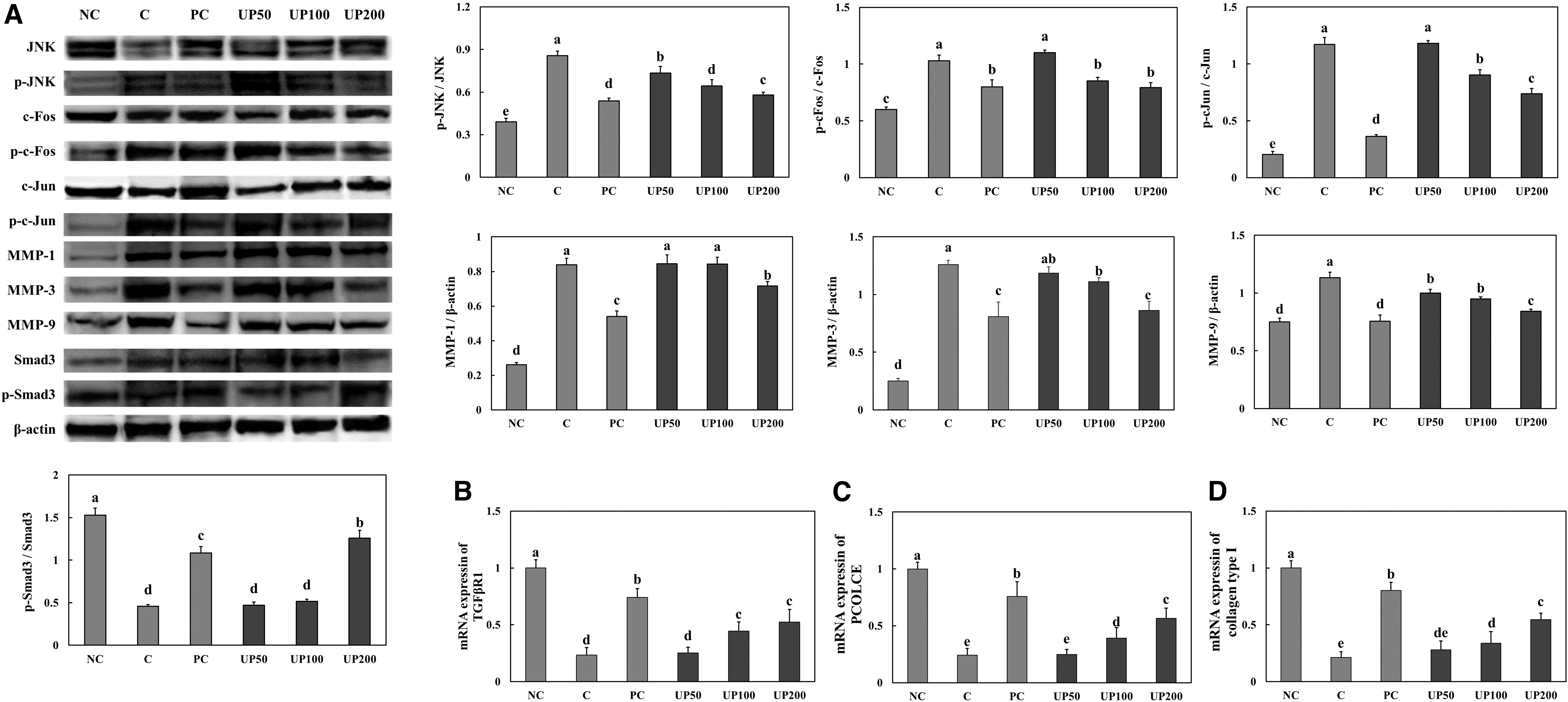
UP suppressed UVB irradiation-induced wrinkle formation pathway in hairless mice. Protein expression of JNK phosphorylation, c-FOS phosphorylation, c-Jun phosphorylation, MMPs, and Smad3 phosphorylation
Effect of UP on moisturizing factors in UVB-irradiated hairless mice
When investigating the effect of UP on moisturizing, we confirmed hyaluronan synthase (HAS) mRNA expression, ceramide synthase 4 protein expression, and LCB1 and DEGS1 mRNA expression in the dorsal skin. Figure 4 shows that HAS mRNA, ceramide synthase 4 protein, LCB1, and DEGS1 mRNA expression, all decreased significantly in the skin of the UVB-irradiated mice compared with the skin of normal mice. Additionally, we observed a rise in these moisturizing factors in both the PC and UP groups in comparison to the control group (Fig. 4) (P < .05).
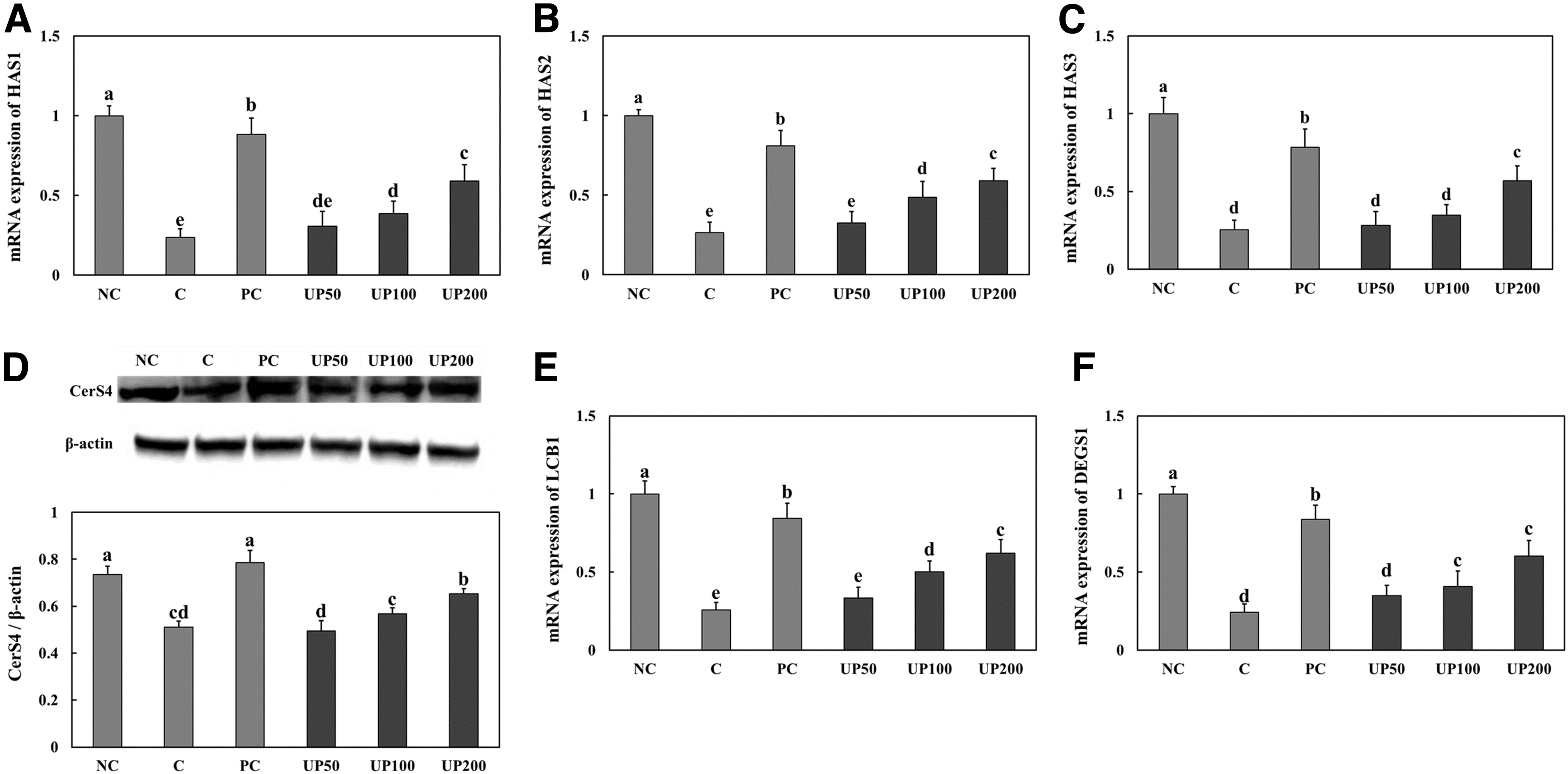
UP increased moisturizing capacity in UVB-irradiated hairless mice. mRNAs expression of HAS1
Effect of UP in UVB-irradiated keratinocytes
We also investigated whether UP can directly protect HaCaT cells against UVB irradiation and found that UP treatment, particularly at UP 400 μg/mL, caused a significant increase in antioxidant enzyme activity (Fig. 5A–C) and suppressed inflammation in the UVB-irradiated cells (Fig. 5D–G). In addition, sphingomyelin and hyaluronic acid production in cells increased in UVB-irradiated cells treated with UP 400 μg/mL (Fig. 6A, B). Furthermore, there was a notable increase in HAS mRNA, ceramide synthase 4 protein, as well as LCB1 and DEGS1 mRNA expression in the UP-treated cells in contrast to the UVB-irradiated cells (Fig. 6C–H) (P < .05).
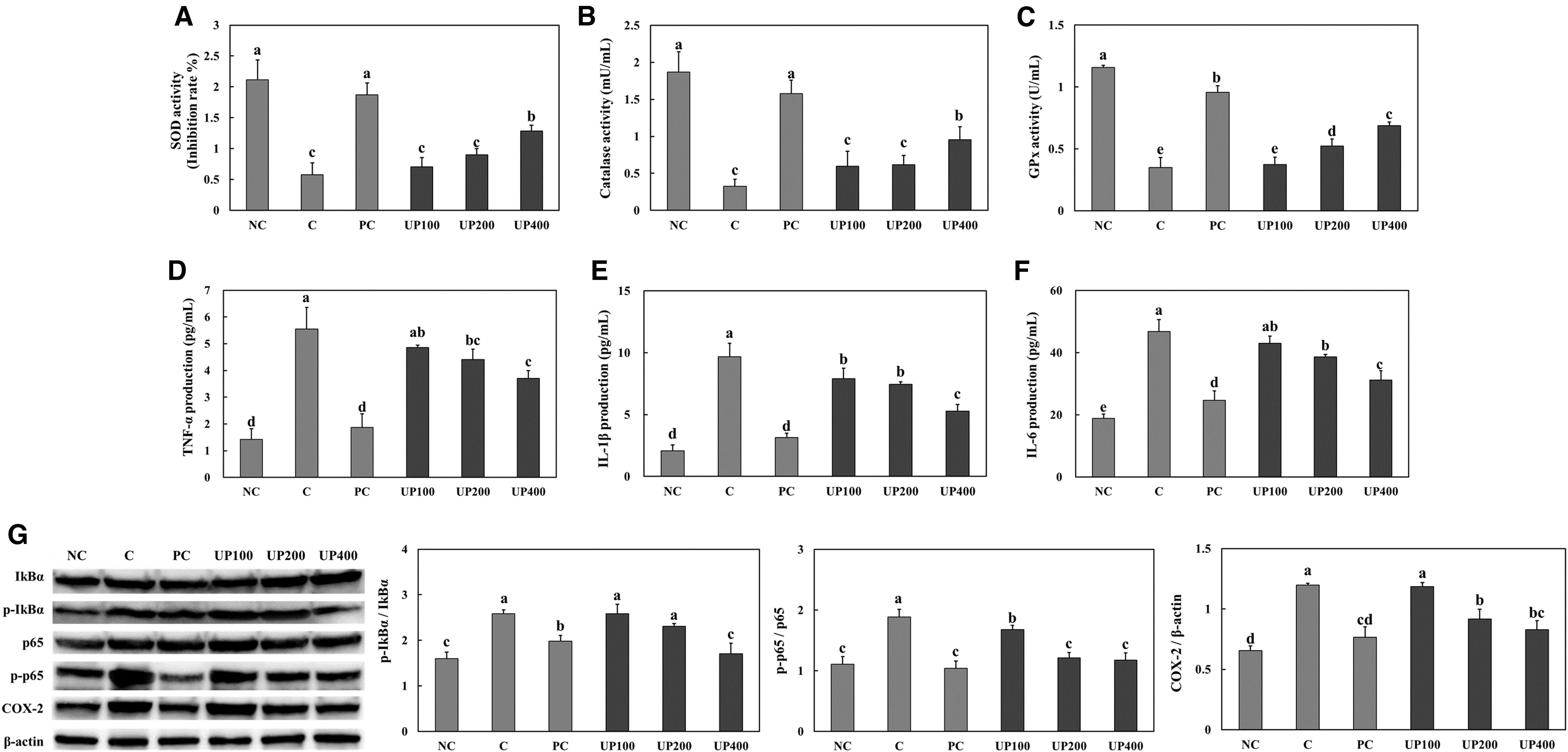
UP suppressed UVB irradiation-induced oxidative stress and inflammation in keratinocytes. Antioxidant activities of superoxide dismutase
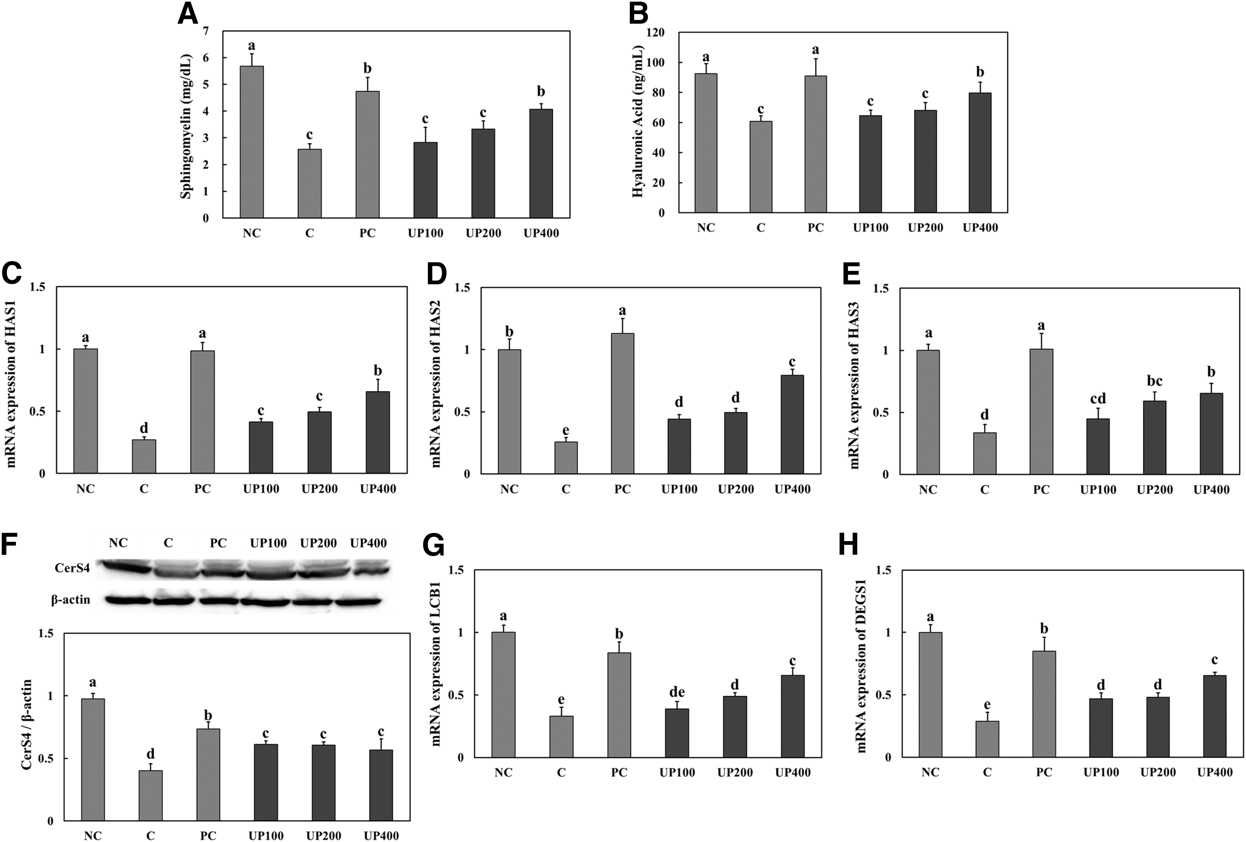
UP increased moisturizing capacity in UVB-irradiated keratinocytes. Levels of sphingomyelin
DISCUSSION
As interest in skin health increases, an increasing number of studies have investigated the positive effects of various natural products on skin aging. Skin aging is a complex process that most intuitively expresses the aging of an organism and is generally divided into aging induced by intrinsic or extrinsic factors. One of the major extrinsic factors is exposure to UV radiation, which plays a role in ROS generation. Recent studies, therefore, have focused on the antioxidative process when developing natural substances for antiskin aging function. 17,18 In this study, we noted that UVB radiation caused oxidative stress, although we also found that dietary supplementation of UP in UVB-irradiated hairless mice and UP treatment of UVB-irradiated keratinocytes were able to suppress oxidative stress. In the study conducted by Lee et al., 19 it was discovered that unripe pears contain substantial levels of chlorogenic acid, a compound renowned for its antioxidant properties and potential health benefits. On the other hand, Cho et al. 20 observed that immature pears contain higher levels of flavonoids and phenolic compounds compared with ripe pears.
These compounds are known to have strong free radical-scavenging abilities, indicating their potential to counteract oxidative stress and cellular damage. These previous studies and our present findings indicate that UP can suppress oxidative stress induced by UVB irradiation through its antioxidant function.
Excessive UVB-mediated ROS levels contribute to pathophysiological conditions by mediating inflammatory signaling pathways in the skin. ROS initiate multiple pathways, including NF-kB and activator protein 1 (AP-1), which produce proinflammatory cytokine in keratinocytes. 17,21,22 In our current research, we established that UVB exposure triggered inflammation in both the dorsal skin and keratinocytes. Notably, UP demonstrated inhibitory effects on inflammation development in both UVB-irradiated dorsal skin of mice and HaCaT cells. Our results suggest that UP directly mitigated inflammation by inhibiting oxidative stress in keratinocytes.
UV radiation also activates c-Jun amino-terminal kinase (JNK), extracellular signal-regulated kinase (ERK), and phosphorylates c-Fos and c-Jun, which leads to the expression of the transcription factor AP-1. AP-1 mediates MMP expression, which induces collagen degradation, thereby degrading the extracellular matrix and contributing to wrinkle formation. 23 –25 However, our study showed that UP inhibited wrinkle formation, MMP expression-associated pathway activation, and pro-collagen type I expression in UVB-irradiated mice. We can conclude, therefore, that UP suppresses wrinkle formation by inhibiting UVB-induced photoaging.
It has been well documented that long-term UVB irradiation reduces hyaluronic acid levels through the downregulation of HAS. Hyaluronic acid, which is abundantly synthesized in keratinocytes by HAS and released into the extracellular space, plays an important role in the structural function of maintaining skin moisture. 6 By contrast, ceramide production in the epidermis is important for maintaining skin health since it plays a role in moisture and barrier function. We found reduced hyaluronic acid, sphingomyelin, HAS, ceramide synthase, LCB1, and DEGS1 expression in both the UVB-irradiated dorsal skin of mice and HaCaT cells. However, UP increased these moisturizing factors in both the dorsal skin of UVB-irradiated mice and in UVB-irradiated HaCaT cells.
It has been demonstrated that Asian pear cultivars contain substantial amounts of phenolics, arbutin, and chlorogenic acid. 13,14,26 Cha et al. 27 conducted a study investigating the effects of chlorogenic acid on UVB-irradiated HaCaT cells, revealing that chlorogenic acid absorbs electromagnetic radiation in the UVB range and effectively prevents DNA damage. While we have not yet identified the exact active compound responsible for the effects of UP, the research findings of Cha et al. 27 lead to speculation that chlorogenic acid might serve as a skin-protective active component. Further research in this direction is required to pinpoint the precise active compound within UP responsible for its effects on skin photoaging.
In conclusion, this study has shown that UP demonstrates anti-inflammatory and antioxidative effects, which protect the skin against photoaging caused by UVB in mice. Moreover, we have shown that UP treatment directly inhibits oxidative stress and stimulates the expression and production of moisturizing factors in keratinocytes. Our findings suggest that UP could potentially mitigate skin photoaging by reducing oxidative stress, alleviating inflammation, and regulating skin hydration.
Footnotes
AUTHORs' CONTRIBUTIONS
J.P. and D.K.: conceptualization (equal), methodology (equal), formal analysis (equal), and writing—review and editing (equal); M.L., G.D.P., S.R.K., Y.J., and WJ.: formal analysis (supporting); O.-K.K. and J.L.: conceptualization (lead), methodology (lead), formal analysis (lead), and writing—review and editing (lead).
AUTHOR DISCLOSURE STATEMENT
No competing financial interests exist.
FUNDING INFORMATION
No funding was received for this article.