
Abstract
This study aims to examine the ameliorative effect of macadamia nut protein peptides (MPP) on acetaminophen (APAP)-induced liver injury (AILI) in mice, and develop a new strategy for identifying hepatoprotective functional foods. The molecular weight distribution and amino acid composition of MPP were first studied. Forty mice were then randomized into four groups: control group (CON), APAP model group, APAP+MPP low-dose group (APAP+L-MPP), and APAP+MPP high-dose group (APAP+H-MPP). The APAP+L-MPP (320 mg/kg per day) and APAP+H-MPP (640 mg/kg per day) groups received continuous MPP gavage for 2 weeks. A 12 h of APAP (200 mg/kg) gavage resulted in liver damage. Pathological alterations, antioxidant index levels, expression of toll-like receptor 4 (TLR4)/nuclear factor-κB (NF-κB), and associated inflammatory factors were determined for each treatment group. The results revealed that the total amino acid content of MPP was 39.58 g/100 g, with Glu, Arg, Asp, Leu, Tyr, and Gly being the major amino acids. The molecular weight range of 0–1000 Da accounted for 73.54%, and 0–500 Da accounted for 62.84% of MPP. MPP ameliorated the pathological morphology and reduced the serum levels of alanine aminotransferase, aspartate aminotransferase, and alkaline phosphatase of AILI in mice. MPP significantly increased the activities of superoxide dismutase and glutathione peroxidase in the liver compared with the APAP group. MPP inhibited the expression of TLR4, NF-κB, interleukin-1β (IL-1β), and tumor necrosis factor-α (TNF-α) genes in AILI mice. MPP also inhibited the expression levels of inflammatory factors (TNF-α and IL-6). Our study concludes that MPP alleviates AILI in mice by enhancing antioxidant capacity and inhibiting TLR4/NF-κB pathway-related gene activation.
INTRODUCTION
As the largest substantive organ and the most metabolically active organ in body, the liver is responsible for maintaining human health through physiological processes, such as bile secretion, metabolism, coagulation, detoxification, and immunity. 1,2 In severe cases, viral infection, oxidative stress, alcohol, and drugs can cause acute liver injury (ALI) and liver failure, posing significant health risks. 3 –5 Recently, the incidence of drug-induced liver injury (DILI) has increased. The direct hepatotoxic effects of drugs and their active metabolites cause DILI. 6
As a widely used analgesic and antipyretic drug in clinical practice, acetaminophen (APAP) has a high incidence of liver injury and serious side effects, and has become one of the most common drugs that cause drug-induced ALI and even liver failure. 7,8 When the body consumes an excessive amount of APAP, cytochrome P450 enzymes generate toxic intermediates that deplete glutathione and trigger a series of biochemical reactions, including liver mitochondrial dysfunction and oxidative stress. 9 The hepatotoxicity of APAP is a growing concern; however, few treatments are available. Therefore, it is crucial to identify novel, safe, and effective liver-protective substances in natural products. 10,11
Macadamia nuts are rich in fats, proteins, and other nutrients, and have multiple biological activities. 12 Long-term consumption significantly reduces the incidence of atherosclerosis as well as cardiovascular and cerebrovascular diseases. 12 Macadamia nut cake (MNC) is a rich source of protein, with a protein content of up to 30% and an amino acid ratio coefficient of up to 86.95. 13,14 Owing to its high amino acid content, it can be used as a potential feed substitute. 15
Macadamia nut residue protein can be effectively processed to yield macadamia nut protein peptides (MPP). Protein peptides have a small molecular weight and are easily absorbed by the human body, as evidenced by their antioxidant, anticancer, blood pressure-lowering, lipid-regulating, and immunity-regulating properties. 16,17 Nut-like protein peptides have become a popular research topic.
Wang et al. reported that walnut protein peptides could activate the nuclear factor E2-related factor 2 pathway protecting HepG2 cells from insulin resistance and oxidative stress induced by high glucose. 18 Walnut protein hydrolysate contains a variety of anti-inflammatory peptides that inhibit oxidative stress and reduce neuroinflammation in mice with lipopolysaccharide-induced cognitive injury. 19 The functional investigation of MPP has far-reaching implications for further realizing the comprehensive utilization of macadamia slag and enhancing the added value of by-products. Developing relevant functional foods has excellent application potential for promoting human health.
Plant protein peptides have been reported to improve pathological changes in liver injury, but whether MPP prevents liver damage is unknown. Animal models of acetaminophen-induced liver injury (AILI) are frequently used to investigate effective interventions to prevent and treat liver injury. Therefore, this study extracted and prepared MPP from MNC and analyzed its amino acid composition and molecular weight range to explore the ameliorative effect and molecular mechanism of MPP on AILI in mice. This study aims to provide a theoretical foundation for developing of novel nut proteins with hepatoprotective effects.
MATERIALS AND METHODS
MPP preparation
Using hydraulic press technology, MNC was extracted from macadamia nuts (Yunnan, China) by a hydraulic cold machine. The MNC powder was ultra-finely ground using a colloid mill (with 20% water), and the ground nut solution was added to the enzyme digestion tank and hydrolyzed using neutral protease (pH 7.0, temperature 55°C, stirring speed 56 g/min, and time 3 h). After enzymatic digestion, the peptide solution was obtained by centrifugal separation, and the solid was enzymatically digested. The peptide solution was dried in a spray drying tower (inlet air temperature of 170–190°C and exhaust air outlet temperature of 70–85°C) to produce MPP.
Detection of MPP amino acid composition and molecular weight distribution
The proportion of acid-soluble proteins in MPP samples was measured using an automatic Kjeldahl nitrogen tester (UDK-159; Velp, Milan, Italy). The composition and mass fractions of 16 amino acids in the MPP samples were determined using an automatic amino acid analyzer (A300; membraPure GmbH, Berlin, Germany). The molecular mass of MPP samples was determined using high-performance liquid chromatography (E2695; Waters, Milford, USA). Chromatographic conditions: TSKgel 2000 SWXL 300 × 7.8 mm column, and acetonitrile/water/trifluoroacetic acid (40/60/0.1, V/V) mobile phase.
Animal experiments
Forty male SPF mice (5 weeks old, 24 ± 2 g) were purchased from Guizhou Medical University (production license number SCXK [Qian] 2018-0001). Feeding conditions were as follows: temperature 25°C ± 2°C, humidity 43% ± 5%, and free access to food and water. After 7 days of acclimatization, 10 mice were randomly assigned to one of the four groups: control (CON), model (APAP), low-dose (APAP+L-MPP), and high-dose (APAP+H-MPP). MPP and APAP were prepared using distilled water. The APAP+L-MPP group received 320 mg/kg per day, the APAP+H-MPP group received 640 mg/kg per day, and the CON and APAP groups received equal amounts of distilled water for 2 weeks.
After 16 h of fasting following the final gavage, the APAP, APAP+L-MPP, and APAP+H-MPP groups were each gavaged with 200 mg/kg APAP once to induce ALI for 12 h. Mice in each group had their blood was drawn by retro-orbital collection and serum isolated, and after dislocation and execution, the liver was quickly dissected and removed. Guizhou University Experimental Animal Ethics Sub-Committee (EAE-GZU-2022-E021) approved all procedures as they met the ethical standards for animal experiments.
Detection of blood biochemical index
Blood was collected from the mouse eyes, and serum was obtained by centrifugation at 2824 g/min for 15 min. Serum levels of alanine aminotransferase (ALT), aspartate aminotransferase (AST), and alkaline phosphatase (ALP) were measured using kit instructions (AbCam, Cambridge, United Kingdom).
Detection of superoxide dismutase and glutathione peroxidase activity
Typically, 0.6 g of mouse liver left lobe tissue was weighed in a glass homogenizer tube, 5.4 mL of saline was added, ground until the tissue was homogenized, centrifuged, and the supernatant was kept obtaining a 10% mass fraction of liver homogenate. The levels of superoxide dismutase (SOD) and glutathione peroxidase (GSH-Px) in the liver tissues were measured following kit instructions (Jiancheng Bioengineering Institute, Nanjing, China).
Histopathological observation
Some right lobes of mouse liver were fixed in 4% paraformaldehyde, dehydrated by gradient alcohol, embedded in paraffin, and sectioned into 5 μm using a microtome (RM2135; Leica, Wetzlar, Germany). The sections were routinely dewaxed, hydrated, and stained with hematoxylin and eosin (H&E), and images were collected under microscope (BX43F; Olympus, Tokyo, Japan) to compare the histopathological changes in the livers of each group.
Detection of toll-like receptor 4/nuclear factor-κB pathway mRNA expression
Total RNA was extracted from the liver tissues using an RNA extraction kit (Takara Bio, Inc., Dalian, China). cDNA was reverse-transcribed from RNA according to the instructions of the reverse transcription kit (Takara Bio, Inc). The mRNA transcript levels of toll-like receptor 4/nuclear factor-κB (TLR4/NF-κB) pathway-related genes in liver tissues were measured by real-time quantitative reverse transcription polymerase chain reaction (qRT-PCR). The primer sequences were synthesized by Sangon Biotech Co., Ltd., Shanghai, China, and are shown in Table 1.
Primer Sequences Used for Real-Time Quantitative Reverse Transcription Polymerase Chain Reaction
IL-1β, interleukin-1β; NF-κB, nuclear factor-κB; TLR4, toll-like receptor 4; TNF-α, tumor necrosis factor-α.
Detection of TLR4, NF-κB p65, and interleukin-1β protein expression
Paraffin sections of liver tissue were dewaxed, hydrated, and repaired with citrate antigen, and immunohistochemical (IHC) staining was performed according to the kit instructions (Maixin Biotech Co., Ltd., Fuzhou, China). TLR4 (Servicebio Co., Ltd., Wuhan, China) and NF-κB p65 antibodies (Bioss Biotechnology Co., Ltd., Beijing, China) were incubated for 1 h at 37°C, DAB-stained, hematoxylin restained nuclei, and neutral gum sealed slices. Images were obtained using an optical microscope (Olympus BX43F, Japan).
Immunofluorescence (IF) staining was performed according to the kit (Beyotime Biotech, Inc., Shanghai, China). Interleukin-1β (IL-1β) antibodies (Bioss Biotechnology Co., Ltd.) were incubated for 1 h at 37°C. Fluorescein isothiocyanate-conjugated donkey anti-rabbit IgG was incubated for 1 h at 37°C, secondary antibody dilution buffer stained, and anti-fade mounting medium-sealed slices. Fluorescence images were captured using an optical microscope (Olympus BX43F, Japan).
Determination of serum levels of tumor necrosis factor-α and interleukin-6
Blood was extracted from the mouse eyes, and serum was obtained by centrifugation at 2824 g/min for 15 min. Serum levels of inflammatory factors tumor necrosis factor-α (TNF-α) and interleukin-6 (IL-6) were determined using an enzyme-linked immunoassay (ELISA) kit (Sangon Biotech Co., Ltd.).
Statistics and analysis
All data were expressed as means ± standard errors (x ± s), and one-way analysis of variance was performed using SPSS 23.0 statistical software. Duncan's method was used for multiple comparison tests. GraphPad prism software was used for graph analysis.
RESULTS
MPP amino acid composition analysis
Table 2 shows the free amino acids content in MPP. The total amino acid concentration was 39.58 g/100 g. Free amino acid contents of up to 2 g/100 g were Glu, Arg, Asp, Leu, Tyr, and Gly, with Glu having the highest concentration of 9.09 g/100 g.
Composition and Content of Free Amino Acids in Macadamia Nut Protein Peptides
MPP peptide molecular weight analysis
Figure 1 depicts the detection of the molecular weight distribution of MPP using liquid chromatography. The retention time was 13.176 min, with the highest Mn value of 460,496 and an area of 0.12%. The retention time was 15.563 min, with an Mn value of 189,612 and an area of 18.24%. The retention time was 23.815 min, with an Mn value of 10,843 and the highest area of 35.40%. The retention time was 27.383 min, with an Mn value of 2100 and an area share of 11.95%.
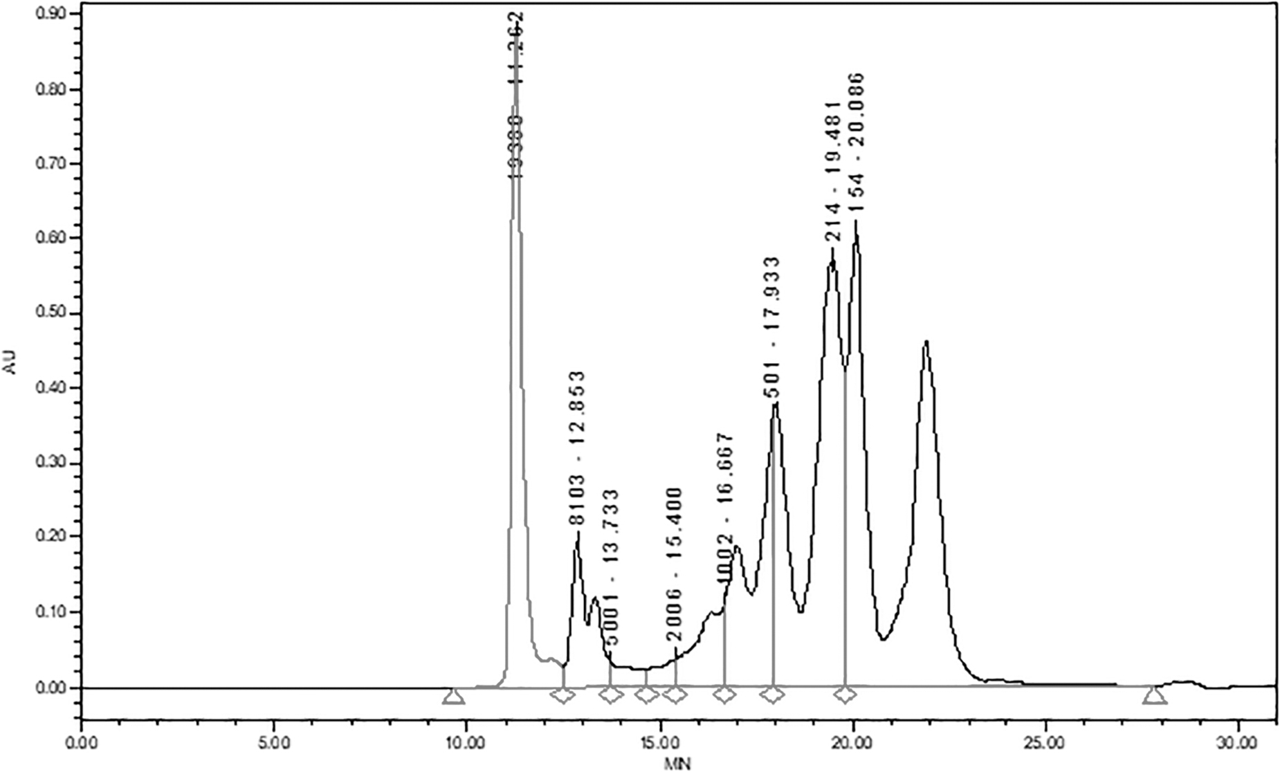
Molecular weight test peak diagram of MPP. MPP, macadamia nut protein peptides.
Table 3 presents the range of molecular weights of MPP. Peptides <1000 Da accounted for 73.54%, and peptides in the range of 0–500 Da accounted for 62.84%.
Molecular Weight Range of Macadamia Nut Protein Peptides
Effect of MPP on serum ALT, AST, and ALP of AILI mice
Figure 2 shows that serum ALT, AST, and ALP activities increased significantly (P < .01) in the APAP model group compared with the CON group. The serum ALT, AST, and ALP vitality of mice in the APAP+L-MPP and APAP+H-MPP groups, decreased relative to the APAP model group, with the decrease in the APAP+H-MPP group being highly significant (P < .01). ALT, AST, and ALP activity decreased by 10.33%, 11.67%, and 3.00%, respectively, in the APAP+H-MPP group compared with the APAP+L-MPP group. The results indicated that AILI was successfully modeled and that MPP had significant concentration-dependent hepatoprotective efficacy.
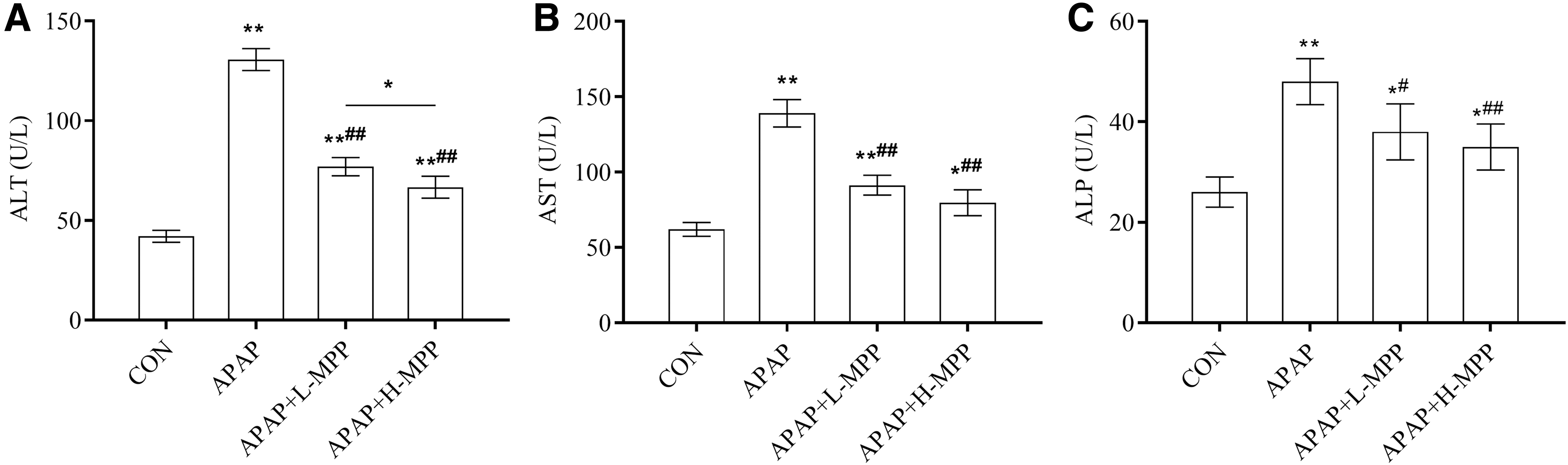
Effects of MPP on the serum levels of ALT
Effect of MPP on histopathological changes in the liver of AILI mice
Figure 3 depicts H&E observed histopathological changes in the mouse livers. In the CON group, the liver lobules were undamaged and clear, the central vein was not stagnant, the hepatocytes were neatly arranged in strips, and there was no cell metamorphic injury or necrosis, and no evidence of inflammatory cell infiltration. Mice with disorganized hepatic cords, dilated central veins with red blood cells, severe hepatocyte swelling, and inflammatory cell infiltration in the confluent area were observed after APAP induction. MPP decreased inflammatory cell infiltration of AILI and improved liver histopathology, with the greatest improvement observed in the APAP+H-MPP group.
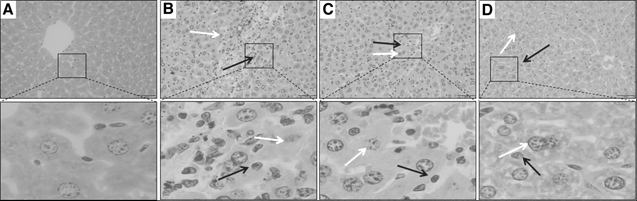
Effects of MPP on histopathological changes in the liver of mice with H&E staining (scale bar = 50 μm). Red arrow: inflammatory cells. Yellow arrow: cell swelling.
Effect of MPP on SOD and GSH-Px in the liver of AILI mice
Figure 4 demonstrates that SOD and GSH-Px activities in the mouse livers in the APAP group were significantly lower than in the CON group (P < .01). The SOD and GSH-Px activities in liver tissues were decreased in the APAP+L-MPP and APAP+H-MPP groups compared with the APAP group, with this decrease in the APAP+H-MPP group (P < .01) being statistically significant. The APAP+H-MPP group had higher levels of SOD and GSH-Px than the APAP+L-MPP group by 12.42% and 33.32%, respectively. These results revealed that MPP significantly increased SOD and GSH-Px activities in the liver tissues and exerted antioxidant effects.
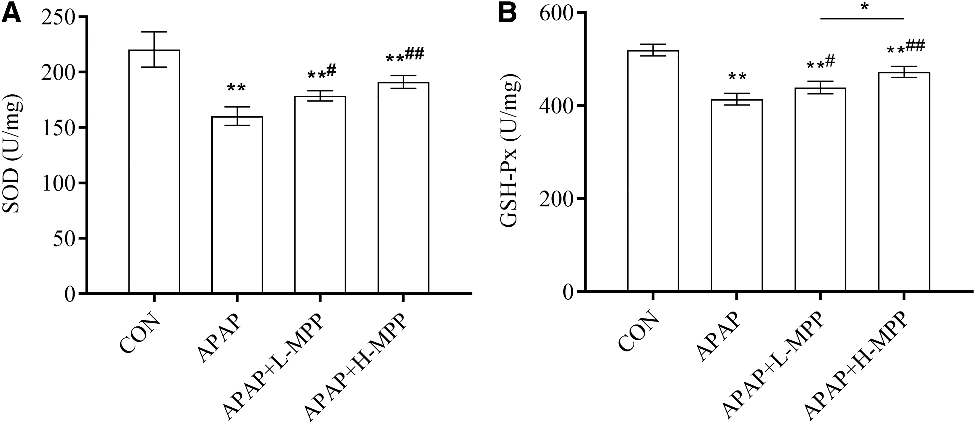
Effects of MPP on the activities of SOD
Effect of MPP on transcript levels of key genes of TLR4/NF-κB pathway in the liver of AILI mice
APAP activates the TLR4/NF-κB inflammatory signaling pathway. qRT-PCR was used to measure the TLR4, NF-κB, TNF-α, and IL-1β mRNA transcript levels in mouse liver tissues (Fig. 5). The mRNA transcript levels of these genes were significantly higher in the APAP liver tissues than in the CON liver tissues (P < .01). However, MPP significantly reduced the mRNA transcript levels of these genes in the APAP-induced liver manipulation (P < .01). TLR4 (P < .01), NF-κB (P < .01), and TNF-α (P < .05) mRNA transcript levels were significantly lower in the APAP+H-MPP group than in the APAP+L-MPP group. These results indicated that MPP can inhibit the transcription of essential genes in the TLR4/NF-κB pathway.
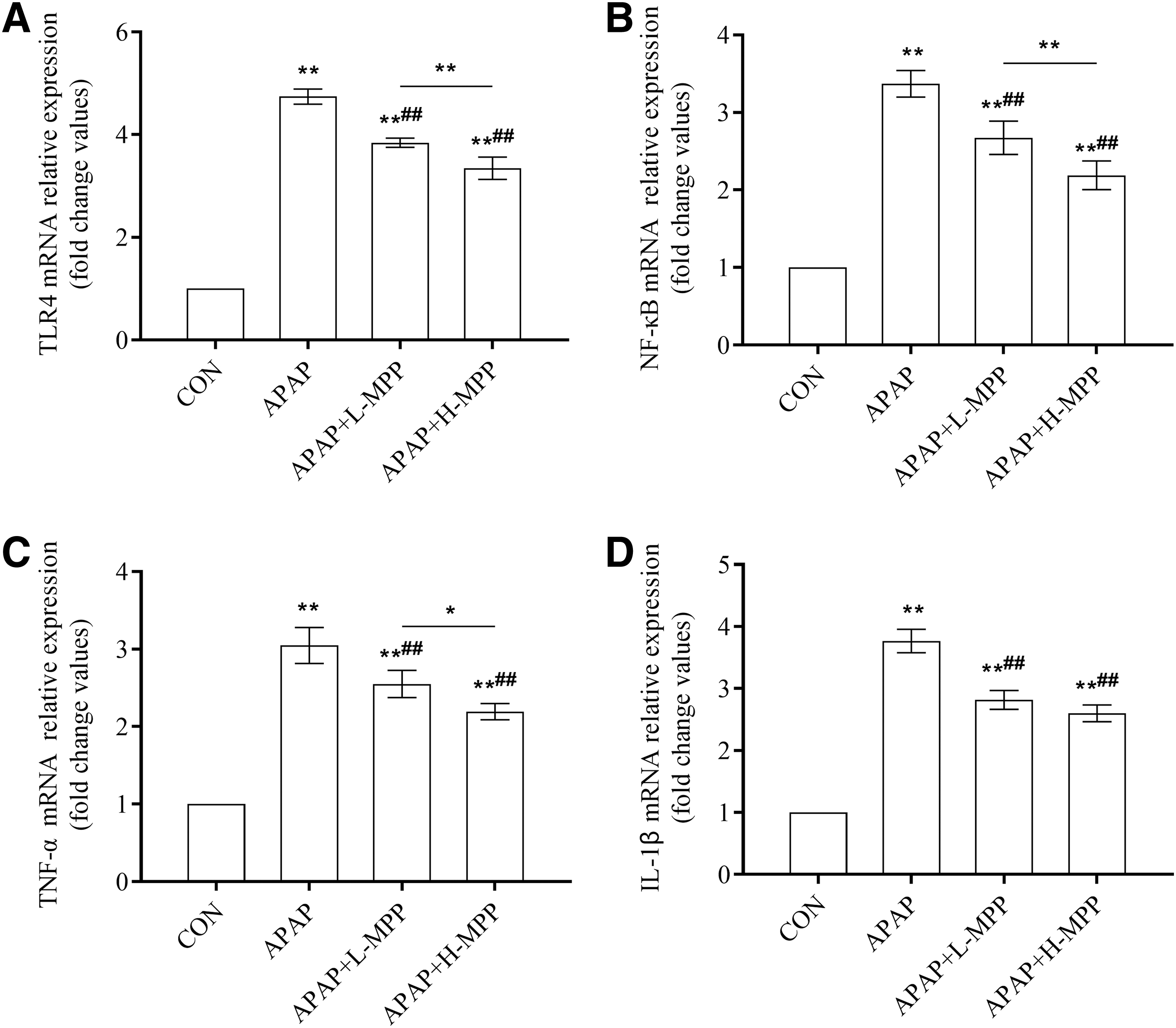
Effect of MPP on transcript levels of TLR4
Effect of MPP on TLR4, NF-κB p65, and IL-1β protein expressions in the liver of AILI mice
The expression of TLR4, NF-κB p65, and IL-1β proteins in mouse liver tissues was detected by IHC and IF (Figs. 6A–C). As shown in Figures 6D–F, the rate of TLR4, NF-κB p65, and IL-1β positive cells in the liver tissues of mice in the CON group was low, whereas the expression of these cells was significantly higher in the APAP group (P < .01). The difference in expression between the APAP+L-MPP and APAP+H-MPP groups versus the APAP group was statistically significant (P < .01). These results indicated that MPP reduced TLR4, NF-κB p65, and IL-1β expression levels in AILI with a positive dose correlation.
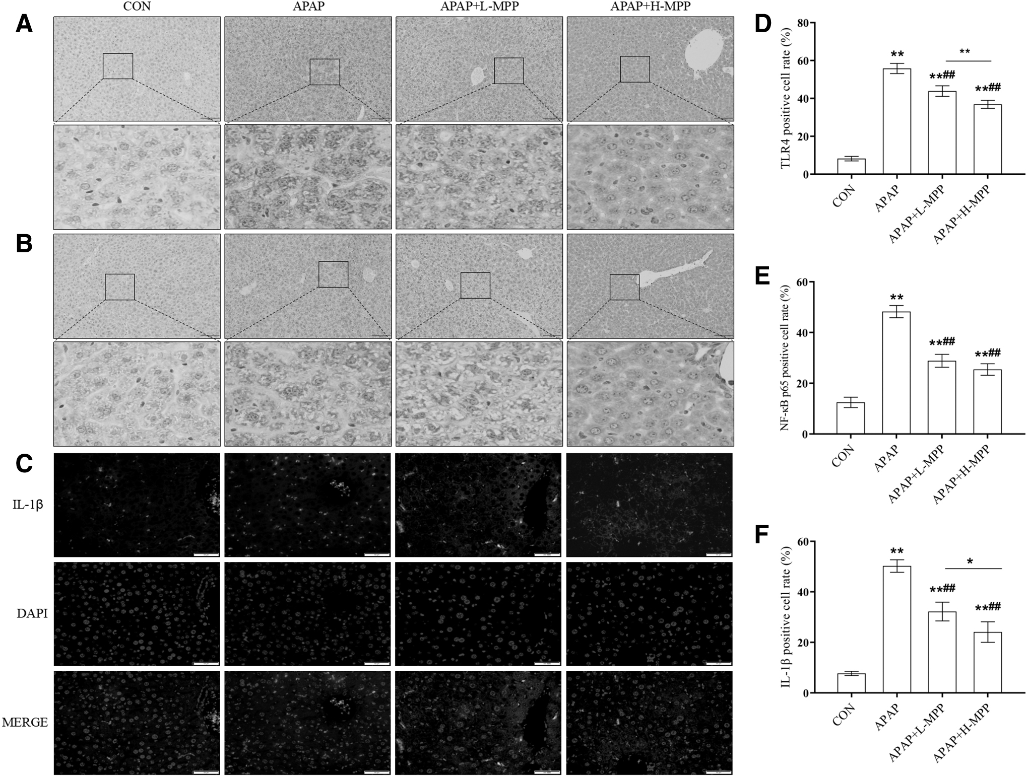
Effects of MPP on the expression of TLR4.
Effect of MPP on serum TNF-α and IL-6 of AILI mice
Figure 7 shows that the serum inflammatory factors TNF-α and IL-6 levels were significantly higher in the APAP group than in the CON group (P < .01). When comparing the APAP group with APAP+L-MPP and APAP+H-MPP groups, TNF-α and IL-6 contents decreased significantly. In addition, the reductions in TNF-α and IL-6 levels in the APAP+H-MPP group were highly significant (P < .01). Compared with the APAP+L-MPP, the APAP+H-MPP group could significantly reduce TNF-α (P < .05) and IL-6 (P < .01) expression levels. The results indicated that MPP decreased the expression levels of inflammatory factors in the serum of AILI mice.
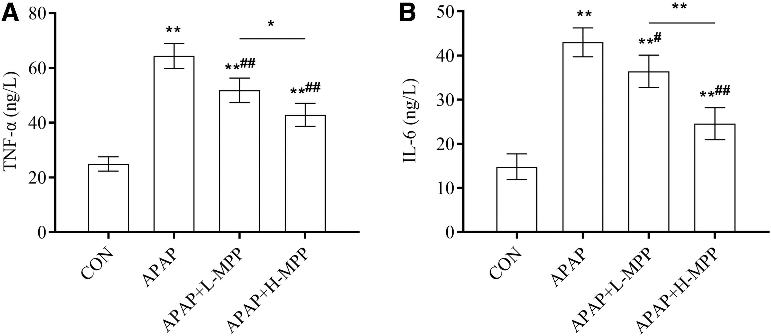
Effect of MPP on the serum levels of TNF-α
DISCUSSION
The liver is essential for converting numerous nutrients and the eliminating of chemicals and drugs. 20 Patients who develop drug-induced ALI and undergo untimely treatment eventually experience liver failure. 21 At reasonable therapeutic doses, APAP is a safe antipyretic analgesic and has been widely used, but overdoses can cause pharmacological ALI, and even lead to liver failure in the body. Oxidative stress and inflammation are important mechanisms of ALI. 22 Establishing animal models of experimental liver injury, screening hepatoprotective drugs, studying the mechanism of liver disease, and exploring the principles of hepatoprotective action are of great practical importance.
Macadamia nuts and other nut products are rich in proteins, amino acids, fatty acids, vitamins, and minerals. Regular consumption of nuts has protective effects against cardiovascular disease and diabetes, making them a popular functional food. 23 Protein peptides have a fast absorption rate, low energy consumption, low saturation, and no competition or inhibition between their operations. The structural properties of peptides, including chain length and amino acid composition, determine their functional activity. 24
Glu, Asp, Arg, Leu, Tyr, and Gly were identified as abundant functional amino acids in MPP. ASP can participate in nucleotide metabolism, promote cell cycle processes, and improve growth performance. 25 The combination of Glu and succinic acid prevented liver metabolic disorder in mice under conditions of normobaric hypoxia, retained the activity of the mitochondrial fast metabolic cluster intact, activated signal transcription factors, and upregulated the expression of transforming growth factors to limit lipid peroxidation, which has a beneficial effect on oxidative stress of hepatocytes. 26
Gly is involved in glutathione synthesis, and studies have confirmed that dietary Gly can effectively prevent pathological liver injury caused by hemorrhagic shock in the rat. 27 Small peptides with molecular weights <1000 Da and amino acid chains between 2 and 20 residues in length have been demonstrated to be the most active. They are more readily absorbed by the intestine than large molecular weight peptides and penetrate cells in a capacity-dependent manner to perform their biologically active functions. 28 According to this study, 73% of the peptides <1000 Da in MPP and the active sequences in the proteins were completely released during enzymatic digestion, which was beneficial for the organism's absorption and presumably could play a better role in liver protection.
In this study, the protein was extracted from MNC meal and converted into MPP by protease hydrolysis, which is currently in the production phase. The animal model of experimental liver injury established by APAP can induce hepatic inflammatory response and oxidative stress, and is commonly employed in the screening and mechanism of action studies of liver-protective drugs. To investigate the biological activity of MPP, the effects of different concentrations of MPP on relevant biochemical indices, oxidative enzymes, histopathological structures, and inflammatory factors during AILI mice were examined. This study aimed to explore the ameliorative effects of MPP on AILI mice.
The AST, ALT, and ALP levels are important markers for assessing liver damage. When liver cells are damaged, cell membrane permeability changes, and AST, ALT, and ALP are released into the blood, causing an increase in AST, ALT, and ALP serum levels. 29 According to this study, MPP pretreatment decreased serum ALT, AST, and ALP levels and improved the structural histopathological changes in the liver, indicating that MPP ameliorated the acute pharmacological damage to mouse hepatocyte membranes by APAP.
SOD is a superoxide radical scavenger, and its concentration indirectly reflects the ability to eliminate free radicals in the body. GSH-Px is a selenium-containing antioxidant enzyme that is widespread in the body. It can remove lipid peroxides induced by hydroxyl free radicals and reactive oxygen species to block the chain reaction of lipid peroxidation and safeguard the structural and functional integrity of cell membranes. 30
APAP can inhibit the antioxidant activities of SOD and GSH-Px, and destroy the dynamic equilibrium between free radical production and scavenging in the body. Excessive free radicals can form covalent bonds with intracellular components to produce lipid peroxidation, attack biological macromolecules, damage cell structure and function, alter cell metabolic activities, exacerbate liver cell damage, and upregulate oxidative stress level. 31
In this study, MPP aided in maintaining the activities of SOD and GSH-Px enzymes in the APAP-induced mouse liver, indicating that it may inhibit oxidative stress and thus protect liver cells. According to previous studies, the amounts of Arg, Glu, Gly, and ASP in peptides correlate closely with their ability to scavenge hydroxyl free radicals. 32 The presence of Asp and Glu in peptides significantly increases antioxidant activity, whereas its absence decreases antioxidant activity. 33 In addition, antioxidant peptides can scavenge free radicals by blocking free radical and reactive oxygen species production by transient metal ion chelation, or by inhibiting electron and hydrogen atom transfer. 33,34
TLR4 is a cell surface signal transduction receptor that plays a key role in inflammatory and immune regulation and is widely expressed in numerous hepatocytes, including hepatic macrophages, hepatic neutrophils, and hepatic dendritic cells. It mediates pathways that are actively involved in liver damage induced by IFN-γ, IL-8, IL-10, IL-6, and TNF-α. 35 Hepatocyte-secreted TNF-α and IL-6 play critical roles in various inflammatory responses. NF-κB is an important transcription factor that regulates cellular immune and inflammatory responses. 36
Chemically active metabolites of APAP trigger hepatocyte injury and inflammatory innate immune responses in the liver, causing progressive and intensifying tissue damage. 37 APAP activates the TLR4/NF-κB pathway in liver tissue, promoting the release of TNF-α, IL-6, NO, and IL-1β pro-inflammatory factors, and exacerbating liver tissue damage. 38
Previous research has revealed that plant peptides administered orally can increase the hepatic antioxidant activity, reduce the inflammatory response, and have a hepatoprotective effect. Yi et al. reported that soy protein-derived peptides ameliorated LPS-induced inflammatory responses in RAW264.7 macrophages by inhibiting TLR4-mediated MAPK-JNK and NF-κB activation and inflammatory factor expression (IL-6, IL-1β, and TNF-α). 39 Ma et al. demonstrated that maize peptide treatment attenuates ethanol-induced liver injury pathology in mice.
This is achieved by inhibiting NF-κB gene expression and reducing the TNF-α and TGF-β1 expression levels. 40 Currently, all peptides with significant anti-inflammatory activity contain positively charged amino acids such as Arg and Lys. 41 In addition, nearly all anti-inflammatory peptides contain at least one glutamine residue, which may be a key amino acid involved in anti-inflammatory activity in the molecular mechanism of protection against cell injury. 42
This study demonstrated that MPP may reduce drug-induced liver inflammatory injury in mice by inhibiting TLR4 and NF-κB protein expression, that is, blocking the TLR-4/NF-κB signaling pathway and subsequently downregulating TNF-α, IL-1β, and IL-6 levels, and by decreasing the inflammatory response to APAP-induced liver tissue. The significant anti-inflammatory activity of MPP may be closely related to its high amino acid content (Glu, Arg, etc.); however, the composition of peptide sequences in MPP and the regulatory mechanism of anti-inflammation require additional study.
In conclusion, in this study, MPP was found to ameliorate ALI induced by APAP and exert anti-inflammatory effects, possibly by modulating the expression levels of genes and proteins related to the TLR4/NF-κB pathway.
Footnotes
AUTHORs' CONTRIBUTIONS
Methodology, writing original draft, and formal analysis by C.S. Project administration, review, and editing by F.M. Resources, visualization, and funding acquisition by G.G.
AUTHOR DISCLOSURE STATEMENT
No competing financial interests exist.
FUNDING INFORMATION
This study was financially supported by the Science and Technology Major Program of Yunnan Province, China (Grant No. 202202AE090006) and Science and Technology Innovation Subsidy Project of Yunnan Forestry and Grassland Administration (Grant No. 2023KJCX002).