
Abstract
Abstract
Purpose:
We aimed at investigating the effect of erdosteine administration on amikacin induced visual evoked potentials (VEPs) alterations in rats.
Methods:
For this purpose, forty male Wistar rats were divided into 4 groups: control, amikacin treated, erdosteine treated, and amikacin + erdosteine treated. Amikacin (600 mg/kg/day) was applied as a single dose of intramuscular injection for 14 days, and 10 mg/kg/day erdosteine was given by gastric gavage for the same period. We recorded all VEP components and measured plasma thiobarbituric acid reactive substance (TBARS) levels in all groups.
Results:
Amikacin increased the latencies of all VEP components (P1, N1, P2, N2, and P3) and elevated plasma TBARS levels compared with control and erdosteine treated rats (p < 0,01). However, prolonged latencies of VEP components in amikacin treated rats returned to control levels after erdostein administration. Treatment of amikacin and erdosteine together significantly decreased plasma TBARS levels (0.05 ± 0.018 nmol/g protein) compared with amikacin group (0.12 ± 0.038 nmol/g protein).
Conclusions:
These results show that erdosteine has a protective effect on amikacin induced changes in the visual system.
Introduction
Many studies have shown the ability of aminoglycosides to facilitate the production of free oxygen radicals both in vivo and invitro and have suggested that this process plays an important role in aminoglycoside induced neprotoxicity and ototoxicity.3–5 Free oxygen radicals cause oxidative stress when there is an imbalance between oxidants and antioxidants production. Oxidative stress is shown indirectly by assaying products of oxidative damage such as thiobarbituric acid reactive substance (TBARS) levels that indicate membrane lipid peroxidation and cellular injury. 6 It has been previously demonstrated that aminoglicoside nephrotoxicity is related to lipid peroxidation. 1 In addition to this, Klemens et al. have indicated that antioxidant enzyme activity and amikacin-induced hearing loss significantly covary. 7 Since retina and brain tissues contain a large quantity of polyunsaturated fatty acids, they are ideal substrates for lipid peroxidation. 8 So, it is likely that aminoglycoside-induced lipid peroxidation may cause alterations in brain and retina functions.
In the last decade, an increasing amount of attention has been focused on free radical scavengers that are able to attenuate tissue damaging capacity of oxidants.2,9,10 One of these free radical scavengers is erdosteine, which has been demonstrated to have free radical scavenging and antioxidant properties in experimental and clinical studies.11–13 Erdosteine is a thiol derivate that has been developed as a mucolytic drug and as an enhancer of respiratory ventilation in the treatment of pulmonery diseases. 13 Erdosteine contains 2 blocked sulphydryl groups that become free only after hepatic metabolisation.12–14 Reducing potentials of these sulphydryl groups explain the free radical scavenging and antioxidant activity of erdosteine.12,13,15 In animals and rats, erdosteine has been successfully applied as a protective substance against various toxic drugs such as cisplatin, doxorubicin, acetaldehyde, halothane, isoniazid, and paracetamol.15–17
However, the effect of amikacin on the visual system has not yet been investigated. Therefore, this study was undertaken to investigate the effects of amikacin on the visual system by means of visual evoked potentials (VEPs), which consist of several components arising from retina, optic pathway, subcortex, and cortex. VEPs are known to be a highly reliable electrophysiological method of detecting the earliest alterations in the visual system.18,19 Additionally, VEP recordings from rats are good models for studying the human visual system. 20 This study aimed at investigating the effect of erdosteine administration on amikacin induced VEP alterations and oxidative damage in rats.
Materıals and Methods
Experimental design
Fourty male Wistar rats, weighing about 190–210 g, were housed at 23°C ± 2°C on a 12 h day-night cycle with a standard diet and water ad libitum. The study protocol was approved by the Akdeniz University Animal Care and Use Committee. We confirm adherence to the Association for Research in Vision and Ophthalmology (ARVO) statement for the Use of Animals in Ophthalmic and Vision Research. Four groups of animals were studied: Group 1: control; Group 2: rats treated with amikacin; Group 3: rats treated with erdostein; Group 4: rats treated with amikacin + erdosteine. Control group received 0.1 mL/day distilled water via gastric gavage accompanied by a single daily injection of 0.5 mL intramuscular saline for 14 days. Erdosteine was given once a day at a dose of 10 mg/kg/day by intragastric gavage (ERDOSTIN 175 mg/5 mL suspension, Ilsan-Iltas, Turkey) for 14 days. A single dose of intramuscular amikacin (600 mg/kg/day, AMİKOZIT 500 mg-amikacin sulphate, Eczacıbaşı, Turkey) was administered to rats in the amikacin treated groups for 14 days.
VEP recordings
VEPs were recorded with stainless steel subdermal electrodes (Medelec 017K024, Medelec Manor Way, Old Woking Surrey, United Kingdom) under ether anesthesia. The reference and active electrodes were placed 0.5 cm in front of and behind the bregma, respectively. A ground electrode was placed on the tails of each animal. After 5 min of dark adaptation, a photic stimulator (Nova-Strobe AB; Biopac System, Inc., Santa Barbara, CA) at the lowest intensity setting was used to provide the flash stimulus at a distance of 15 cm, which allowed lighting of the entire pupilla from the temporal visual field. Repetition rate of flash stimulus was 1 Hz, and flash energy was 0.1 J. VEP recordings from both right and left eyes were obtained, and throughout the experiments the eye not under investigation was occluded with appropriate black carbon paper and cotton. Body temperature was maintained between 37.5°C and 38°C by a heating pad. 21 The averaging of 100 responses was accomplished with the averager of Biopac MP100 data acquisition equipment (Biopac System, Inc.). Analysis time was 300 ms. The frequency bandwidth of the amplifier was 1–100 Hz. The gain was selected as 20 mV/div. The microprocessor was programmed to reject any sweeps contaminated with larger artifacts, and at least 2 averages were obtained to ensure response reproducibility. Peak latencies of the components were measured from the stimulus artifact to the peak in milliseconds. Amplitudes were measured as the voltage between successive peaks.
Chemical analysis
After VEP recordings, heparinized blood was collected from the abdominal aorta of rats under urethane anesthesia. The animals were then killed by exsanguination. The blood samples were centrifuged at 4500 g for 10 min, and plasma was separated. The plasma was used for the determination of plasma TBARS levels.
TBARS levels were measured by a fluorometric method described by Wasowicz et al., using 1,1,3,3-teraethoxypropane as a standard. 22 Briefly, 50 μL of plasma was added to 1 mL distilled water, which was then mixed with equal volumes of 29 mM TBA in acetic acid. After 1 h incubation at >95°C, samples were cooled, and 25 μL of 5 mM HCl was added. The final reaction mixture was extracted with 3.5 mL of N-butanol, and the butanol phase was separated via centrifugation at 1500 g for 5 min. TBARS levels were fluorometrically determined with excitation and emission wavelengths of 532 and 547 nm, respectively.
Determination of protein
Protein concentrations in plasma were spectrophotometrically measured (Shimadzu RF-5500, Kyoto, Japan) by a protein assay reagent kit (Pierce, Rockford, IL) via a modified Bradford method. 23 Bovine serum albumin was used as a standard.
Statistical analysis
The statistical analysis of the obtained data was performed by Sigma Stat (version 2.03) and SPSS (version 13.0) software for windows. The results were expressed as mean ± standard error of the mean. The latencies and amplitude of VEPs were performed by using the analysis of variance test. Tukey test was used for post hoc analysis. The differences in TBARS levels were analyzed via Kruskal–Wallis 1-way analysis of variance on ranks, and all pairwise multiple comparisons were performed by Dunn's Method. P values less than 0.05 were considered significant.
Results
Characteristic waveforms of VEPs for all groups are presented in Fig. 1. In all groups, 3 positive (P1, P2, P3) and 2 negative (N1, N2) potentials were used for measurements. The mean latencies of each VEP component recorded from all experimental groups are shown in Table 1. There was no significant difference between left and right eyes. The mean value of each component was determined by averaging the data of both eyes. The latencies of P1, N1, P2, N2, and P3 components were significantly prolonged in rats treated with amikacin compared with the others groups. Amikacin + erdosteine treated rats significantly decreased all recorded values of VEP components compared with the amikacin treated group (P < 0.05). Table 2 shows the mean VEP amplitude changes of each group over the course of the study. No significant difference was observed in the recorded amplitudes among the different experimental groups. TBARS content of plasma:
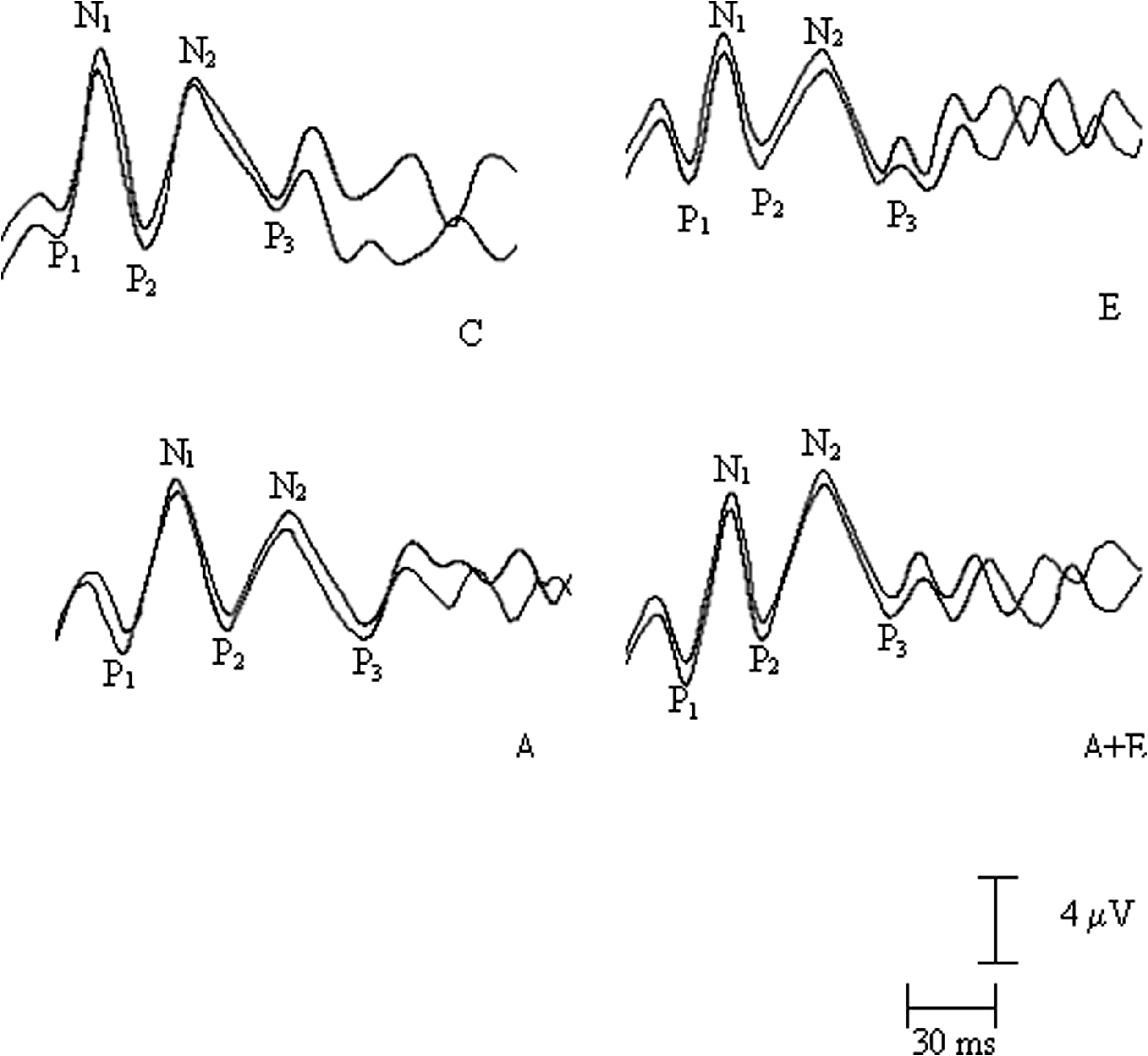
Characteristic visual evoked potentials of the 4 groups. Three positive (P1, P2, P3) and 2 negative (N1, N2) were seen in all groups. C, control; A, amikacin; E, erdosteine; A + E, amikacin + erdosteine.
P < 0.001, **P < 0.01 versus control group. #P < 0.001, ##P < 0.01, ###P < 0.05 versus amikacin group.
Amikacin treatment increased plasma TBARS levels (0.12 ± 0.038 nmol/g protein), compared with the control group (0.026 ± 0.005 nmol/g protein). The increase induced by amikacin was completely prevented by erdosteine in the amikacin + erdosteine group (0.05 ± 0.018 nmol/g protein). Erdosteine treatment alone did not make any significant changes in lipid peroxidation (0.012 ± 0.005 nmol/g protein) compared with the control group (Fig. 2).
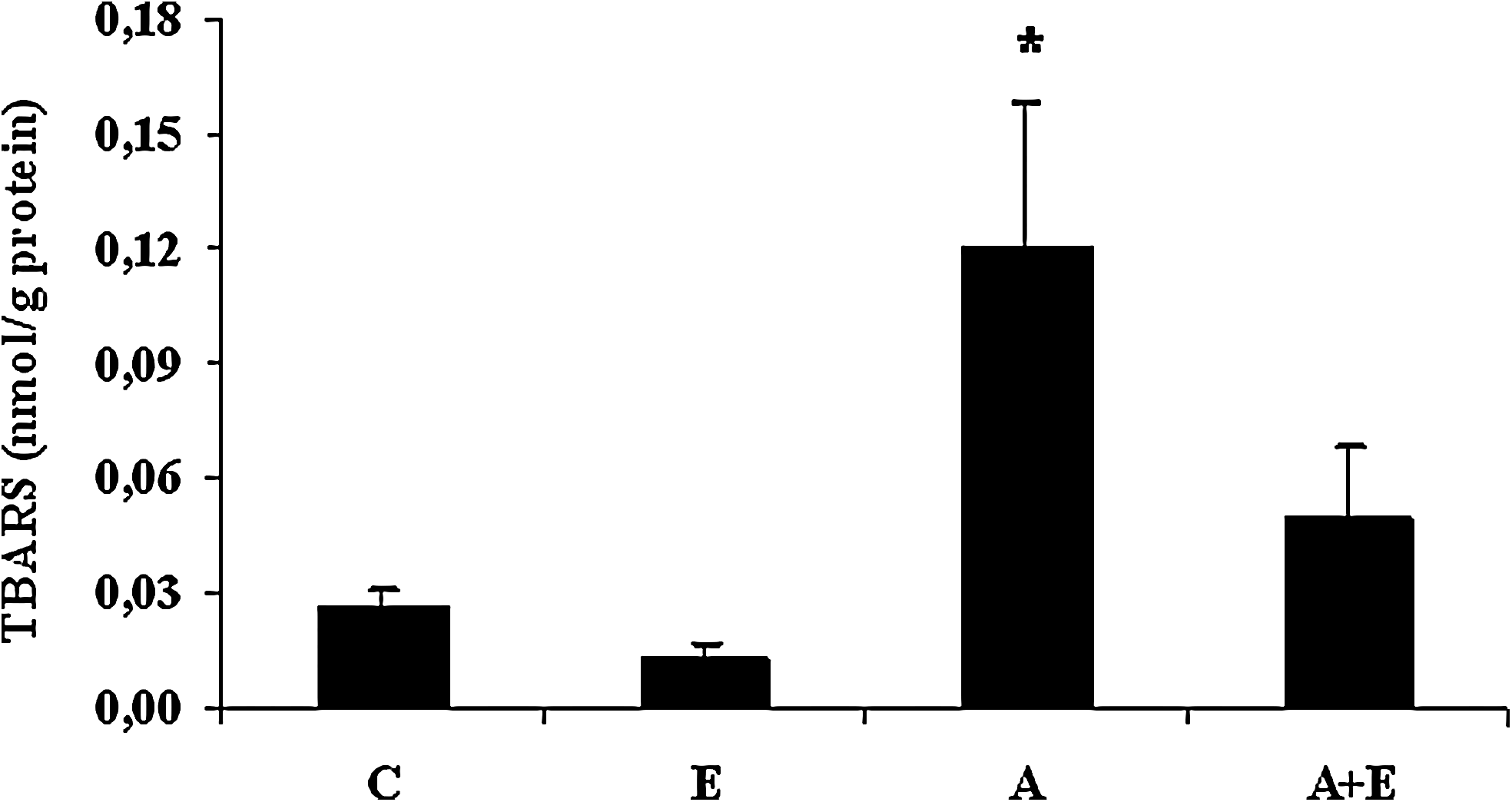
TBARS levels of all groups. Bars represent the group means ± standard error of the mean. C, control; E, erdosteine; A, amikacin; A + E, amikacin + erdosteine. *P < 0.001 versus control, amikacin, amikacin + erdosteine. TBARS, thiobarbituric acid reactive substance.
Dıscussıon
When analyzing the toxic effects of amikacin treatment, it is important to evaluate both the duration and dosage of the treatment. In an experimental study conducted on rats, 600 mg/kg/day of amikacin was administered for 14 days, and the ototoxic effects of amikacin were determined. 2 In another study, rats were injected with a single dose of amikacin (1.2 g/kg i.p.), and electron microscopic changes were apparent in the kidney. 24 We selected 600 mg/kg/day of amikacin from the previous studies that have reported to produce experimental ototoxicity in rats.2,3
Intravitreal injection is the most effective route for administration of antibiotics in intraocular infections. As previously stated, amikacin has long been used in antibacterial therapy of severe gram-negative infections. Thus, intraocular infections caused by gram-negative bacteria can be treated via intravitreal amikacin injections. 25 However, macular infarction, preretinal haemorrhages, and leukocyte infiltration of vitreous have been reported as side effects of intravitreal amikacin administration.25–27 Presented data herein clearly show that erdosteine can decrease the ophthalmic toxicity of systemic amikacin treatment. To our knowledge, there is no study in the literature showing the toxic effects of systemic amikacin therapy in the visual system. In this study, we recorded the VEPs of rats that were treated with amikacin and observed that all latency of VEPs in the amikacin treated rats were prolonged. In view of the fact that VEPs have been shown to be a sensitive and reliable method to evaluate the earliest changes in the visual system,18,19 these results probably indicate that amikacin markedly affects the visual system. As a result, it could be concluded that lipid peroxidation might have a role in the prolongation of all VEP components. Prolonged latencies of VEP components in amikacin treated rats returned to control levels after erdosteine administration (Table 1). These findings can be related to amikacin-induced lipid peroxidation, which is decreased by erdosteine administration (Fig. 2).
It is well known that oxygen-free radicals are important mediators of tissue injury and these radicals may change antioxidants and TBARS levels within the plasma. Monitoring of oxidative stress in experimental research can be done indirectly by assaying products of oxidative damage as TBARS levels and malondialdehyde (MDA) that indicate membrane lipid peroxidation and cellular injury. 6 Increased plasma TBARS levels observed herein are in agreement with earlier studies that have documented amikacin incrased TBARS levels in different tissues such as serum,liver, and kidney.1,24,28 It has also been shown that antioxidant enzymes including superoxide dismutase, catalase, glutathione peroxidase, glutathione-S-transferase, and glutathione reductase had significantly lower activity after amikacin treatment as compared with controls. 7 In summary, an imbalance between prooxidant and antioxidant factors play an important role in the pathophysiology of amikacin induced toxicity.
Oxidation of lipids in cell membranes lead to altered membrane structure and neuronal functions.29,30 This study is the first to report that erdosteine treatment restores altered VEP parameters induced by amikacin. Erdosteine changes in a mucolytic agent used for chronic pulmonary diseases for more than 10 years was used as an antioxidant agent in our study. 31 Erdosteine's metabolites, particularly those with sulphydryl groups, have free radical scavenging and antioxidant activity.17,32,33 It was observed that erdosteine prevented the reduction of endogenous antioxidant enzymes, enhancement of TBARS formation and nitric oxide levels in brain ischemia-reperfusion injury. 31 Moretti and Marchioni shown that erdosteine prevents the accumulation of ROS when their production is accelerated and increases antioxidant cellular protective mechanisms. 14 Our results are in accordance with these studies. Erdosteine significantly attenuated the increase of TBARS levels in plasma and depleted the prolonged latencies of VEP components; these effects are probably due to its elimination capacity of free oxygen radicals.
Although previous studies have shown that amikacin induces nephrotoxicity and ototoxicity, this is the first study to assess the effect of amikacin treatment in the rat visual system. Further, erdosteine provides a marked protection against amikacin induced oxidative stress.
Footnotes
Author Disclosure Statement
This study was supported by a grant from Akdeniz University Research Foundation, Turkey. Akdeniz University Research Foundation had no involvement in the study design, collection, analysis, interpretation of data, writing of the report and in the decision to submit the paper for publication.