
Abstract
Abstract
Purpose:
This investigation evaluated the ocular and systemic pharmacokinetics of besifloxacin in African green monkeys compared with cynomolgus monkeys following topical ocular dosing.
Methods:
A suspension formulation containing 0.6% besifloxacin was administered to African green and cynomolgus monkeys. Animals were euthanized at predetermined time intervals, and ocular tissue and systemic blood samples were collected and analyzed by LC/MS/MS.
Results:
In both African green and cynomolgus monkeys, high concentrations of besifloxacin were detected in anterior segment tissues, while levels in posterior segment tissues and plasma were low. Mean concentration versus time profiles of besifloxacin were generally similar between species, with rapid absorption into ocular tissues after a single dose. In anterior segment tissues, concentrations of besifloxacin were measurable throughout the 24-h sampling period in both species. Quantitatively, concentrations were consistently higher in the conjunctiva of African green monkeys compared with cynomolgus monkeys. Besifloxacin levels were also higher during the first 3 h following dosing in the tear fluid of African green monkeys, but lower in the iris/ciliary body during this timeframe. However after the 3-h time point, concentrations in the tear fluid and iris/ciliary body were similar between species. Exposure in cornea tended to be higher in African green monkeys, but the difference was less pronounced than for conjunctiva. Exposure in aqueous humor was comparable between species. In posterior segment tissues, exposure to besifloxacin tended to be higher in cynomolgus monkeys. Systemic exposure also tended to be higher in cynomolgus monkeys, but measurable levels were present in the plasma of both species throughout the 24-h sampling period. With the exception of iris/ciliary body and vitreous humor, mean ocular tissue weights were generally similar between species although a small, but statistically significant, difference was also observed in the choroid.
Conclusions:
African green monkeys may be a suitable model for preclinical ocular pharmacokinetic studies. Additional studies using a variety of compounds would be useful in determining whether the quantitative differences in ocular exposures and ocular tissue weights observed in the present investigation reflect slight variations in the procedures used in these separate experiments, or true physiological and anatomical differences between species.
Introduction
The most commonly used nonhuman primate species for PK research are cynomolgus (Macaca fascicularis) and rhesus (Macaca mulatta) monkeys. 8 However, with increasing cost and limited supply of cynomolgus and rhesus monkeys, it is important to consider alternative nonhuman primate species for PK research. One species that shows promise as a suitable alternative is the African green monkey or vervet (Chlorocebus aethiops), which is already widely used in other areas of biomedical research. 9 There are 6 subspecies of African green monkeys, including Chlorocebus sabaeus, which was introduced to several Caribbean islands in the West Indies, including St. Kitts.10–12 The African green monkey is not considered to be an endangered species and is classified as “Least Concern” on the International Union for Conservation of Nature's (IUCN) Red List of Threatened Species (www.iucnredlist.org/apps/redlist/details/136265/0). Additionally, the African green monkey is small and easy to handle, and unlike cynomolgus and rhesus monkeys, it is not an asymptomatic carrier of the Herpes B virus, making it a potentially safer species for investigators to work with.13,14
African green monkeys have been used extensively in biomedical research in areas including, but not limited to, neurodegeneration (Parkinson's15–17 and Alzheimer's disease18,19), neurobehavioral (attention deficit disorder20,21 and alcoholism22–24 ), cardiovascular disease (hypertension25–27 ), and immune system and infectious disease (HIV,28–30 leishmaniasis,31,32 Hendra, 33 and Dengue 34 ). Additionally, the African green monkey genome has recently been sequenced by the Washington University Genome Center, providing new opportunities for genetic research (www.genomequebec.mcgill.ca/compgen/vervet_research/genomics_genetics/). Reference ranges for several physiological parameters have also been recently characterized and compared with reference values for other Old World primates and humans for potential use in preclinical models for therapeutic efficacy and safety.35,36 Recently, African green monkeys have been shown to have predictivity similar to cynomolgus and rhesus monkeys for estimating systemic PK in humans.8,37 However, no ocular PK data in this species are reported in the literature. Therefore, the purpose of this investigation was to evaluate the ocular and systemic PK of besifloxacin, a fluoroquinolone antibiotic used for the treatment of bacterial conjunctivitis, in African green monkeys compared with cynomolgus monkeys.
Methods
General experimental design
This investigation was conducted as two separate experiments: one using African green monkeys, and the other using cynomolgus monkeys. The besifloxacin concentration data from cynomolgus monkeys were reported separately in a previous publication from Proksch et al. 38 and have been reanalyzed and included here for comparative purposes.
Materials
The formulation used in these experiments was identical to the marketed product Besivance™ (besifloxacin ophthalmic suspension, 0.6%), and was prepared and supplied by Bausch & Lomb, Inc. (Tampa, FL). All other materials were purchased from commercial sources.
Animals
Adult male and female African green (n=18) and cynomolgus (n=21) monkeys weighing between ∼3–7 kg and 2–5 kg, respectively, were used for this investigation. All in-life procedures involving African green monkeys were conducted by RxGen, Inc., at the St. Kitts Biomedical Research Foundation (St. Kitts, West Indies), and procedures using cynomolgus monkeys were conducted by Avanza Laboratories (previously Bridge Global Pharmaceutical Services, Inc., Gaithersburg, MD). African green monkeys were individually housed in open-air enclosures at ambient temperature, humidity, and light, whereas cynomolgus monkeys were individually housed in an animal room that was maintained under controlled environmental conditions. All animals were handled according to generally accepted practices. Both studies were conducted in accordance with the Association for Research in Vision and Ophthalmology statement for the Use of Animals in Ophthalmic and Vision Research, and the Institute of Laboratory Animal Resources Guide for the Care and Use of Laboratory Animals. The study protocols were approved by the Institutional Animal Care and Use Committee of the test facility prior to the start of each study.
Test substance administration
Prior to assignment on the studies, a complete ophthalmic examination was performed on both eyes of each animal using slit-lamp biomicroscopy and indirect ophthalmoscopy to verify that there were no preexisting ocular abnormalities that would interfere with the outcome of the study. Only animals in good physical condition with no overt ocular abnormalities were included in the study.
On the day of dosing, animals were anesthetized with an intramuscular injection of a cocktail containing commercially available anesthetics: Telazol® (tiletamine hydrochloride 50 mg/mL, zolazepam hydrochloride 50 mg/mL; Fort Dodge, Iowa) for cynomolgus monkeys, and Ketaset® (ketamine hydrochloride 100 mg/mL; Fort Dodge, Iowa), and AnaSed® (xylazine hydrochloride 20 mg/mL; Shenandoah, IA) for African green monkeys prior to being dosed with besifloxacin. Animals then received a single 50-μL topical dose of 0.6% (6 mg/mL) besifloxacin suspension into the conjunctival sac of each eye using a positive-displacement pipette. This dose volume resulted in a target dose level of 300 μg/eye. To minimize any runoff of the administered dose after instillation and to facilitate the distribution of test substance over the surface of the eye, the eyelids of each animal were gently held closed for several seconds after dosing. Following dose administration, the animals were observed for general health and appearance.
Ocular tissue and plasma collection
Over a 24-h period after dosing, all animals (n=3 per collection time) were humanely euthanized at predetermined time intervals. Immediately following euthanasia, samples of tear fluid were collected by inserting Schirmer tear strips into the lower conjunctival sac. The amount of tear fluid collected was determined by measuring the difference in the weight of the Schirmer strip before and after sample collection. Next, aqueous humor samples were collected in situ via paracentesis. The eyes were then enucleated, snap-frozen in liquid nitrogen, and dissected while frozen. Individual tissues were collected separately for each eye and placed into pre-tared vials and weighed. With the exception of tear fluid and conjunctiva, where representative samples were taken, an attempt was made to collect each ocular tissue in its entirety.
Serial blood samples (∼1 mL each) were collected from a femoral vein from one cohort of 3 monkeys each. The blood was transferred into tubes containing K3EDTA and stored on wet ice until being centrifuged to obtain plasma. The entire plasma volume from each sample was transferred to pre-labeled tubes. All ocular tissue and plasma samples were maintained at or below −20°C until being thawed for bioanalysis.
Bioanalysis
Concentrations of besifloxacin in ocular tissues and plasma were quantitated using HPLC/MS/MS. The analytical equipment used consisted of two Shimadzu LC-20AD pumps (Shimadzu Co., Colombia, MD); a SIL-20A column temperature controller (Shimadzu Co.); a CTC HTC PAL autosampler (LEAP Technologies, Carrboro, NC); a Phenomenex Gemini C6-phenyl column with C18 guard column, 2.1×50 mm (Phenomenex, Torrance, CA); and a Sciex API 4000 triple quadrupole mass spectrometer (Applied Biosystems/MDS Sciex, Concord, Ontario, Canada) with a TurboIonSpray™ interface with positive ion detection. Deuterium-labeled besifloxacin (d8) was used as the internal standard.
Calibration standard samples were prepared at a minimum of 10 concentration levels by spiking known amounts of each analyte into appropriate blank tissues for each type of tissue studied. Quality control samples were prepared in a like manner at 3 concentration levels in blank tissue. Blank plasma specific to each species was used to prepare the calibration standards and quality control samples for plasma bioanalysis. Blank ocular tissues from African green monkeys were used to prepare calibration standards and quality control samples for the analysis of African green monkey ocular tissues. However, due to the limited availability of blank cynomolgus monkey ocular tissues at the time of bioanalysis, a surrogate matrix (pigmented rabbit tissue) was used to prepare the calibration standards and quality control samples for analysis of cynomolgus monkey ocular tissues. The bioanalytical methods were evaluated for precision and accuracy, as assessed by the back-calculated concentrations for quality control samples included within each bioanalytical run.
With the exception of vitreous humor, the preparation methods used for ocular tissue and plasma samples were similar between experiments. In the African green monkey experiment, the entire vitreous humor sample was processed and quantitated, whereas in the cynomolgus monkey experiment an aliquot of the vitreous humor sample was used for analysis. Based on the sample preparation methods used, the lower limit of quantitation for ocular tissues was in the range of 0.0001 μg/mL (aqueous humor) to 0.235 μg/g (tear fluid), and the lower limit of quantitation for plasma was 0.0500 ng/mL.
Pharmacokinetic analysis
PK analysis of ocular tissue and plasma data was performed using noncompartmental methods in WinNonlin Professional® (version 5.3; Pharsight Corporation, St. Louis, MO). For plasma, individual concentration versus time data from serial sampling was used for PK analysis. However, for ocular tissues, it was not possible to collect more than one sample of each tissue per eye. Therefore, composite (mean) concentration versus time data were used for PK analysis of ocular tissue data. To perform a statistical comparison between AUC(0–24h) values for African green and cynomolgus monkeys, the sparse data option in WinNonlin was used to calculate AUC(0–24h) values and the corresponding standard error (SE) estimates for ocular tissues. Because the sparse data method was not used for PK analysis of the cynomolgus monkey data reported in an earlier publication from Proksch et al., 38 some of the PK parameter values reported here differ slightly from what was previously reported.
AUC(0–24h) and mean residence time (MRT) were calculated using the linear trapezoid method for ocular tissues and the log/linear trapezoid method for serial plasma samples. PK parameters including maximum concentration (Cmax) and the time at which the maximum concentration was observed (Tmax) were determined directly from the concentration versus time profiles. For tear fluid analysis, concentration at time zero was determined by extrapolating to time zero using log-linear regression analysis of the first two data points (intravenous bolus model). For all other ocular tissues and plasma, the concentration at time zero was assumed to be zero (extravascular model).
The resulting estimates of AUC(0–24h) and SE for African green monkey tissues were compared with the values obtained for cynomolgus monkey tissues using Welch's t-test as demonstrated by Schoenwald et al. 39 and Tang-Liu and Burke. 40 A two-tailed Student's t-test was used to determine significant differences in Cmax after an F-test was used to determine equal or unequal variance between individual concentration values at Cmax. Differences were considered statistically significant when the calculated P value was less than or equal to 0.05. All statistical calculations were performed using Microsoft Excel (2002).
Results
Clinical assessment
No clinical or ocular abnormalities were observed during the study.
Ocular Pharmacokinetics
Considerable interanimal variability was observed in besifloxacin concentrations in all ocular tissues examined, with coefficients of variation (CV%) for each tissue between 35%–76% for African green monkeys and 58%–91% for cynomolgus monkeys. In general, the concentration versus time profiles of besifloxacin in ocular tissues were similar between African green and cynomolgus monkeys following a single, topical ocular dose. In both species, besifloxacin was rapidly absorbed and distributed throughout the eye with maximum concentrations in the majority of tissues achieved within 30 min of dosing. Based on Cmax and/or AUC(0–24h) values, the highest concentrations of besifloxacin were observed in the anterior segment tissues, in the order of tear fluid≫ conjunctiva>cornea>aqueous humor≈iris/ciliary body (Figs. 1 and 2). Concentrations of besifloxacin were measurable in these tissues throughout the 24-h sampling period in both species. In the posterior segment tissues (vitreous humor, retina, and choroid), concentrations of besifloxacin were considerably lower than what was measured in the anterior segment tissues, and while concentrations were measurable throughout the 24-h sampling period in cynomolgus monkeys, concentrations were below the limit of quantitation in African green monkeys within 3 to 6 h after dosing (Fig. 3).
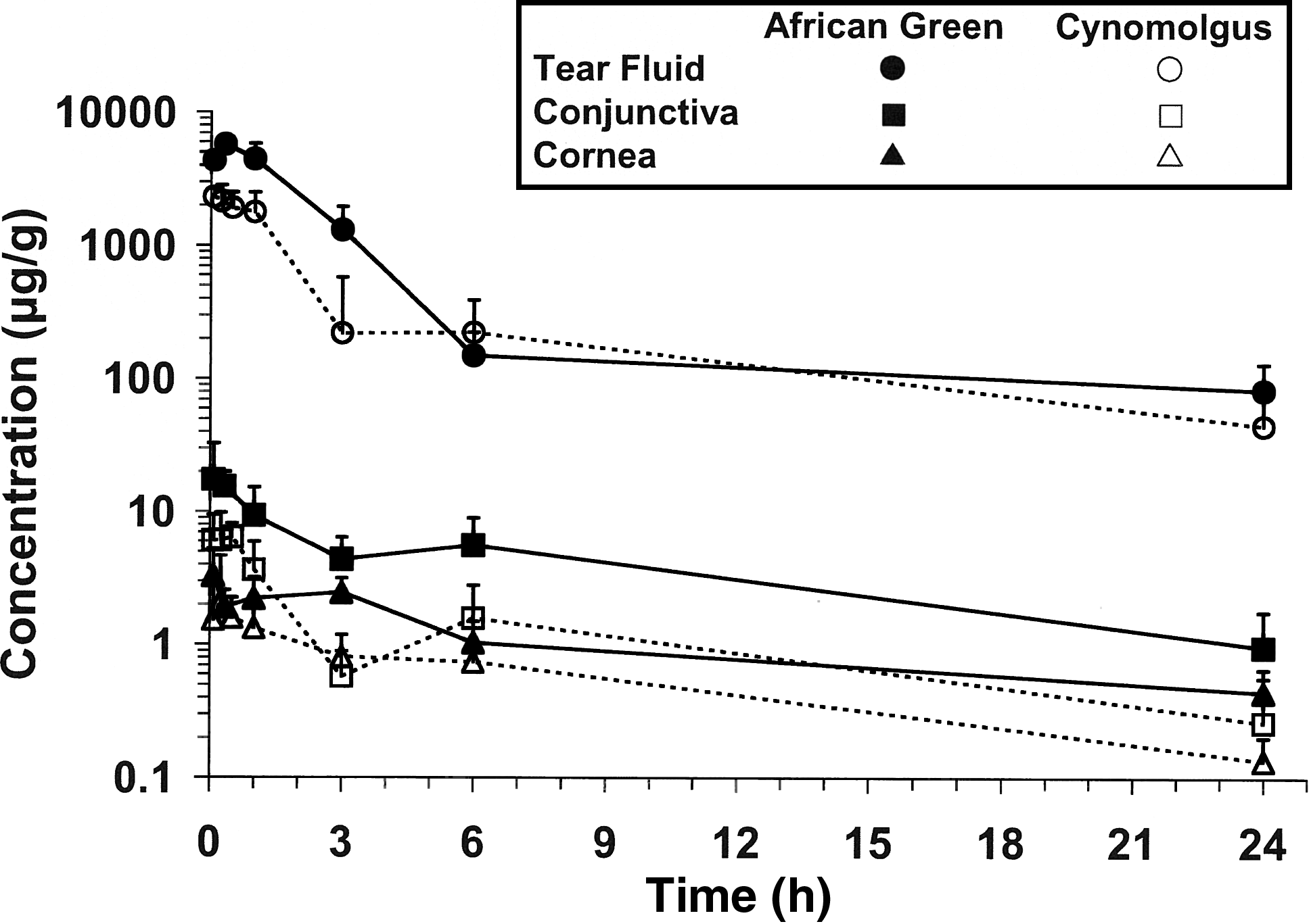
Besifloxacin concentration versus time profiles in the tear fluid, conjunctiva, and cornea of African green (solid symbols) and cynomolgus (open symbols) monkeys following a single topical ocular administration of besifloxacin ophthalmic suspension (0.6%). Data represent mean [+ standard deviation (SD)] besifloxacin concentrations. Data from cynomolgus monkeys were included in a previous publication from Proksch et al. 38
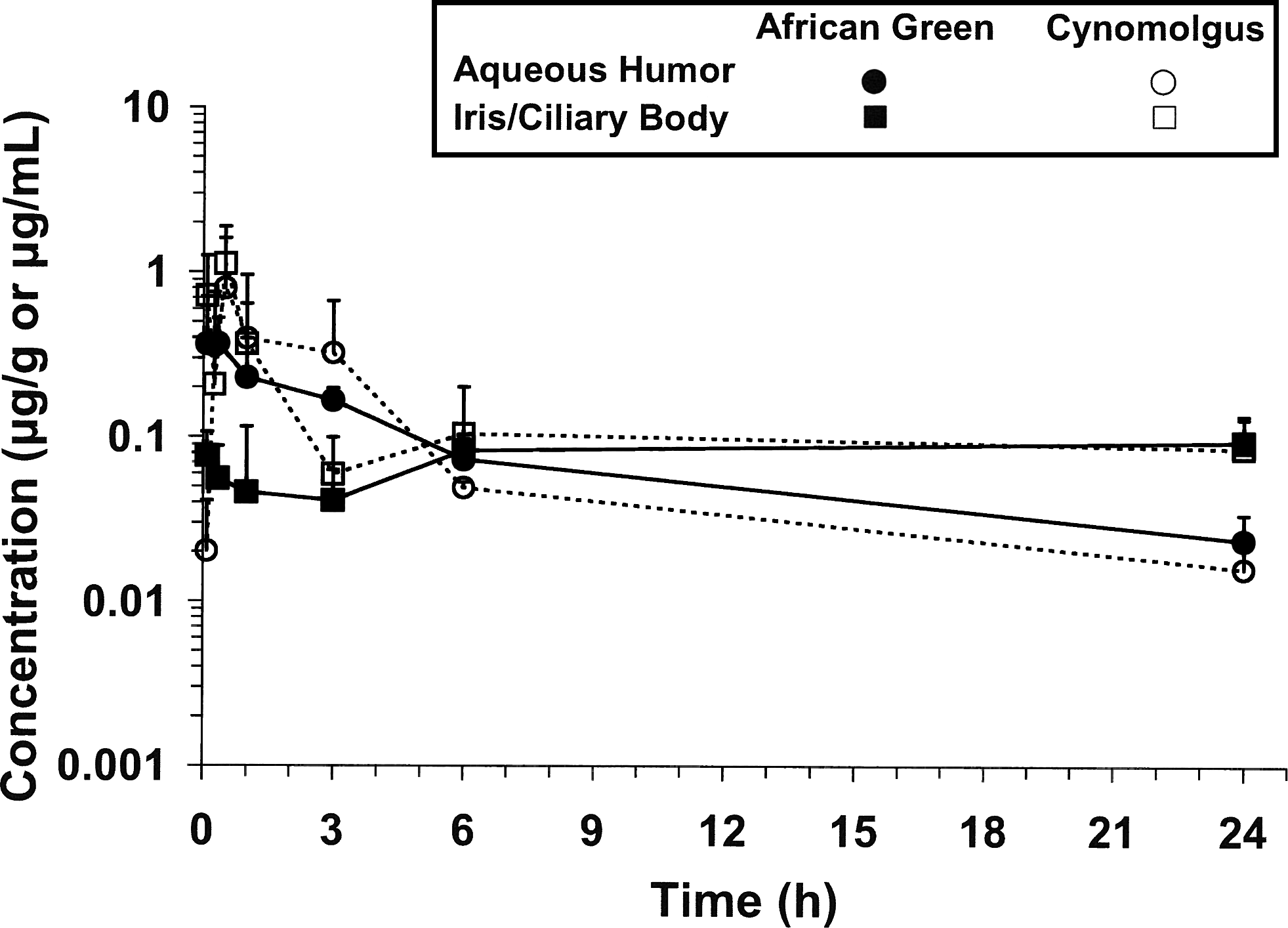
Besifloxacin concentration versus time profiles in the aqueous humor and iris/ciliary body of African green (solid symbols) and cynomolgus (open symbols) monkeys following a single topical ocular administration of besifloxacin ophthalmic suspension (0.6%). Data represent mean (+SD) besifloxacin concentrations. Data from cynomolgus monkeys were included in a previous publication from Proksch et al. 38
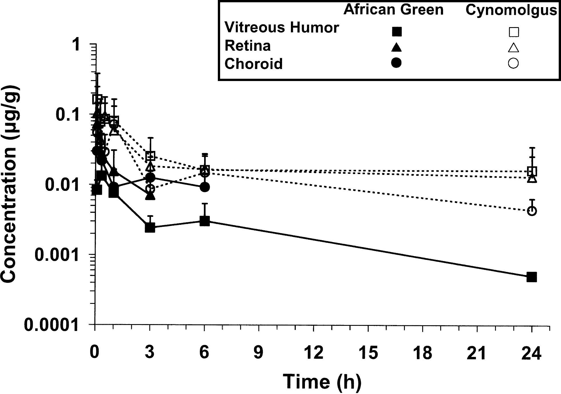
Besifloxacin concentration versus time profiles in the vitreous humor, retina, and choroid of African green (solid symbols) and cynomolgus (open symbols) monkeys following a single topical ocular administration of besifloxacin ophthalmic suspension (0.6%). Data represent mean (+SD) besifloxacin concentrations. Data from cynomolgus monkeys were included in a previous publication from Proksch et al. 38
Although the ocular PK profiles of besifloxacin in African green and cynomolgus monkeys were similar, some quantitative differences in exposure were observed (Table 1). Based on Cmax and/or AUC(0–24h) values, exposure to besifloxacin in tear fluid, conjunctiva, and cornea was significantly higher in African green monkeys when compared with cynomolgus monkeys. Overall, exposure did not vary by more than 3.5-fold between species in these tissues. This difference was most pronounced in conjunctiva where concentrations were consistently higher in African green monkeys throughout the 24-h sampling period, whereas in cornea, the difference was less distinct. In tear fluid, the difference in exposure was greatest during the first 3 h after dosing; however, AUC values were nearly identical between species from 6 to 24 h. In aqueous humor, exposure to besifloxacin was comparable between species, based on Cmax and AUC(0–24h) values. Exposure to besifloxacin in the remaining ocular tissues tended to be higher in cynomolgus monkeys when compared with African green monkeys, but the differences were statistically significant for only some of the tissues. In retina and vitreous humor, for example, AUC(0–24h) values were significantly higher for cynomolgus monkeys compared with African green monkeys. However, the total amount of drug present in these tissues was minimal, with maximum concentrations <0.2 μg/g in both tissues. In the iris/ciliary body of cynomolgus monkeys, exposure was notably higher during first few hours after dosing, but was comparable between species by 3 h. Although exposure to besifloxacin tended to be higher in the choroid of cynomolgus monkeys, the difference was not statistically significant. For comparative purposes, the ratio between African green and cynomolgus monkey Cmax and AUC(0–24h) values for ocular tissues was calculated and the data are presented in Fig. 4. Mean residence times were similar between species for the majority of anterior segment tissues. In some tissues, such as conjunctiva, the difference in MRT values was negligible (6%), while in other tissues, such as iris/ciliary body, the difference (36%) was more apparent. In posterior segment tissues, MRT values were larger in cynomolgus monkeys.
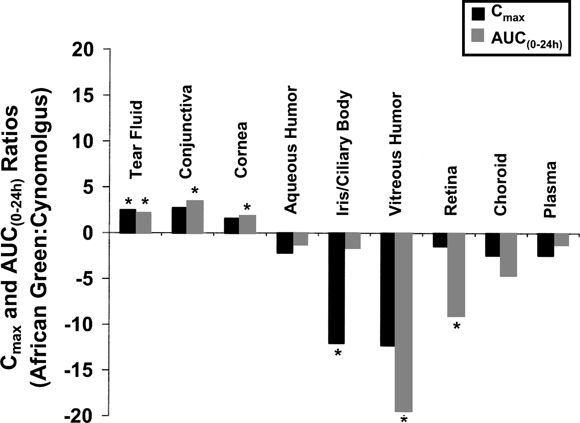
Ratio of African green:cynomolgus monkey Cmax and AUC(0–24h) values for besifloxacin in ocular tissues and plasma. A ratio of 1 indicates that the values are identical between species. Ratios greater than 1 indicate higher Cmax and AUC(0–24h) values for African green monkeys. For tissues where exposure was higher in cynomolgus monkeys, the ratio is plotted as a negative number. Asterisk (*) denotes statistical significance (P≤0.05) between species.
Some of the cynomolgus monkey data were reported previously in a publication from Proksch et al. 38
AUC(0–24h) values are presented as mean (±standard error) for ocular tissues and mean (±SD) for plasma (n=3).
Units for aqueous humor are μg/mL and μg * h/mL for Cmax and AUC(0–24h), respectively.
Units for plasma are ng/mL and ng * h/mL for Cmax and AUC(0–24h), respectively.
Median Tmax reported for plasma (n=3).
Mean (±SD) MRT reported for plasma (n=3).
Statistically significant (P≤0.05) from corresponding values for cynomolgus monkeys.
Systemic Pharmacokinetics
Low, but measurable, levels of besifloxacin were detected in the plasma of both species throughout the 24-h sampling period, with maximum concentrations ≤9.2 ng/mL. While systemic exposure tended to be higher in cynomolgus monkeys, based on Cmax and AUC(0–24h) values, the difference was not statistically significant. The ratio between African green and cynomolgus monkey Cmax and AUC(0–24h) values for plasma is presented in Fig. 4. The MRT for besifloxacin in plasma was significantly longer in African green monkeys due to a spike in concentration between 12 and 24 h, which was not observed in cynomolgus monkeys (Table 1).
Ocular tissue weights
To gain insight into potential differences in ocular anatomy between species, a comparison of ocular tissue weights was conducted, and the data are presented in Table 2. Because only partial samples of tear fluid and conjunctiva were collected, these tissues were not included in the comparison. The values obtained for African green monkey tissues include weights for blank tissues that were used for the preparation of calibration standards and quality control samples for bioanalysis.
Representative samples of tear fluid and conjunctiva were collected. Data not reported. Values represent the mean (±SD).
n=56 for each tissue except for choroid where n=55 (1 value was excluded due to a weighing error).
n=42 for each tissue.
Statistically significant (P≤0.05) from corresponding values for cynomolgus monkeys.
There was considerable variability between individual ocular tissue weights for both species. In African green monkeys, mean iris/ciliary body and vitreous humor weights were 2.9-fold and 2.6-fold higher, respectively, and mean choroid weights were 1.3-fold lower when compared with cynomolgus monkey tissue weights. These differences were statistically significant. All other ocular tissue weights were comparable between species. For comparative purposes, the ratio between mean tissue weight values for African green and cynomolgus monkeys was calculated and the data are presented in Fig. 5.
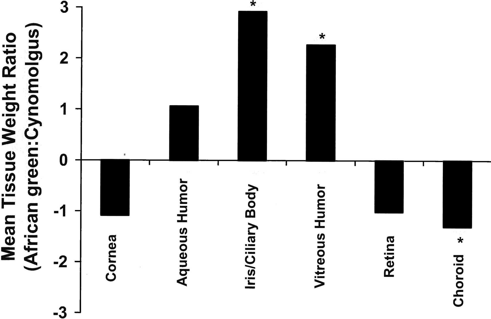
Ratio of mean ocular tissue weight values for African green:cynomolgus monkeys. A ratio of 1 indicates that the values are identical between species. Ratios greater than 1 indicate higher mean tissue weight values for African green monkeys. For tissues where the tissue weight was higher in cynomolgus monkeys, the ratio is plotted as a negative number. Asterisk (*) denotes statistical significance (P≤0.05) between species.
Discussion
African green monkeys have been extensively used in many areas of biomedical research, but their use in preclinical drug development has remained limited. For PK studies in particular, cynomolgus and rhesus monkeys have historically been the nonhuman primate species of choice. However, due to the limited availability and high costs associated with these species, there is an interest in identifying other nonhuman primates that can reliably predict human PK. Recently, Ward et al. demonstrated that African green monkeys exhibit similar predictivity for human PK as cynomolgus monkeys following oral and intravenous dosing.8,37 The present investigation sought to determine whether the same would hold true following topical ocular dosing. To our knowledge, this is the first report that compares the ocular PK of a compound in African green monkeys with cynomolgus monkeys.
The concentration versus time profiles of besifloxacin in ocular tissues of African green monkeys were similar to those observed in cynomolgus monkeys, although quantitative differences in exposure were observed. In both species, regional differences in exposure were observed with much higher concentrations observed in anterior tissues compared with posterior tissues. This pattern of regional distribution is consistent with that seen with other ophthalmic drugs administered topically, as delivery to posterior ocular tissues is limited by nasolacrimal drainage, corneal permeability, conjunctival and scleral blood flow, and blood ocular barriers. 5
In the anterior segment tissues, the most significant difference in exposure was seen in the conjunctiva where concentrations were consistently higher in African green monkeys throughout the 24-h sampling period. Differences in Cmax and/or AUC values were also seen in the tear fluid and iris/ciliary body, but only during the first 3 h after dosing. The same trend was also seen in the cornea, but the difference was much less pronounced. It is likely that higher concentrations of drug available on the surface of the eye contributed, at least in part, to increased exposure in the anterior segment tissues. It is unclear why concentrations of besifloxacin in tear fluid were higher in the African green monkey, but it may be related to differences in study design. For example, in the present investigation, African green and cynomolgus monkeys were given different anesthetic agents prior to being dosed. Investigations on the effects of anesthetic agents on tear dynamics in rabbits, dogs, and humans reveal that tear production and drainage are reduced following both local 41 and general anesthesia.42–46 In addition to the loss of palpebral and corneal reflexes that can increase the rate of evaporation of tear fluid from the surface of the eye and/or reduce nasolacrimal drainage, some anesthetic agents inhibit the trigeminal nerve which is responsible for stimulating the lacrimal gland to produce tears. 42 These effects can potentially increase the retention of drug on the surface of the eye. However, because of a lack of published data for the particular anesthetic agents used in these experiments, it is unclear how they affect tear fluid dynamics in primates. Furthermore, the studies were not controlled for how often the eyes were manually blinked while the animals were under anesthesia, and the exact duration of sedation was not recorded. These potential differences in study design between experiments also could have influenced the retention of drug on the surface of the eye. Nevertheless, the possibility of species differences in anatomy, basal tear production and turnover rate, ocular blood flow, tissue penetration, or melanin content in the retina, choroid, or iris/ciliary body, or a combination of these cannot be ruled out as potential factors contributing to the interspecies differences in besifloxacin levels in conjunctiva.
Although besifloxacin levels in tear fluid, conjunctiva, and cornea were higher in African green monkeys, exposure in the aqueous humor was comparable between species. Given the lipophilicity and low aqueous solubility of besifloxacin, it is possible that the concentration differences in tear fluid, conjunctiva, and cornea relative to cynomolgus monkeys were not sufficiently high to drive additional drug into the aqueous humor.
In the pigmented tissues (iris/ciliary body, retina, and choroid), concentrations of besifloxacin tended to be higher in cynomolgus monkeys. Since fluoroquinolone antibiotics have been shown to bind to melanin, 47 it is possible that the difference in exposure to besifloxacin in these tissues may be related to inherent differences in melanin content between the species. To our knowledge, the melanin levels in these tissues have not been published for either of these species.
As Old World primates, African green and cynomolgus monkeys share substantial homology in ocular anatomy and physiology. Although there is limited published data on the ocular biometry of either species, 48 the present investigation suggests that there is little difference in ocular tissue weights between groups. The most pronounced differences were seen in the iris/ciliary body and vitreous humor samples where the mean weight of these tissues was ∼2 to 3-fold higher for African green monkeys. Considering that these experiments were conducted at separate test facilities, it is possible that the observed differences in tissue weight are the result of slight variations in the dissection procedure, and do not reflect anatomical differences. In both experiments, the eyes were dissected after being flash-frozen. While this procedure often facilitates accurate separation of ocular tissues along natural tissue planes, delineation of the ora serrata, which defines the junction between the ciliary body and retina, can be the least reproducible step in this method of dissection, making it difficult to consistently collect the iris/ciliary body in its entirety. This may, in part, explain the large interanimal variability that was observed for iris/ciliary body weights, as coefficients of variation were ∼50% and 80% for African green and cynomolgus monkeys, respectively. Similarly, the apparent interspecies differences in vitreous humor weights may be related to possible inconsistencies in the time taken to freeze the eyes or the time taken to dissect the eyes, resulting in partial melting of vitreous humor at the time of dissection and consequently, incomplete collection of this tissue.
As reported previously, in ocular tissues for which comparative human data are available, the PK profiles of besifloxacin in the tear fluid and conjunctiva of cynomolgus monkeys were similar to that observed in humans following a single topical dose.38,49 Based on the results from the present investigation, the same general conclusion holds true for African green monkeys. Although cynomolgus monkeys were somewhat better predictors of exposure in these tissues, both species over-predicted human exposure. This over-prediction in exposure may be the product of inherent differences between nonhuman primates and humans. Alternatively, it may be the result of differences in study design. For example, in the animal experiments, monkeys were dosed while under anesthesia, while human subjects were dosed while fully awake. As mentioned previously, this may have altered tear flow dynamics and allowed the drug to remain on the surface of the eye longer, providing increased exposure in these tissues. To further evaluate the potential correlation between altered exposure on the surface (i.e., in tear fluid) and corresponding tissue levels, the apparent partitioning between tear fluid and conjunctiva was calculated as the ratio of AUC(0–24h) in tear/conjunctiva. The observed AUC(0–24h) ratios for African green monkeys, cynomolgus monkeys, and humans were 150, 240, and 185, respectively. The comparability of these partition coefficients across species suggests that the relative penetration of besifloxacin into conjunctiva is consistent across species, with human falling between the two nonhuman primate species.
In both species, mean concentrations of besifloxacin in aqueous humor 1 h after dosing were similar to concentrations observed in humans undergoing cataract surgery, 50 with mean values of 0.23±0.17, 0.40±0.25, and 0.13±0.58 μg/mL for African green monkeys, cynomolgus monkeys, and humans, respectively. When normalized for body weight, systemic exposure to besifloxacin also tended to be similar between nonhuman primates and humans, based on exposure through 6 h, the sampling interval in human subjects with bacterial conjunctivitis. 38
Taken together, the results from this preliminary investigation look very promising for the use of African green monkeys as an alternative nonhuman primate species to cynomolgus monkeys for topical ocular PK studies. Although some quantitative differences in exposure were noted, the ocular and systemic PK profiles were generally similar between species and each provided similar predictivity of human exposure. It is unclear whether the observed quantitative differences were species-related or due to other factors such as study design, as this investigation highlighted the potential impact of study design details, such as choice of anesthetic and tissue collection procedures, on experimental results. Additional studies using a wide range of ophthalmic drugs and alternate routes of ocular administration would be useful for further characterization of the utility of African green monkeys for ocular PK studies.
Footnotes
Acknowledgments
The authors would like to thank Tim Ordway for the bioanalysis of besifloxacin levels in ocular tissues and plasma samples.
Author Disclosure Statement
Shellise Glogowski, Joel W. Proksch, and Keith W. Ward were employees of Bausch & Lomb at the time the work was conducted. Matthew S. Lawrence and Robin J. Goody are both employees of RxGen, Inc.