
Abstract
Abstract
Purpose:
To investigate the effect of topical motesanib, an inhibitor of receptor tyrosine kinase, on experimental choroidal neovascularization (CNV).
Methods:
CNV was induced in 46 nine-week-old male C57BL/6 mice using fundus laser photocoagulation. The right eye of each mouse was treated with motesanib eye drop (4 times daily) and the left eye with vehicle eye drop (4 times daily) for 14 days. To evaluate changes in the CNV lesions, fluorescein angiography, immunofluorescence staining with CD34, and histological examinations were performed 14 days after CNV induction. The expression of phosphorylated extracellular signal-regulated kinase (ERK1/2) in choroidal tissues was determined using western blot analysis to demonstrate the inhibitory effect of topically administered motesanib on intracellular signaling pathways involved in CNV development.
Results:
Fluorescein angiography showed that fluorescence leakage in eyes treated with topical motesanib was significantly less than in mice treated with vehicle (P=0.01). On immunofluorescence staining, the CD34-labeled area was smaller in topical motesanib-treated eyes (P<0.001). The expression level of phosphorylated ERK1/2 relative to that of total ERK1/2 decreased in eyes treated with topical motesanib compared with eyes treated with vehicle.
Conclusion:
Topical motesanib significantly reduced laser-induced CNV in the experimental mouse model.
Introduction
C
Therefore, the VEGF signaling pathway is an ideal therapeutic target for CNV. Anti-VEGF therapies using ranibizumab (Lucentis; Genentech, San Francisco, CA) and aflibercept (Eylea; Regeneron, Tarrytown, NY) have emerged as standard treatments in clinical practice. Bevacizumab (Avastin; Genentech), which is another anti-VEGF antibody, has been used as “off-label” treatment for AMD. The reported clinical results with these anti-VEGF treatments are satisfactory and they are widely used in clinical practice. 12 However, these therapies require repeated intravitreal injections. Frequent ocular injections are generally associated with risks of endophthalmitis development, retinal detachment, and vitreous hemorrhage. Moreover, regaining of vision lost to AMD is not always achieved and a considerable number of patients are regarded as “non-responders” to these treatment options. 13 Accordingly, a new drug and delivery system are needed to reduce treatment-related complications and to identify an alternative therapeutic modality, respectively.
Motesanib is a novel nicotinamide, oral small-molecule multiple tyrosine kinase inhibitor (TKI), which inhibits all VEGFRs (VEGFR-1, -2, and -3), PDGF receptor (PDGFR), and stem cell factor receptor/CD117 (c-KIT). 5 Previous clinical and experimental evidence suggested that motesanib has potent antiangiogenic effects and suppresses tumor angiogenesis.5,14,15 It functions mainly by blocking the VEGF signaling pathway, which is associated with endothelial cell proliferation, migration, and survival. Therefore, this drug may be a new candidate agent for inhibiting CNV development in AMD. Previous study showed that when VEGF protein expression was induced, phosphorylation of extracellular signal-regulated kinase (ERK1/2) protein levels was upregulated. 16 Another study reported that ERK1/2 and NOS were elements of different signaling pathways in VEGF-induced hyperpermeability. 17 We think that laser-injury-induced increased levels of VEGF and motesanib inhibited VEGFR to decrease the subsequent phosphorylation of ERK1/2 protein.
Topical drug administration is widely used in glaucoma treatments. This route is noninvasive in comparison with intravitreal injection and is associated with lower rates of systemic side effects compared to oral administration. In this study, we investigated the antiangiogenic effects of motesanib in an animal model of CNV using a topical administration.
Methods
Animals
A total of 46 nine-week-old male C57BL/6 mice (weight range 20–22 g) were used as follows: fluorescein angiography (n=20), immunofluorescence staining (n=20), western blot analysis (n=3), and histology (n=3). All study protocols were approved by the Institutional Animal Care and Use Committee of Deajeon St. Mary's Hospital of the Catholic University of Korea, College of Medicine. The mice were handled in accordance with the Association for Research in Vision and Ophthalmology Statement for the Use of Animals in Ophthalmic and Vision Research.
Topical motesanib eye drop
Motesanib powder was purchased (Selleck Chemicals, Houston, TX), dissolved in 100% dimethyl sulfoxide (DMSO; Sigma, St. Louis, MO), and diluted with 0.5% carboxymethylcellulose solution (Allergan, Waco, TX). The final solution was 5 mg/mL of motesanib in 1:19 solution of DMSO:carboxymethylcellulose solution. Solutions of 5 mg/mL were prepared and stored at 4°C. Both eyes of each mouse were used; the right eye was treated with motesanib eye drop (4 times daily) and the left eye was topically treated with vehicle eye drop (4 times daily) for 14 days since experimental CNV induction. Eye drops were instilled with micropipette and 1 eye drop volume was 10 μL.
Induction of experimental CNV
Mice were anesthetized using intraperitoneal injections of 30-mg zolazepam (Zoletil; Virbac, Carros, France) and 10-mg xylazine hydrochloride (Rompun; Bayer, Leuverkeusen, Germany) per kg of body weight. After topical dilation of both pupils with 0.5% tropicamide and 0.5% phenylephrine (Mydrin-P; Santen Pharmaceutical Co., Osaka, Japan), 5 to 7 laser photocoagulation spots were applied close to the optic disc, to obtain at least 5 effective disruption of Bruch's membrane, avoiding major retinal vessels. Laser parameters were 50-μm spot size, 200-mW power, and 0.1-s duration. Only mice that experienced central bubble formation, indicating rupture of the Bruch's membrane, were included in the study. The brightest 5 lesions were included in the statistical analysis.
Fluorescein angiography
Fluorescein angiography was performed 14 days after laser application to quantitate the degree of leakage from CNV lesions, as described previously. 18 Each mouse was injected intraperitoneally with 0.5-mL 10% sodium fluorescein (Fluorescite; Alcon, Tokyo, Japan). Fluorescein angiography was performed 10-min postinjection using confocal scanning laser ophthalmoscopy (Spectralis; Heidelberg Engineering, Inc., Heidelberg, Germany). The images of late-phase angiograms were taken and analyzed using the Image J software (National Institutes of Health, Bethesda, MD). The brightness of the CNV lesion with leakage and the signal intensity were measured as described previously 18 and presented as an arbitrary unit from 0 (darkest) to 1 (brightest). As a reference, the signal intensity within a nonphotocoagulated capillary area was defined as 0 and the signal intensity at the major branch of the retinal vein was defined as 1.
Quantification of CNV by immunofluorescence staining
The mice were sacrificed 14 days after laser photocoagulation and the eyes were enucleated and processed for labeling of the endothelial cells in the CNV lesions. For immunofluorescence staining, enucleated eyes were immediately fixed in 4% paraformaldehyde for 1 h. The cornea and crystalline lens were removed, and the retina was softly peeled and separated from the optic nerve head. The remaining eye cup was washed with a buffer containing 0.5% bovine serum albumin and 0.2% polysorbate 20 (Tween20; Sigma) diluted in phosphate-buffered saline (PBS). A 1:1,000 dilution of 10 mg/mL solution of 4′,6-diamidino-2-phenylindole (DAPI), a 1:50 dilution of 0.2 mg/mL CD34 (endothelial cell marker) conjugated with phycoerythrin (BD Pharmingen, San Diego, CA), and a 1:100 dilution of 0.2 U/L phalloidin conjugated with Alexa Fluor 488 (Invitrogen-Molecular Probes, Eugene, OR) were prepared. The eye cups were incubated with prepared fluorescent dyes overnight at 4°C and washed with cold PBS. The eye cups were flattened and mounted with the sclera facedown and the choroid faceup. Images were obtained using fluorescent microscopy (Eclipse TE300; Nikon, Tokyo, Japan) and evaluated using imaging-analysis software (NIS Elements BR; Nikon).
Western blotting of the choroid and retinal pigment epithelium layer
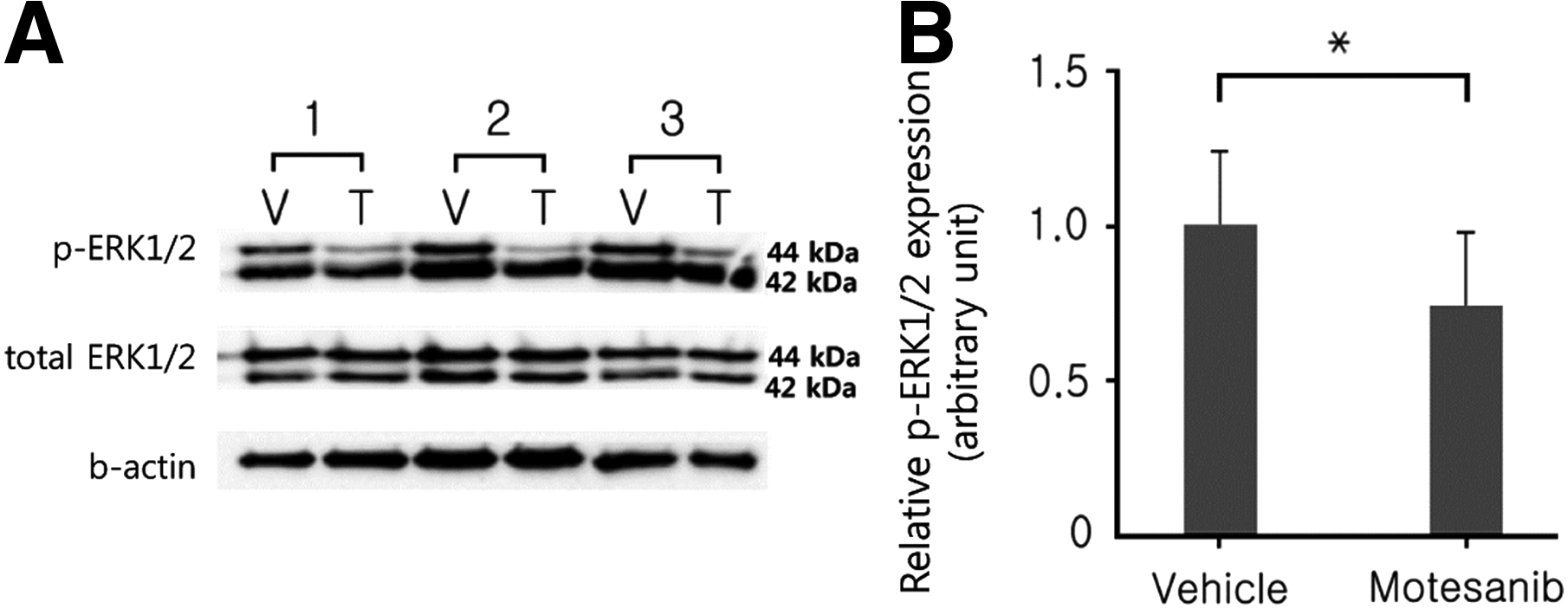
The phosphorylation of ERK1/2 was evaluated by western blot analysis of the choroid and retinal pigment epithelium (RPE). Protein extracts were obtained from RPE-choroid tissues homogenized using the TissueLyser II (Qiagen, Hilden, Germany) 7 days after laser photocoagulation and the protein concentrations were determined using the Bradford assay. Equal amounts of protein were electrophoresed in a 10% sodium dodecyl sulfate–polyacrylamide gel and transferred electrophoretically onto a nitrocellulose membrane. Phosphorylated ERK1/2 (p-ERK1/2) and total ERK1/2 were detected using an anti-p-ERK1/2 (pThr202/pTyr204) antibody (Cell Signaling, Danvers, MA) and a p44/42 mitogen-activated protein kinase antibody (Cell Signaling), respectively.
Histologic procedure
After fluorescein angiography analysis on day 14, the eyes were enucleated for histological evaluation. The right eyes treated with motesanib eye drop and the left eyes treated with vehicle eye drop were fixed for 2 h in 4% paraformaldehyde and embedded in paraffin. Serial sections of 4-μm thickness were cut through the center of the laser photocoagulation site and stained with hematoxylin–eosin.
Statistical analysis
The CD34-labeled area from choroidal flat mounts and the signal intensity from fluorescein angiography were analyzed by Mann–Whitney U-tests using SPSS for Windows, version 17.0 (SPSS, Chicago, IL). The expression level of ERK was analyzed by nonparametric test using SPSS. Values are given as means±standard deviation.
Results
We found no ocular toxicity or side effects on cornea or lens with repeated topical administration during this study. Plasma motesanib level after topical instillation was below the concentration to show systemic effects and to cause side effects (Figure S1, Supplementary Table S1, and Supplementary Table S2; Supplementary materials are available online at http://www.liebertpub.com/jop).
Fluorescein angiography
The average signal intensity scores of angiographic analysis 14 days after laser induction of CNV of the left eyes treated with vehicle (Fig. 1A) and of the right eyes treated with motesanib (Fig. 1B) were 0.86±0.63 and 0.59±0.35, respectively. Topically administered motesanib significantly reduced the fluorescein leakage of CNV lesions (P=0.01) (Fig. 1C). No abnormalities were found in normal retinal blood vessels as determined by the use of fluorescein angiography.
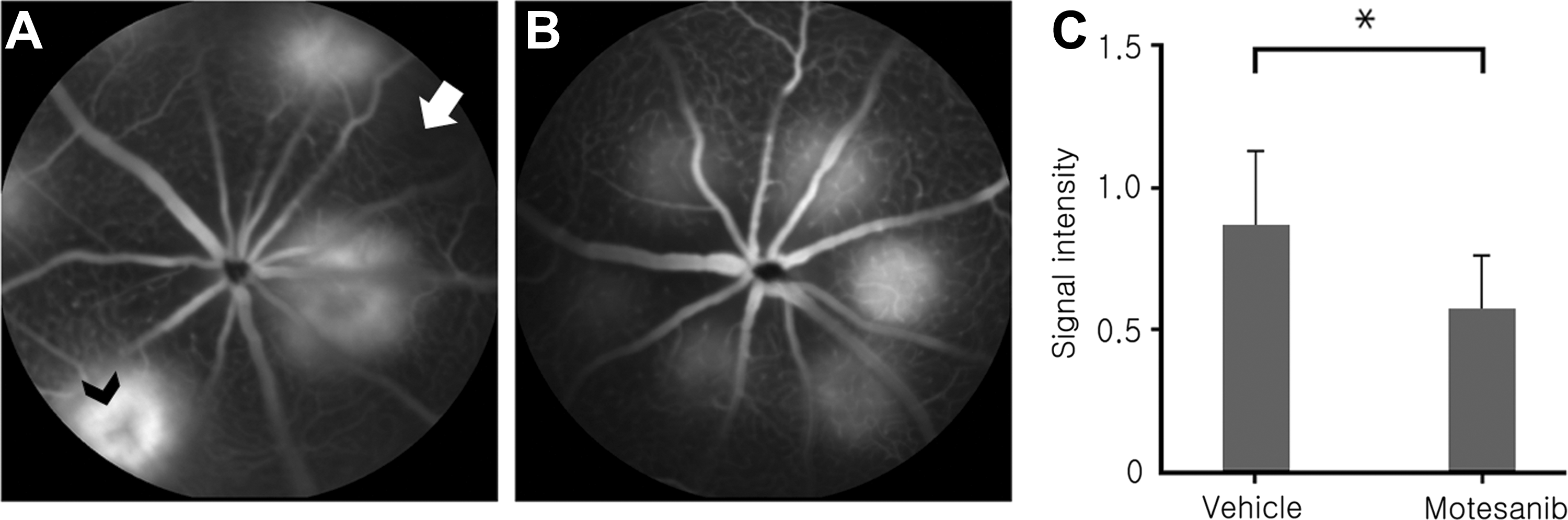
Fluorescein angiograms (n=20, n: the number of animals in each group) displayed leakage of fluorescein in the photocoagulated lesions of
Quantitative assessment of CNV by immunofluorescence staining
Figure 2 shows the images of RPE-choroid flat mounts immunofluorescently stained with phalloidin:phalloidin-labeled F-actin bundles of the RPE cells (green channel) (Fig. 2A, B); CD34:CD34-labeled endothelial cells in the RPE layer (red channel) (Fig. 2C, D); DAPI:DAPI-labeled nuclei of the RPE cells (blue channel) (Fig. 2E, F); and merged images (Fig. 2G, F). On day 14 after laser injury, CD34-labeled areas were visible in both motesanib and vehicle-treated eyes (Fig. 2C, D red channels). However, the CD34-labeled areas were much smaller in the motesanib-treated eyes compared with the vehicle-treated eyes (P=0.001, Fig. 3). The mean CD34-labeled area in motesanib and vehicle-treated eyes was 11,737±11,584 μm2 and 21,185±16,208 μm2, respectively.
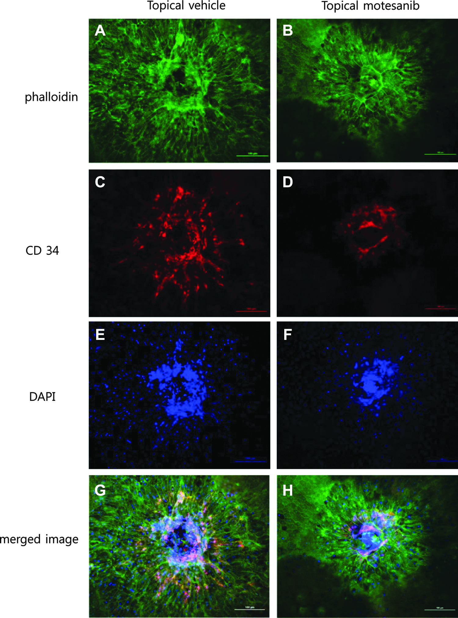
Immunofluorescence staining of CNV lesions 14 days after laser photocoagulation. Choroidal flat mounts were fluorescently labeled with F-actin-specific marker phalloidin (green channel), endothelial cell marker CD34 (red channel), and nuclear marker DAPI (blue channel). CNV lesions in topical vehicle-treated eye
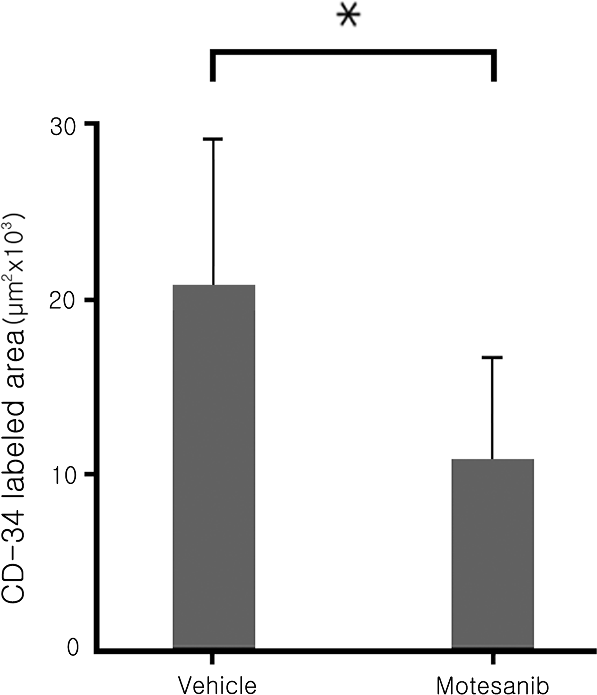
The size of endothelial cell marker CD34-labeled area of topical motesanib-treated eye was smaller than that of the vehicle-treated eye (*P=0.001).
Expression of p-ERK1/2 in topical motesanib-treated eye
The expression level of p-ERK1/2 relative to total ERK1/2 three days after laser injury was decreased in eyes treated with motesanib compared with eyes treated with vehicle (Fig. 4).
Histological evaluation of CNV
Figure 5 shows representative hematoxylin–eosin-stained sections from CNV lesions 14 days after laser application. Proliferative fibrovascular membranes and PRE clumping were observed in the central areas of laser lesions (Fig. 5A). The thickness of CNV lesions in eyes treated topically with motesanib was thinner than in vehicle-treated eyes (Fig. 5B).
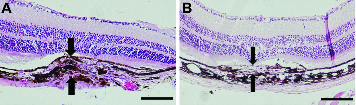
Effect of topical motesanib on CNV development 14 days after laser induction of CNV. Light micrographs of hematoxylin–eosin-stained sections of the CNV lesions in
Discussion
CNV is the major cause of vision loss associated with AMD. Although intravitreal anti-VEGF therapy currently remains the mainstay of CNV treatment, the limitation of this type of treatment is the necessity for frequent intraocular injections, which always carries the risk of side effects. Therefore, a topical eye drop that can effectively inhibit CNV is preferable, and the data of our experiments showed that topical administration of motesanib could inhibit CNV in the experimental CNV model.
Multiple kinases are involved in angiogenic processes, and their antagonism of angiogenesis-related receptor tyrosine kinase (RTK) is an important cancer treatment strategy. 19 Such pharmacological approaches using RTK have also been applied to inhibition of CNV. Many investigators have reported that orally or intravitreally administered RTK inhibitors have therapeutic potential in CNV treatment by inhibiting RTK-related signaling pathways.20,21
Motesanib is a multiple receptor TKI that selectively targets and inhibits VEGFR-1 (IC50=2 nM), VEGFR-2 (IC50=3 nM), VEGFR-3 (IC50=6 nM), PDGFR (IC50=84 nM), and c-Kit (IC50=8 nM), thereby inhibiting angiogenesis. Oral administration of motesanib potently inhibited VEGF-induced angiogenesis in the rat corneal model and induced regression of A431 tumor xenografts. 22 This drug is currently being studied in clinical trials for the treatment of cancer and has displayed antiangiogenic and antitumor activity in patients with advanced solid tumors, including breast and thyroid cancers. Recently, a pivotal Phase 3 trial that evaluated motesanib administered together with paclitaxel and carboplatin to 1,090 patients with advanced nonsquamous nonsmall cell lung cancer did not meet its main goal of improving survival.
To our knowledge, this is the first study to demonstrate the antiangiogenic activity of motesanib in a mouse CNV model using a topical administration strategy. Our experimental model does not exactly represent every aspect of human AMD. However, this model does imitate human CNV in that the laser disrupts the Bruch's membrane and neovascularization from choroidal vessels follows. 23 Fluorescence angiographic analysis showed that the average signal intensity score decreased by 31% in motesanib-treated eyes compared with vehicle-treated eyes. CD34 is expressed in the endothelial cells of blood vessels.24–26 In choroidal flat mounts using CD34, topically applied motesanib significantly suppressed the development of laser-induced CNV. The motesanib-treated eyes displayed a 44.6% reduction in area compared with vehicle-treated eyes. Additionally, we demonstrated that motesanib decreased ERK1/2 activation, which could indicate an attenuated VEGF/VEGFR pathway. Taken together, these findings suggest that topical motesanib treatment inhibited CNV development.
Similarly, another RTK inhibitor, pazopanib, was reported to significantly reduce CNV via topical application as well as systemic administration.27–30 Topical pazopanib treatment in rats with induced CNV reduced VEGF release in retina and decreased VEGF-induced signaling and chemotaxis. 29 Clinically, pazopanib eye drop was administered topically to patients with subfoveal CNV secondary to AMD and resulted in improvement in best corrected visual acuity. 29 However, improvement in macular thickness was found only in subset of patients with the CFH Y402H TT genotype. To explain this perplexing result, Danis et al. proposed that the study drug could have a favorable effect on neuronal/photoreceptor function without influencing the exudative process. 30
Previous study showed suppression of VEGF-induced ERK1/2 phosporylation in cultured choroidal endothelial cells by motesanib. 29 In line with this result, we demonstrated that motesanib has an inhibitory effect on CNV formation, suppressing phosphorylation of ERK1/2 in vivo (Fig. 4).
The most suitable method of drug delivery is theoretically topical administration because this route can avoid potential ocular and systemic side effects. Potential side effects of systemic motesanib include hypertension, fatigue, anorexia, diarrhea, and nausea. 5 Moreover, angiogenesis is required for vascular maintenance under normal conditions. It may be unsafe to administer multiple TKI systemically in older patients. Although current intravitreal injections are an effective and direct approach to delivery of drugs to the posterior segment, frequent drug administration via this route can lead to ocular discomfort, cataract, endophthalmitis, retinal detachment, and increased intraocular pressure. In clinical practice, topical eye drops have not been approved to treat ocular diseases in the posterior segment of the eye, and it was thought that topical ocular medications would not reach the posterior segment. However, according to recent studies, trans-scleral delivery allows drug transfer to the posterior segment because the sclera has a larger surface area than the cornea, a high degree of hydration, and a low number of cells, and is permeable even to large-molecular-weight compounds.31,32
The limitations of the current investigation should be noted. One is that the size of mouse eye is much smaller than human eye, and it is not plausible to extrapolate the current result directly to human retina. A second limitation is using CD34 as an endothelial marker. As CD34 can stain immune cells as well as endothelial cells, some immune cells in CNV lesions could also be stained. However, we found crowding vessels of CNV lesion in Fig. 5.
In summary, topical motesanib application suppressed CNV, and none of the experimental animals showed any visible side effects. Taken together, our findings suggest that topical motesanib may be a strong candidate for the treatment of CNV in clinical practice. Future studies of the optimal dosage, and combinations with/without bevacizumab and ranibizumab are needed.
Footnotes
Acknowledgments
This work was supported by a Clinical Research Institute Grant funded by the Catholic University of Korea, Daejeon St. Mary's Hospital. None of the authors has any financial interest in any of the products described.
Author Disclosure Statement
No financial conflict of interest exists between the authors.
References
Supplementary Material
Please find the following supplemental material available below.
For Open Access articles published under a Creative Commons License, all supplemental material carries the same license as the article it is associated with.
For non-Open Access articles published, all supplemental material carries a non-exclusive license, and permission requests for re-use of supplemental material or any part of supplemental material shall be sent directly to the copyright owner as specified in the copyright notice associated with the article.