
Abstract
Abstract
Purpose:
The effects of cidofovir were investigated against canine herpesvirus-1 (CHV-1) in vitro and in dogs with experimentally induced recurrent ocular CHV-1 infection, a host-adapted pathogen animal model of ocular herpes simplex virus-1 (HSV-1) infection.
Methods:
The cidofovir EC50 was determined for CHV-1 and HSV-1. A randomized, masked vehicle-controlled trial was performed using beagles with experimentally induced recurrent ocular CHV-1 infection. Dogs received 1 drop of 0.5% cidofovir solution or 0.9% sodium chloride solution (vehicle) in both eyes 2 times daily for 14 days. Dogs were monitored at intervals for 30 days by a clinical ophthalmic examination, in vivo confocal microscopy of the cornea and conjunctiva, ocular sample CHV-1 polymerase chain reaction assay, hemogram, and serum biochemistry panel. Clinical ocular disease scores were calculated and infiltrating leukocytes detected by in vivo confocal microscopy quantified.
Results:
Cidofovir displayed similar in vitro antiviral activity against CHV-1 and HSV-1. Clinical ocular disease scores were significantly higher in the cidofovir group compared to the vehicle group. Marked conjunctival pigmentation and blepharitis were also detected in the cidofovir group, but not the vehicle group. Conjunctival and corneal leukocyte infiltration scores determined by in vivo confocal microscopy were significantly higher in the cidofovir group. Dogs administered cidofovir had significantly reduced durations of ocular viral shedding compared to the vehicle group. Hemogram and serum biochemistry panel values were unremarkable.
Conclusions:
Twice-daily application of topical 0.5% cidofovir ophthalmic solution reduced the duration of ocular viral shedding in dogs with experimentally induced recurrent ocular CHV-1 infection, but was associated with local ocular toxicity.
Introduction
C
Following primary ocular CHV-1 infection, viral latency is established in ocular sensory ganglia.8,9 Latent CHV-1 can be reactivated in some dogs under experimental conditions by the systemic administration of an immunosuppressive dosage of corticosteroids.10,11 Experimental recurrent ocular CHV-1 infection, in otherwise healthy mature dogs, is most commonly associated with a clinically mild-to-moderate self-limiting conjunctivitis, brief period of ocular viral shedding, and conjunctival and corneal leukocyte infiltrates, as detected by in vivo confocal microscopy.10,12 Experimental ocular CHV-1 infection in dogs represents a unique host-adapted pathogen, large animal model of HSV-1 infection.13,14
Cidofovir is an acyclic nucleoside monophosphate analog of cytosine that selectively inhibits viral DNA polymerase. 15 Cidofovir exhibits a broad-spectrum in vitro antiviral activity against many DNA viruses, including a variety of adenovirus and herpesvirus ocular pathogens.16,17 Cidofovir is currently commercially available only as an intravenous compound that is labeled for the treatment of cytomegalovirus retinitis; however, it has been suggested that there could be additional clinical applications for topical ophthalmic cidofovir formulations in the treatment of viral keratoconjunctivitis.18,19 There are clinical reports of ophthalmic cidofovir solutions being used for the therapy of ocular surface viral infections, including CHV-1 keratitis in dogs, feline herpesvirus-1 keratoconjunctivitis in cats, and adenovirus keratoconjunctivitis in humans; however, there are limited experimental studies in host-adapted pathogen animal models supporting this practice.20–22 The majority of previous experimental studies evaluating topical cidofovir therapy were performed using nonhost-adapted pathogen animal models of viral infection, and aspects of viral behavior may differ in these models from infections in the natural hosts for which the virus is adapted.23–31
Cidofovir is an appealing topical antiviral agent as the long intracellular half-life of its active metabolites may provide clinical efficacy with relatively infrequent dosing schedules. 32 The purpose of the present study was to determine the effects of twice-daily topical application of 0.5% cidofovir ophthalmic solution in dogs with experimentally induced recurrent ocular CHV-1 infection. Topical ocular cidofovir therapy has not been previously evaluated in a large animal model of recurrent ocular HSV-1 infection. Therapeutic effects in the present study were assessed using clinical, virological, and in vivo confocal microscopic outcome measures.
Methods
In vitro efficacy of cidofovir against CHV-1
Madin–Darby Canine Kidney or Vero cells were plated into 96-well plates at 1 × 10⁴ cells/well and maintained in Dulbecco's minimal essential medium with 1 g/L glucose,
Animals and induction of latent CHV-1 infection
All protocols were approved by the Animal Care and Use Committee of Cornell University and were conducted in accordance with the ARVO Statement for the Use of Animals in Ophthalmic and Vision Research. Ten 2.5-year-old, specific pathogen-free laboratory beagles were used. Dogs were maintained individually in runs, and direct contact between dogs was prevented during the study. Strict bioisolation was maintained throughout the study for all personnel in contact with dogs, and the dogs were maintained in the isolation facilities for the duration of the study. Dogs were acclimated to housing facilities for a minimum of 12 weeks before the beginning of the study.
Latent CHV-1 infection was experimentally induced in each dog by topical ocular inoculation 12 months before the beginning of the study, using the ocular drop method as previously described. 33 Briefly, dogs that were seronegative for CHV-1 were topically inoculated in both eyes with 2 × 10⁵ TCID50 of a field strain of CHV-1, isolated from corneal samples of a dog with dendritic ulcerative keratitis treated at the Cornell University College of Veterinary Medicine Hospital for Animals (Ithaca, NY), followed immediately by gentle manual massage of the closed eyelids for 60 s. To confirm the development of primary ocular CHV-1 infection, dogs were monitored for 30 days after inoculation by a clinical ophthalmic examination performed at 3-day intervals and CHV-1 virus neutralization titers 12 performed at 15-day intervals.
Study design
A randomized, masked vehicle-controlled study design was used. Total study duration was 30 days. Recurrent ocular CHV-1 infection was experimentally induced as previously described10,12 in all 10 dogs by administration of systemic prednisolone (3.0 mg/kg PO q24 h) for 7 consecutive days beginning on study day 1. Study dogs were randomly assigned into 1 of 2 groups: cidofovir group (n = 5 dogs) or vehicle group (n = 5 dogs). Dogs received either 1 drop of 0.5% cidofovir ophthalmic solution or 0.9% sodium chloride solution (vehicle) in both eyes 2 times daily for 14 days beginning on study day 4. Study agents were administered at 8:00 am and 8:00 pm. Clinical ophthalmic examinations, in vivo confocal microscopic examinations, ocular sample CHV-1 polymerase chain reaction (PCR) assays, hemograms, and serum biochemistry panels were performed at intervals.
Study agents
Study agents were prepared by a licensed pharmacist in a manner that masked the administrator and investigators. The cidofovir 0.5% ophthalmic solution was prepared from cidofovir 75 mg/mL intravenous solution (Vistide®; Gilead Sciences, Foster City, CA) and sterile 0.9% sodium chloride solution using the aseptic technique under a laminar flow hood. Aliquots of 2.5 mL cidofovir and vehicle solutions were placed in identical sterile plastic drop containers and stored at 4°C until use.
Clinical examination and sample collection
Complete physical and ophthalmic examinations, including slit-lamp biomicroscopy (Kowa SL-15; Kowa Co, Tokyo, Japan), indirect ophthalmoscopy, Schirmer I tear testing (Schirmer tear test standardized sterile strips; Intervet, Inc., Summit, NJ), and corneal application of lissamine green stain (Lissamine green ophthalmic strips; Contacare Ophthalmics and Diagnostics, Vadodara, Gujarat, India), were performed on each dog before study initiation. Ophthalmic examination of both eyes with slit-lamp biomicroscopy, before and after application of lissamine green stain, was then performed at 2-day intervals for the duration of the study. A modified ocular surface disease clinical scoring system was used to quantify ophthalmic examination findings. 33 The following ocular parameters were scored: blepharospasm, ocular discharge, conjunctival hyperemia, chemosis, conjunctival ulceration, and corneal epithelial ulceration. All parameters, except conjunctival ulceration and corneal epithelial ulceration, were scored: 0, none; 1, mild; 2, moderate; 3, severe. Conjunctival and corneal epithelial ulceration were scored: 0, none; 1, punctate ulcerations; 2, ≥1 linear or dendritic ulcerations; 3, geographic ulcerations. Both eyes were scored and a single cumulative ocular surface disease clinical score was calculated for each dog on each examination day. When clinical disease was not symmetrical between eyes, the dog was assigned the score from the eye with the highest clinical score for the statistical analysis. The minimum total clinical score possible with the system was 0 and the maximum total clinical score possible was 18. Schirmer I tear tests were repeated on study days 15 and 30 before ophthalmic examination or study agent administration. Ophthalmic examinations were repeated in all dogs after the conclusion of the study at 30-day intervals for 4 months to monitor for resolution of any ophthalmic lesions that persisted after the initial study period.
Samples for hemograms and serum biochemistry panels were collected by peripheral venipuncture from each dog immediately before the study and on study days 15 and 30. Following clinical ocular disease scoring, but before lissamine green application, conjunctival swab specimens were collected from both eyes for CHV-1 PCR assay by brushing sterile polyester-tipped swabs across the conjunctival fornices. Ocular swab samples were collected at 3-day intervals for the duration of the study and they were stored in sterile tubes at −80°C until analysis.
In vivo confocal microscopic examination
In vivo confocal microscopic examinations of the cornea and conjunctiva were performed with a Heidelberg Retina Tomograph II and Rostock Cornea Module (Heidelberg Engineering, Heidelberg, Germany) using a 63× objective (Carl Zeiss Meditec AG, Jena, Germany) and 400 μm field lens. Confocal microscopic examinations were performed on both eyes of each dog immediately before study initiation and on study day 10. Examination of both eyes was performed after the application of a single drop of topical anesthetic (proparacaine 0.5% ophthalmic solution). Several drops of contact gel (GenTeal tear gel; Novartis Pharmaceuticals Corp, East Hanover, NJ) were applied to the front of the microscope lens and ocular surface. A sterile, single-use polymethyl methacrylate cap (TomoCap; Heidelberg Engineering, Heidelberg, Germany) mounted on the microscope lens was positioned perpendicular to, and in slight contact with, the ocular surface. The polymethyl methacrylate caps were changed between each dog examined.
Multipoint corneal and conjunctival imaging was performed with a combination of manual and automated image acquisition modes. Following examination, digitized images were analyzed for pathology. Images of standardized anatomic locations were acquired and used for leukocyte infiltrate scoring by a masked investigator. For keratitis scores, images of the basal corneal epithelium 1.0 mm anterior to the 12 o'clock limbal position were collected. For conjunctivitis scores, images of the surface conjunctival epithelium 1.0 mm posterior to the 12 o'clock limbal position were collected. Leukocytes were quantified (cells/mm2 of tissue) in 3 corneal images from each eye using semiautomated cell counting software (Rostock Cornea Module Software Version 1.3.3; Heidelberg Engineering, Heidelberg, Germany). Leukocytes were also quantified in 3 conjunctival images from each eye; however, distinct leukocyte cellular borders were more difficult to distinguish in the conjunctival images, and therefore, a semiquantitative scoring system was used to describe conjunctival infiltrates: 0, absent; 1, mild; 2, moderate; 3, marked.
CHV-1 real-time PCR analysis
CHV-1 real-time PCR was performed using CHV-specific primers and probes as described previously. 12 Samples were held at −80°C until processed. DNA was extracted using a 96-well magnetic bead-based process (Mag Max 96; Life Technologies) with a commercial DNA extraction kit (AM 1840; Life Technologies).
Statistical analysis
The Student's t-test was used to compare the in vitro antiviral activity of cidofovir against CHV-1 and HSV-1. The duration and frequency of viral shedding as detected by CHV-1 PCR assay were compared between the vehicle and cidofovir groups using the Student's t-test and Fischer's exact test, respectively. Regression analysis was performed to assess the effects of study group on clinical ocular disease scores. The Student's t-test was used to compare Schirmer tear test values from study days 15 and 30 with baseline values in each study group. Mean keratitis and conjunctivitis confocal microscopy scores were compared between the vehicle and cidofovir group using the Student's t-test. The analysis was performed using a software package (Statistix 10.0; Analytical Software, Tallahassee, FL) and a P ≤ 0.05 was considered significant for all comparisons.
Results
In vitro efficacy of cidofovir against CHV-1
Twofold serial dilutions of cidofovir were made to evaluate the antiviral effects of cidofovir against CHV-1 in vitro. Based on these results, an EC50 of 4.4 ± 2.3 μM was calculated (Fig. 1). For comparative purposes, we repeated the experiments using HSV-1, for which the in vitro antiviral effect of cidofovir has been reported previously. 35 For HSV-1, an EC50 of 6.4 ± 2.1 μM was calculated, which was not statistically significantly different from CHV-1 (P = 0.32; Fig. 1), indicating that the antiviral effect of cidofovir is comparable for these 2 alphaherpesviruses.
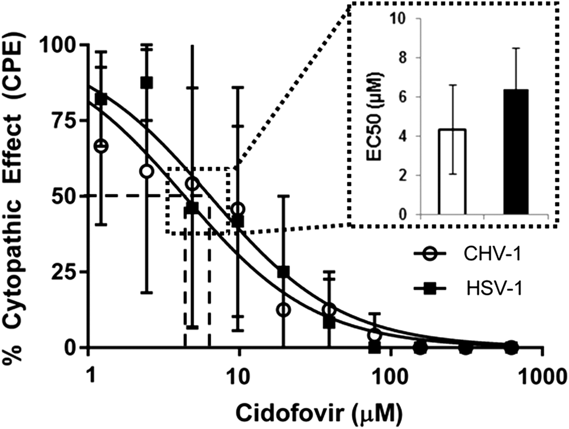
EC50 determination of cidofovir for CHV-1 and HSV-1. Cells were infected with CHV-1 (white circles and bars) or HSV-1 (black squares and bars), treated with cidofovir at varying concentrations, and fixed after CPE was observed in the infected nontreated controls. EC50 values were calculated based on the percent of wells showing CPE. Data are expressed as the mean ± standard deviation of at least 3 independent experiments. CHV-1, canine herpesvirus-1; CPE, cytopathic effect; HSV-1, herpes simplex virus-1.
Primary infection, prestudy, and baseline examinations
Following ocular CHV-1 inoculation, all study dogs developed bilateral ocular disease typical of primary ocular CHV-1 infection. 33 The ocular disease was characterized by bilateral conjunctival hyperemia, chemosis, and mucopurulent ocular discharge that slowly resolved over a 2–3-week period in each dog. All dogs seroconverted and had detectable CHV-1 virus neutralization antibody titers by 15 days after viral inoculation.
No abnormalities were detected in any dog during the physical and ophthalmic examination performed immediately before the initiation of the study. Schirmer I tear test results were within reference range values (>15 mm/min) in both eyes of all study dogs. Corneal and conjunctival retention of lissamine green stain was not present. Results of hemograms and serum biochemistry panels were unremarkable for each dog. No conjunctival or corneal abnormalities, including leukocyte infiltrates, were detected by in vivo confocal microscopy.
Clinical ophthalmic examinations
Recurrent ocular CHV-1 infection was detected by clinical examination in all dogs from the cidofovir and vehicle groups by study day 6. The clinical ocular disease was characterized by intermittent blepharospasm, conjunctival hyperemia, chemosis, and ocular discharge (epiphora or mucopurulent discharge). The clinical ocular disease findings were typically symmetrical between both eyes of individual dogs on specific examination days. Lissamine green-positive conjunctival ulcerations and petechial conjunctival hemorrhages were detected in 2 dogs (1 from each study group) between study days 6 and 10. No corneal epithelial ulcerations were detected by lissamine green application in any dog during the study.
In the vehicle group, ocular disease was clinically characterized by mild-to-moderate conjunctivitis. Ocular disease scores in the vehicle group peaked on study days 6 and 8 (mean ± standard deviation: 3.6 ± 0.5 and 3.6 ± 0.8, respectively) and then rapidly declined, returning to near baseline values by study day 16 (Fig. 2). In the cidofovir group, ocular disease was clinically characterized by relatively severe conjunctivitis and ocular disease scores peaked on study days 14 and 16 (mean ± standard deviation: 5.6 ± 1.4 and 5.6 ± 2.6, respectively) and remained elevated from baseline values until study conclusion (Figs. 2 and 3). Regression analysis revealed significantly (P ≤ 0.001) higher clinical scores in the cidofovir group relative to the vehicle group after study day 10 (Fig. 2). On average, the mean daily group score for the cidofovir group was 2.96 higher (in absolute clinical score value) than the vehicle group between study days 10 and 30.
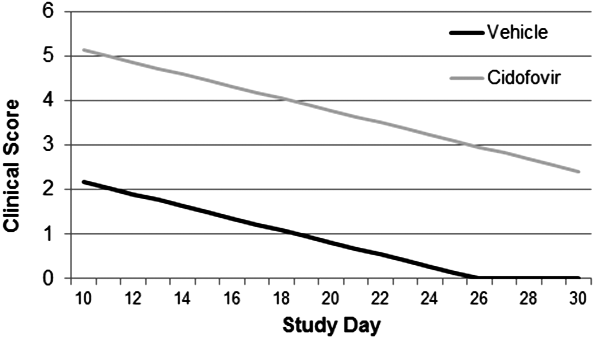
Plot of the cumulative ocular surface disease clinical score regression lines for dogs with experimentally induced recurrent ocular CHV-1 infection. Recurrent ocular CHV-1 infection was experimentally induced by administration of systemic prednisolone for 7 consecutive days beginning on study day 1. Dogs were administered either 0.5% cidofovir solution or vehicle solution for 14 days beginning on study day 4, and clinical scores were calculated every 2 days for 30 days. Regression analysis revealed significantly (P ≤ 0.001) higher clinical scores in the cidofovir group relative to the vehicle group after study day 10.

Clinical photographs of dogs with experimentally induced recurrent ocular CHV-1 infection and administered 0.5% cidofovir solution:
In addition to conjunctivitis, conjunctival pigmentation (n = 5 dogs) and ulcerative blepharitis (n = 3 dogs) were detected in the cidofovir group. Conjunctival pigmentation was first noted clinically on study day 10 as multifocal bulbar conjunctival pigment and then rapidly progressed to diffuse bulbar, palpebral, and nictitans conjunctival pigmentation in all affected dogs by study day 30 (Fig. 3). Ulcerative blepharitis of the inferior eyelid was first detected on study day 14 and persisted in some dogs until study day 26. Conjunctival pigmentation and ulcerative blepharitis were not present at any time in dogs of the vehicle group. Although it became slightly less dense, the conjunctival pigmentation persisted in all dogs in the cidofovir group during the ophthalmic examinations performed for 4 months following study conclusion.
Schirmer I tear test values (mean ± standard deviation in mm/min) on study days 0, 15, and 30 for dogs in the vehicle group were as follows: 17.5 ± 1.6, 16.7 ± 1.6, and 17.2 ± 2.9, respectively. Schirmer I tear test values (mean ± standard deviation in mm/min) on study day 0, 15, and 30 for dogs in the cidofovir group were as follows: 20.5 ± 1.3, 15.0 ± 2.8, and 21.4 ± 2.2, respectively. There were no significant changes in the reflex aqueous tear production relative to baseline values for dogs in the vehicle group; however, Schirmer I tear test values were significantly (P ≤ 0.0001) reduced on study day 15 relative to study day 0 for dogs in the cidofovir group. The reflex aqueous tear production had returned to baseline levels on study day 30 in the cidofovir group.
Ocular viral shedding
Ocular viral shedding was detected by CHV-1 PCR in study dogs between study days 6 and 15 (Fig. 4). In all dogs that shed virus for multiple days, the assays were positive on consecutive sampling days. In the vehicle group, CHV-1 PCR assays were positive for 12 samples collected from 4 separate sampling days and 5 individual dogs had one or more positive assays. The mean (±standard deviation) number of shedding days for dogs in the vehicle group was 2.4 ± 1.2 days and the mean (±standard deviation) shedding duration was 5.2 ± 3.6 days. Viral shedding durations for individual dogs were 1 day for 2 dogs, 7 days for 2 dogs, and 10 days for 1 dog. In the cidofovir group, CHV-1 PCR assays were positive for 3 samples collected from 2 separate sampling days and 2 individual dogs had one or more positive assays. The mean (±standard deviation) number of shedding days for dogs in the cidofovir group was 0.6 ± 0.8 days and the mean (±standard deviation) shedding duration was 1.0 ± 1.5 days. Viral shedding durations for individual dogs were 1 day for 1 dog and 4 days for 1 dog. The frequency of viral shedding was not significantly different between study groups, but the duration of viral shedding was significantly (P ≤ 0.04) shorter in the cidofovir group relative to the vehicle group.
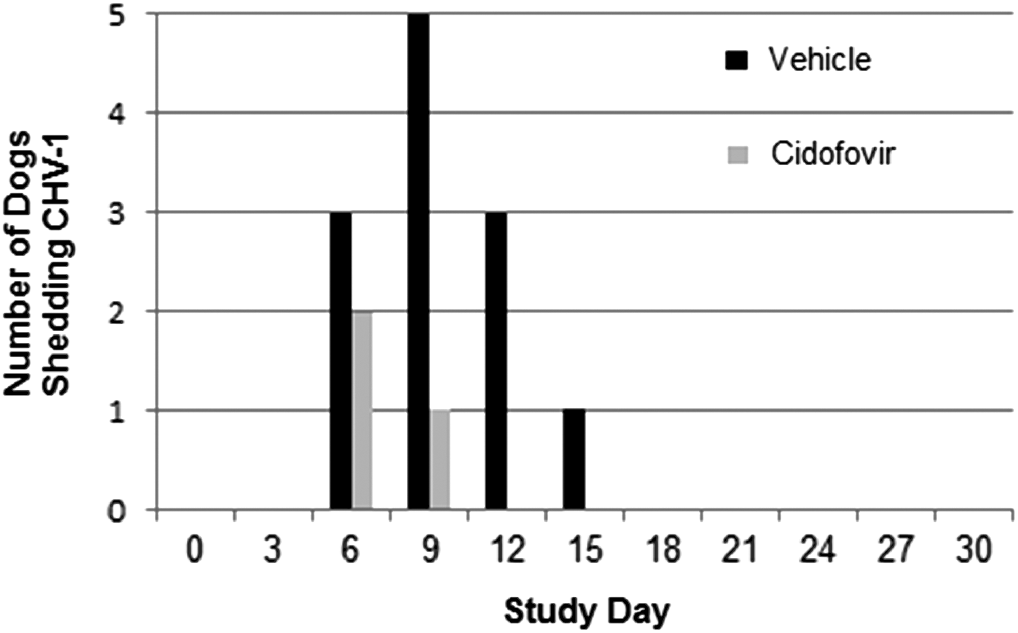
Number of study dogs with ocular CHV-1 shedding as detected by polymerase chain reaction assay. Dogs were administered either 0.5% cidofovir solution (n = 5 dogs) or vehicle solution (n = 5 dogs) for 14 days beginning on study day 4, and ocular viral shedding was evaluated every 3 days for 30 days.
In vivo ocular confocal microscopy examinations
Variable amounts of epithelial leukocyte infiltration in the bulbar conjunctiva and peripheral cornea were detected by in vivo confocal microscopy in all dogs on study day 10. The mean ± standard deviation in vivo confocal microscopy keratitis score from study day 10 was 971 ± 402 leukocytes/mm2 for dogs in the cidofovir group and 145 ± 116 leukocytes/mm2 for dogs in the vehicle group (Fig. 5). The mean ± standard deviation in vivo confocal microscopy conjunctivitis score from study day 10 was 2.5 ± 0.5 for dogs in the cidofovir group and 1.2 ± 0.6 for dogs in the vehicle group (Fig. 5). The keratitis (P ≤ 0.0001) and conjunctivitis (P ≤ 0.004) scores were significantly higher in the cidofovir group relative to the vehicle group.
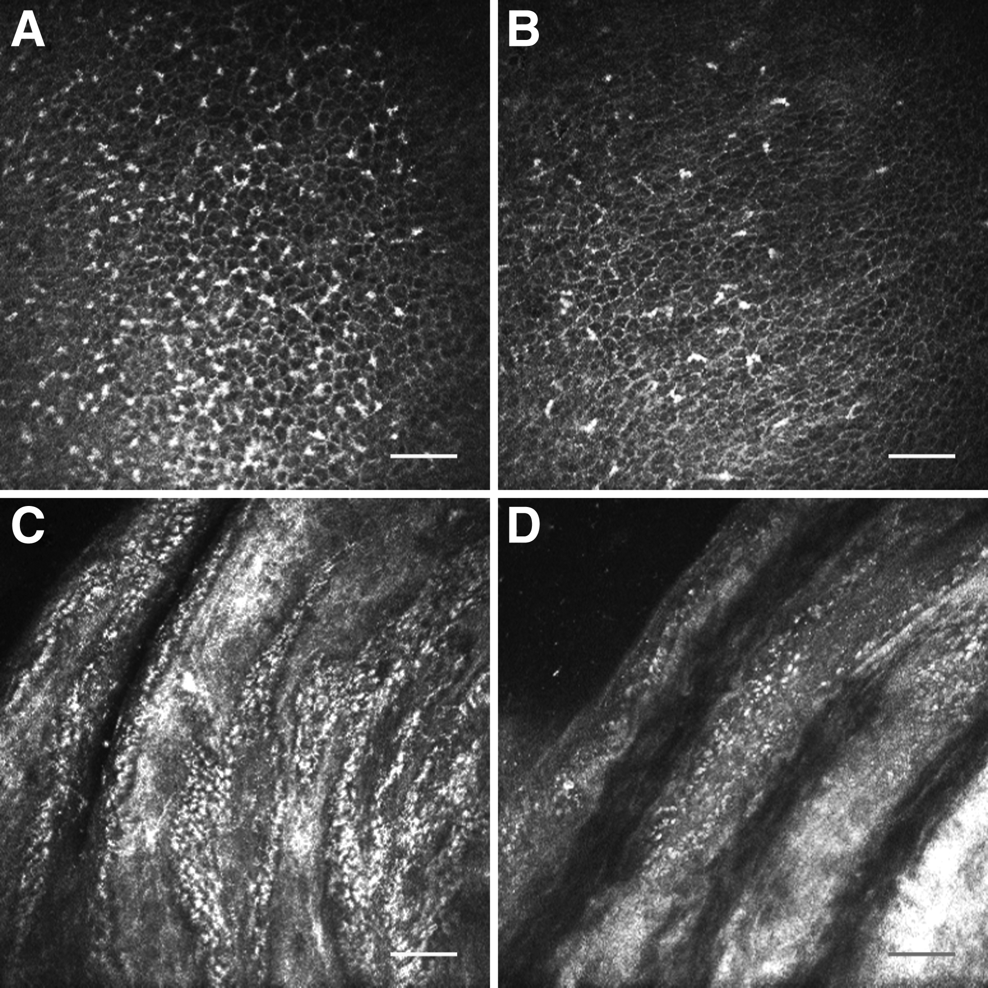
Representative in vivo confocal photomicrographs of dogs with experimentally induced recurrent ocular CHV-1 infection. Dogs were administered either 0.5% cidofovir solution
General diagnostic assays and examinations
Results of hemograms and serum biochemistry panels collected on study days 15 and 30 were unremarkable for each dog. No overt systemic clinical abnormalities were observed in any dog at any time during the study.
Discussion
Cidofovir displayed in vitro antiviral activity against CHV-1 comparable to its activity against HSV-1. When evaluated in dogs with experimentally induced recurrent ocular CHV-1 infection, twice-daily ocular application of 0.5% cidofovir ophthalmic solution effectively reduced the duration of ocular viral shedding, but was associated with marked local ocular toxicity. The ocular toxicity was characterized by exacerbation of conjunctivitis, ulcerative blepharitis, and extensive conjunctival pigmentation. Similar lesions were not found in the vehicle control group where only self-limiting mild-to-moderate conjunctivitis, typical of experimentally induced recrudescent CHV-1 ocular disease, was detected in the dogs.
Although the duration of viral shedding was reduced, CHV-1 was detected in ocular samples of 40% of study dogs administered cidofovir (compared to 100% of dogs administered vehicle). Previous descriptions of experimental recurrent ocular CHV-1 infection reported detection of ocular viral shedding in 37.5%–50% of dogs; however, these studies utilized sampling schedules with longer intervals between collections than the current study.10,12 As experimentally induced ocular CHV-1 shedding is brief in most dogs, the design of the present study may have resulted in the detection of more dogs shedding virus. The study results suggest that initiation of cidofovir therapy early in the disease course can reduce, but not eliminate, ocular viral shedding. Alternatively, the differences in viral shedding detected between the cidofovir and vehicle groups might reflect natural variation between the dogs.
Ocular toxicity is reported in humans associated with the clinical use of topical ophthalmic cidofovir formulations. In a human clinical trial performed to determine the efficacy of a 4–10 times daily dosage regimen of cidofovir 1.0% ophthalmic solution for the treatment of acute adenoviral keratoconjunctivitis, severe local ocular toxicity was observed in a dose-dependent manner. 22 In these patients, severe conjunctivitis, blepharitis, and conjunctival pseudomembranes developed and necessitated the discontinuation of the cidofovir administration in many study subjects and the premature cessation of patient recruitment. 22 When cidofovir was used in a human for the treatment of conjunctival papillomatosis and squamous cell carcinoma, prolonged application of a topical 0.25% ophthalmic solution was associated with cicatricial changes of the inferior punctum. 36 In addition to toxicity associated with topical ophthalmic administration, topical gel formulations of cidofovir used in the treatment of genital and mucocutaneous herpes simplex infection were reported to cause frequent application-site reactions, including ulcerative dermatitis.37,38
Cidofovir has displayed therapeutic efficacy, without overt systemic or local toxicity, in several animal studies evaluating experimental primary ocular herpetic infections. In cats with experimental primary ocular feline herpesvirus-1 infection, twice-daily application of 0.5% cidofovir ophthalmic solution significantly decreased ocular viral shedding loads and the severity of clinical disease compared to a study group treated with placebo. 23 In New Zealand rabbits with experimental HSV-1 keratitis, 7-day treatment courses of both 0.5% and 1.0% cidofovir solutions administered twice daily reduced ocular viral titers, number of eyes shedding HSV-1, duration of viral shedding, keratitis scores, and time to keratitis resolution. 24 Six times-daily application of a 0.2% cidofovir solution for 4 days, followed by 4 times daily for 6 days, in a New Zealand rabbit HSV-1 keratitis model was associated with reduced corneal ulcer healing times, lower ocular viral titers, and shortened HSV-1 shedding durations relative to vehicle. 25 In a separate rabbit study, administration of 0.2% cidofovir solution once or twice daily reduced the severity of experimental HSV-1 keratitis. 26 Cidofovir 0.2% solution administered 1, 3, or 9 times daily for 5 days was effective in healing corneal epithelial disease during experimental herpetic keratitis in rabbits. 27 Cidofovir 0.5% and 1.0% solutions administered twice daily were also effective in preventing HSV-1 stromal keratitis in experimentally infected rabbits. 28
In contrast to reports where cidofovir was not associated with toxic effects, adverse ocular reactions attributed to topical ocular application of cidofovir are described in several other experimental animal studies. In healthy New Zealand white rabbits, the use of cidofovir ophthalmic solution was associated with blepharitis, conjunctivitis, blockage of the nasolacrimal duct, and superficial punctate keratitis in a dose-dependent manner. 29 When administered in concentrations ranging from 1.0% to 5.0% for 10 days, local ocular toxicity was consistently observed when total dosages of ≥10 mg were applied. 29 In another study using healthy Japanese albino rabbits, 1.0% cidofovir solution was administered 4 times daily for 14 days and associated with conjunctival hyperemia, eyelid erythema, and significant narrowing of the lacrimal canaliculus, as measured by ultrasonography. 30 Histopathologic evaluation of the rabbits that received cidofovir revealed eosinophilic inflammation of the conjunctiva and lacrimal canaliculus and markedly reduced conjunctival epithelial and goblet cell numbers. 30 In guinea pigs with experimentally induced superficial corneal defects, dose-dependent ocular toxicity was induced by once-daily application of 0.04%, 0.4%, and 4.0% cidofovir ophthalmic solution for 10 days. 31 Histopathological lesions in the guinea pigs included corneal leukocyte infiltrates, corneal vascularization, corneal edema, bullous keratopathy, and iridocyclitis. 31
Significantly reduced aqueous tear production relative to baseline values was measured in the present study on study day 15 in the dogs of the cidofovir group. This may have contributed to the observed ocular and adnexal pathology in the dogs, but was unlikely to be the primary pathophysiologic mechanism as the mean Schirmer tear test value remained within reported canine reference range values and no dogs had values <10 mm/min in either eye.39–41 In the study dogs, transient reduced aqueous tear production may have occurred secondary to lacrimal ductal occlusion associated with severe conjunctivitis or a direct pharmacological effect on the lacrimal gland. 42 A previous histologic study of topical ophthalmic cidofovir toxicity in rabbits demonstrated eosinophilic inflammation in ocular tissues without evidence of direct cytotoxicity, suggesting an allergic mechanism for the local ocular reaction. 30
The apparently successful clinical use of cidofovir ophthalmic solution is reported for a dog with naturally acquired CHV-1 dendritic ulcerative keratitis. 20 In this report, 0.5% cidofovir ophthalmic solution was applied topically twice daily for 4 weeks to both eyes of a dog with unilateral CHV-1 dendritic corneal ulcers. The corneal ulcerations healed after 1 week of antiviral therapy. Although adverse ocular effects specifically attributed to cidofovir administration were not clinically appreciated, the dog did manifest with persistent conjunctival hyperemia and ocular discharge of both eyes during cidofovir treatment. 20 These findings may represent a local ocular toxicity similar to the dogs of this report. Alternatively, these persistent ocular abnormalities may have been unrelated to cidofovir administration and dogs may display individual levels of susceptibility to the local ocular toxic effects of cidofovir similar to that reported for humans. 22
Cidofovir displayed similar in vitro antiviral activity against both CHV-1 and HSV-1. Twice-daily application of topical 0.5% cidofovir ophthalmic solution reduced the duration of ocular viral shedding in dogs with experimentally induced recurrent ocular CHV-1 infection, but was associated with marked local ocular toxicity and increased corneoconjunctival leukocyte infiltrates, as determined by in vivo confocal microscopy. Future studies are required to determine if there are effective, but clinically tolerable, concentrations or formulations of cidofovir for use in dogs and other species with ocular surface viral infections.
Footnotes
Acknowledgment
This study was partially funded by the Merial Veterinary Scholars Program.
Author Disclosure Statement
No competing financial interests exist.