
Abstract
Abstract
Purpose:
Biohcly is a type of acidic nanoclustered water shown to exert an antimicrobial effect and play a role in the tissue-healing process in previous experiments. This study was performed to evaluate the in vivo effects of Biohcly treatment on mechanical corneal wound healing.
Methods:
Rabbit cornea “acute” mechanical wounds were created with an 8-mm hydraulic pressure trephine. The corneal wounds were treated with either Biohcly (left eye) or sterile saline (right eye) 4 times per day. To determine the state of the wounds, the wound healing rate (WHR), inflammatory index (IF), neovascularization, and anterior segment optical coherence tomography (AS-OCT) were evaluated. The expression of inflammatory factors was examined by quantitative real-time reverse transcriptional polymerase chain reaction, enzyme-linked immunosorbent assay, and immunohistochemical staining.
Results:
Biohcly was more effective than saline in healing corneal ulcers as demonstrated by the WHR calculated on the 9th and 14th days after surgery (P < 0.05). The histological and AS-OCT examinations revealed better regeneration and reduced corneal scars in the Biohcly-treated corneas. No neovascularization formed in the corneas treated with Biohcly, whereas 25% of the saline-treated wounds developed this complication. In addition, the IF scores of the Biohcly-treated wounds were significantly lower than those of the saline-treated wounds. Compared with the right-eye corneas, the left-eye corneas had much lower protein and mRNA levels of matrix metalloproteinase-9 (MMP-9) after the injury.
Conclusions:
Biohcly plays a role in wound healing and anti-inflammation in the treatment of corneal wounds. The downregulation of MMP-9 in the Biohcly-treated corneas might have been responsible for this effect.
Introduction
Corneal disease is one of the main causes of blindness worldwide. Ocular trauma and corneal ulceration cause corneal blindness and may be responsible for 1.5–2.0 million new cases of monocular blindness every year. In 1992, Thylefors focused on ocular trauma, which is a significant cause of monocular blindness and is responsible for 5% of all cases of bilateral blindness. 1 Ocular trauma imposes a severe social burden involving medical treatment, economics, and other factors, especially in developing countries that are war torn and experiencing civil unrest. 2 In addition, the usual cause of monocular and bilateral blindness in young adults and children is corneal scarring. 3 A hospital-based study revealed that corneal scarring has become the chief cause of blindness in children (40%). 4 Therefore, drug therapies for ocular trauma and corneal ulceration should be given increasing attention to decrease the social burden of visual disability.
Biohcly, which is an ocular solution based on Tehclo and a type of acidic nanoclustered water (ANW), exerts an antimicrobial effect by creating a local microenvironment and can induce variation in the expression of some genes relevant to the tissue healing process. Along with its mechanical and wound-cleansing effects, the physicochemical properties of the solution may help control the bioburden of the wound and, thus, further facilitate healing. In preclinical tests, Biohcly exhibited no significant toxicity. Topical applications do not irritate the skin, mucosal membranes, and eyes and are not sensitizing.5,6
It is well known that matrix metalloproteinase-9 (MMP-9) and tumor necrosis factor-α (TNF-α) make effects during the course of corneal wound healing. 7 Extracellular matrixes (ECMs) not only represent the main structural components of the cornea but also perform special functions in corneal wound healing.8,9 ECMs are degraded mainly by MMPs. MMP-9 affects the formation of corneal ulcers and neovascularization.10,11. Therapy with medications that inhibit the production of MMP-9 further prevent recurrence in cases of recurrent corneal erosions that do not respond to traditional therapies. 12 In addition to MMPs, the role of TNF-α, which is one of the major proinflammatory cytokines, has been verified in numerous ocular inflammatory diseases, including Behcet's disease, 13 diffuse subretinal fibrosis syndrome, 14 posterior scleritis, 15 retinal vascular tumors, 16 and photoreceptor degeneration in retinal detachment. 17 Therefore, we believed that the question of whether Biohcly plays a role in the course of wound healing after corneal injury warranted further investigation.
The aims of this study were to (1) evaluate the effects of Biohcly in treating corneal wounds and (2) examine the expression of different inflammatory factors in Biohcly versus saline-treated wounded corneas. Findings from this study may potentially broaden our knowledge of the critical mechanisms in corneal wound healing.
Methods
Animals
All experimental procedures were approved by the Institutional Animal Ethics Committee of the Zhongshan Ophthalmic Center, Sun Yat-sen University (Acceptance number: 2017–103). Forty-one 3-month-old male New Zealand rabbits (weight, 2,300–2,500 g) were purchased from Guangdong Medical Animal Center and housed under a 12-h light/12-h dark cycle. During the period of light, 100 lx of illumination was maintained in the cage, and the temperature was continuously maintained at 24°C. All steps followed the Association for Research in Vision and Ophthalmology Statement for the Use of Animals in Ophthalmic and Vision Research and institutional ethical guidelines. Before the treatment, we confirmed that none of the rabbits had ocular diseases.
Corneal wound induction
The mechanical corneal wounds were performed based on the method described by Akyol–Salman with some modification. 18 In brief, after anesthetization with intramuscular ketamine (30 mg/kg), a superficial keratectomy was performed in the central area of the rabbit cornea of both eyes by using a corneal trephine that was 8 mm in diameter. The epithelium and superficial lamellae of the stroma were scraped approximately one-third of the way through the stromal thickness using a scalpel blade. Then, the corneal wounds were treated with 100 μL of either Biohcly (left eye) or sterile saline (right eye) 4 times a day. To determine the state of the wounds, the wound healing rate (WHR), inflammatory index (IF), neovascularization, and corneal scarring were evaluated in 8 rabbits. In addition, the levels of MMP-9, MMP-2, vascular endothelial growth factor (VEGF), TNF-α, and nuclear factor-kappa B (NF-κB) were examined by quantitative real-time reverse transcriptional polymerase chain reaction (qRT-PCR), and the expression of the MMP-9 protein was examined by immunohistochemistry and an enzyme-linked immunosorbent assay (ELISA) on days 4, 9, and 14 after the injury. At each time point, 3 rabbits were used for qRT-PCR, immunohistochemistry, and ELISA, respectively. Six normal rabbits used as controls were as follows: 2 rabbits were used for PCR, 2 rabbits were used for immunohistochemistry, and 2 rabbits were used for ELISA.
WHR and IF scoring after corneal injury
Wound regeneration was observed under a slit lamp microscope once every day. The daily record included the wound area, the depth of the wound, the presence or absence of inflammation, and the area of scarring at 14 days. In addition to the daily observation of each wounded under a slit lamp microscope, at days 4, 9, and 14 after the initiation of the experiment, images of the cornea were obtained using fluorescent dye to display the wounded area under a slit lamp microscope. The following calculations were performed: WHR = (initial wound area − actual wound area at time point)/initial wound area = 1 − [actual wound area at time point/(3.14 × 16)]. The initial wound area was 3.14 × 16 (mm2) because the radius of each wound in each cornea was 4 mm at the beginning. The actual wound area at each time point was calculated using the software Imagenet 2000 (Topcon, Ltd.) based on the delineated boundary of the wound area at a specific time point. In addition, IF was recorded and evaluated on days 4, 9, and 14 after injury. The IF scores were analyzed by a previously described method19–21
with some modification as follows:
ciliary hyperemia (0, absent; 1, present but <1 mm; 2, present between 1 and 2 mm; 3, present and >2 mm) central corneal edema (0, absent; 1, present with visible iris details; 2, present without visible iris details; 3, present without a visible pupil) peripheral corneal edema (0, absent; 1, present with visible iris details; 2, present without visible iris details; 3, present with no visible iris).
The final IF was obtained by summing the crosses of the different parameters divided by a factor of 9.
Examination of the depth of the wounds and corneal scarring
The depth of the corneal wounds and scarring areas were determined by anterior segment optical coherence tomography (AS-OCT; Zeiss, Germany) as previously described. 22 In brief, the scans were obtained using the 4-line scanning method, the obtained images were saved, and the corneal thickness and distance from the deepest corneal wound to the endothelium were determined by the analysis system. In addition, the value of the corneal thickness minus the distance from the deepest corneal wound represented the depth of the wound. The changes in the wound depth were detected on days 4, 9, and 14 of healing.
Using the 4-line scanning method, the areas of wound scarring in the images were analyzed and automatically calculated by ImageJ (Version 1.48; National Institutes of Health, Bethesda, MD) 14 days after injury.
Corneal histological examination
At 14 days after injury, the histology of the corneas was examined to evaluate corneal scarring and inflammation. Animals were killed by an intraperitoneal (i.p.) injection of at least 140 mg/kg sodium pentobarbital (SCI Pharmtech, Taoyuan, Taiwan) in a considerate and painless manner (Scientific Procedures Act 1986). Then, the eyeballs were fixed in formalin. The corneal specimens were embedded in paraffin, serially sectioned (4 μm in thickness), and stained with hematoxylin and eosin. Then, the sections were analyzed using standard light microscopy (Eclipse 200; Nikon). Under 100 × magnification, the 5 most severe corneal scarring and epithelium defects were identified by 2 observers independently, and photographs were obtained.
Quantitative real-time reverse transcriptional polymerase chain reaction
Using a corneal scissors, each whole cornea of each rabbit was carefully incised following the corneal limbus under an operating microscope, and the corneas were then carefully excised through the corneal limbus. Trizol (Invitrogen) was used to extract and purify total RNA. Reverse transcription into cDNA was performed with a reverse transcriptase kit (Fermentas, St. Leon-Rot, Germany) according to the manufacturer's instructions. The primer sequences used for MMP-9, MMP-2, VEGF, TNF-α, NF-κB, and glyceraldehyde-3-phosphate dehydrogenase (GAPDH) amplification are given in Table 1. qRT-PCR was performed on a StepOne plus Real-Time PCR System (Applied Biosystems, Alameda, CA) according to the manufacturer's instructions. For real-time PCR, the parameters consisted of predenaturation at 95°C for 60 s followed by 40 cycles of denaturation at 95°C for 10 s, annealing, and extension at 60°C for 30 s. Then, a melt curve analysis was conducted to check amplification specificity. The threshold cycle (CT) values were calculated for each well using real-time cycler software. Gene expression and fold changes were calculated using the ΔΔCt method. ΔCt data were calculated using GAPDH as a housekeeping gene, and the fold changes were calculated using the 2−ΔΔCt method. Similarly, normal corneal button samples were processed and analyzed as the control.
Primer Sequences for Quantitative Real-Time Reverse Transcriptional Polymerase Chain Reaction
Enzyme-linked immunosorbent assay
The whole cornea and limbus were collected from each rabbit, homogenized in 500 μL phosphate-buffered saline with 0.1% Tween-20 and a protease inhibitor cocktail (Cwbiotech, Beijing, China) and centrifuged at 5,000 g for 10 min. The serum supernatants were finally collected and stored at −80°C before the assay. The levels of MMP-9 in the supernatant were measured using rabbit ELISA kits (USCN Business CO., Ltd., Wuhan, China) by ELISA, and the absorbance was read using an ELISA reader (Thermo) at 450 nm according to the manufacturer's instructions. The assay was repeated twice per sample.
Immunohistochemistry
After they were fixed in 10% neutral formalin for 24 h, embedded in paraffin, serially sectioned at a thickness of 4 μm, and rehydrated through graded ethanol–water mixtures, the excised corneal segments were washed with distilled water. Endogenous peroxidase activity was blocked after the tissues were incubated with 30 mL/L hydrogen peroxidase for 20 min. For antigen retrieval, tissue sections were then autoclaved at 121°C in 10 mM citrate buffer (pH 6.0) for 10 min. Then, the sections were allowed to cool at room temperature for 30 min. Subsequently, sections were incubated overnight at 4°C with mouse anti-rabbit MMP-9 monoclonal antibodies (1:100; Abcam PLC, Cambridge, United Kingdom) and biotin-labeled goat anti-mouse immunoglobulin (1:500; Abcam PLC) as the secondary antibody. Streptavidin–biotin complex peroxidase was used as the immune check system. The slides were visualized for peroxidase activity with diaminobenzidine and counterstained with hematoxylin.
Statistical analysis
The analysis of the significant differences in the WHR, depth of the wound, and area of scarring between the left and right eyes was performed using a paired Student's t-test. The nonparametric Wilcoxon test was used to analyze the differences in the PCR and ELISA results between the left and right corneas. Pearson's analysis was used to analyze the correlation between WHR and IF (SPSS 23.0 statistical software; SPSS, Inc., Chicago, IL). Values are given as mean ± SD. All reported P-values are 2-tailed, and statistical significance was defined at α = 0.05 level.
Results
Evaluation of the corneal wounds
The data showed that the corneal epithelium and stroma rapidly repaired and that the wound area decreased dramatically in both eyes in the first 4 days after injury. Although the WHR in the left corneas (77.78% ± 9.06%) was moderately faster than that in the right corneas (71.84% ± 19.38%), the differences between the 2 groups were not significant (P > 0.05). However, the difference in the depth of the wound between the 2 groups during the first 4 days was significant (P < 0.05) (Table 2). Then, corneal wounds developed forward toward healing in the corneas of left eyes, whereas the areas of the corneal ulcers in the right eyes had enlarged on day 9 after injury. The differences in the WHR (P < 0.05) and depth of the wound (P < 0.05) between the 2 groups gradually became much more significant. On day 14 after injury, half the corneal ulcers had healed in the cases treated with Biohcly; however, some corneas treated with saline still exhibited a large ulcer (Fig. 1A). Furthermore, despite the fact that only 2 of the right corneas recovered (appeared clear), there were some stained points with localized scattering in the healed corneas, indicating that the recovery of the corneal epithelium was incomplete with the saline treatment (Fig. 1B). In addition, we compared the WHR of unhealed ulcers between the left and right eyes and found that the WHR of the unhealed ulcers was much higher in cases treated with Biohcly than in cases treated with saline (Fig. 1C).

Evaluation of the Corneal Wounds
Although the mean WHR was moderately increased at 4th day in group A than that in group B, the difference between 2 groups were not significant. And there were significant differences in WHR between the group A and group B in 9th, and 14th day after the injury. As for the depth of wound, the difference between 2 groups was significant at all time points.
WHR, wound healing rate.
Evaluation of corneal scarring
The histology of the corneas was examined to evaluate the corneal scarring, and the AS-OCT images were automatically calculated to quantify the scars 14 days after injury. Compared with the corneas treated with saline, the corneas treated with Biohcly showed less epithelium loss and more regular collagen. There were no or few fibroblasts localized below the proliferative epithelium in the cases treated with Biohcly, whereas many fibroblasts had infiltrated into the stroma and localized irregularly in the cases treated with saline. The following comparison of both corneas in the same rabbit demonstrates that although in some cases, both the left and right corneas exhibited epithelium deficiency and scarring formation, the Biohcly-treated cornea had less epithelium loss and less scarring (Fig. 2A). In some other cases, although the epithelium recovered in both the left and right cornea, the scarring was much smaller in the left cornea treated with Biohcly (Fig. 2B). In fact, there were some cases in which the scarring was obvious in the right cornea treated with saline, whereas no corneal scarring appeared in the left cornea treated with Biohcly (Fig. 2C). The above histological comparison reveals that Biohcly inhibits corneal scarring during the course of wound healing. In addition, based on the AS-OCT images, compared with the left cornea, the right cornea displayed obviously larger scars (Fig. 3A). The areas of corneal wound scarring in the group treated with saline (12.42 ± 5.13 mm 2 ) were significantly larger than those in the group treated with Biohcly (7.11 ± 4.06 mm 2 ) (P < 0.05, Fig. 3B).
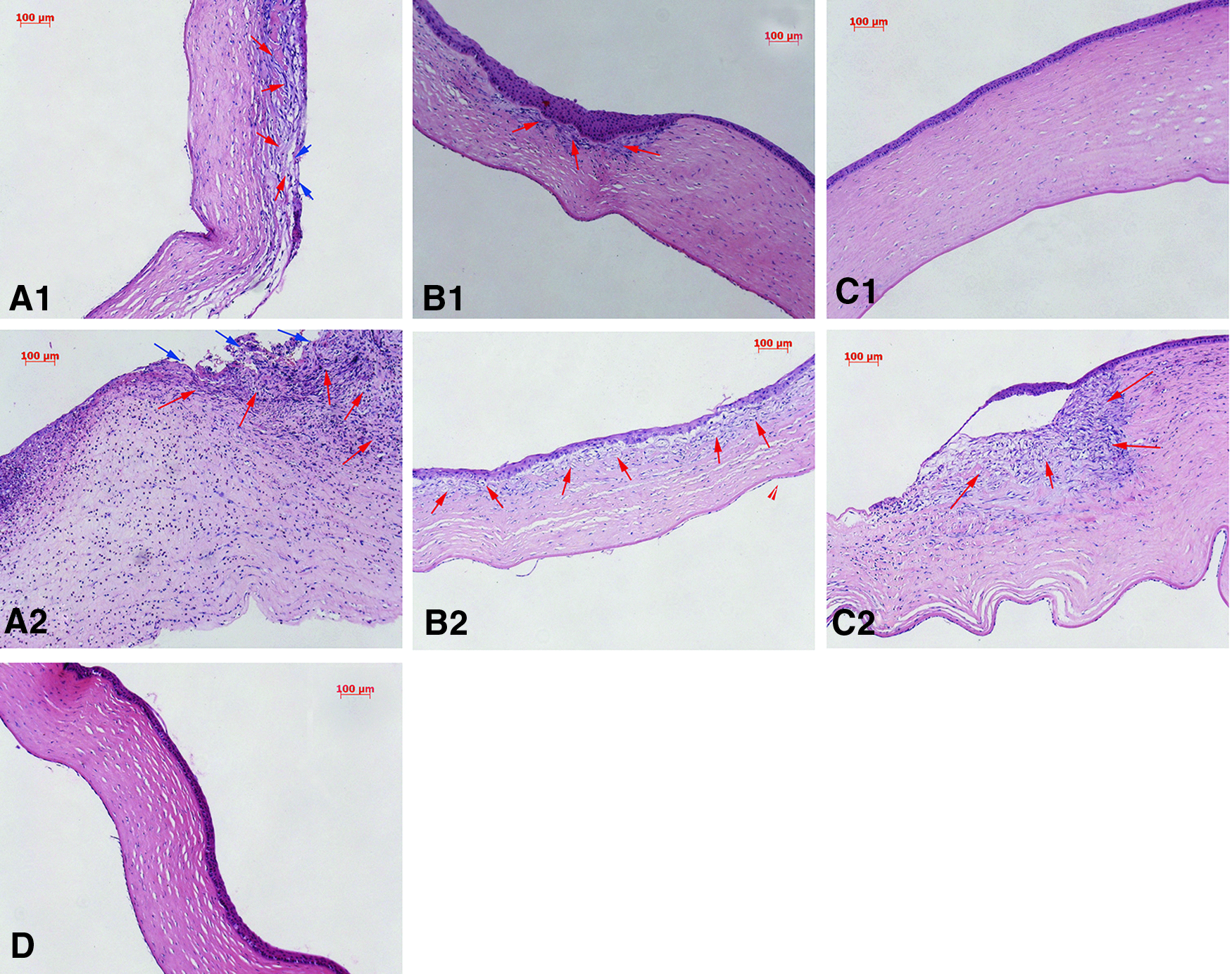
Histological images of both corneas in rabbits. In some rabbits, corneal epithelium deficiency and scar formation were found in both eyes. Compared with the left eye
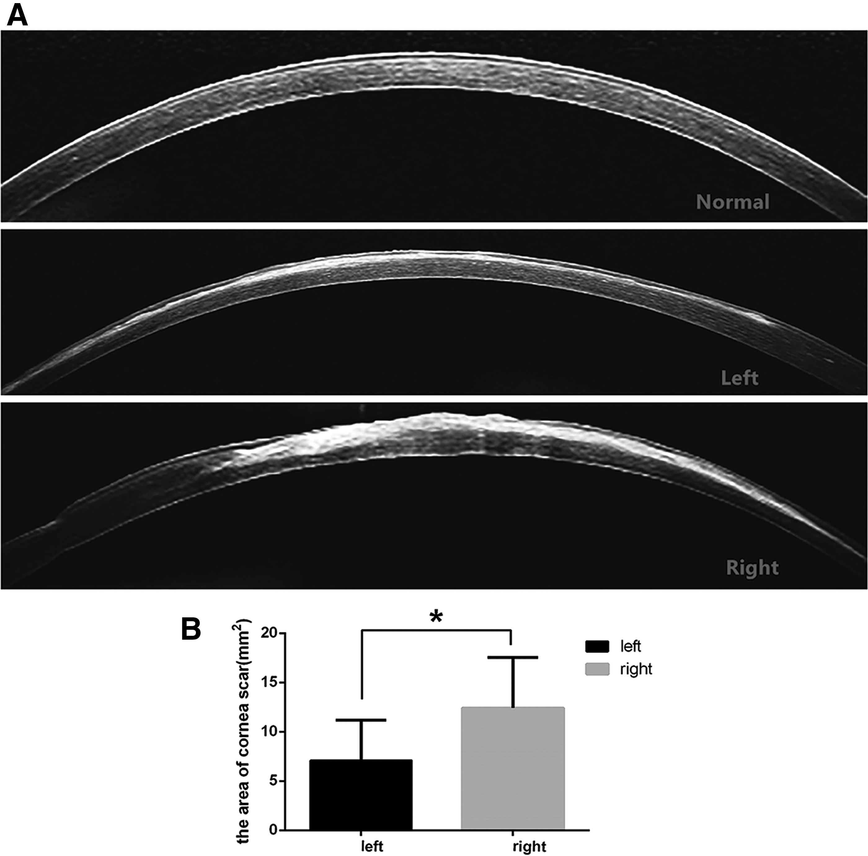
AS-OCT imaging of both corneas on day 14. It showed no corneal scarring in the normal corneal. Compared with the left cornea, the right cornea displayed obviously larger scars
Evaluation of corneal neovascularization
There were no blood vessels in any of the corneas treated with Biohcly during the course of wound healing, but corneal hemangiogenesis occurred and developed in 2 corneas (25%) treated with saline, in which new blood vessels, such as a hairbrush from the limbus toward the central cornea, reached the apex and extended to 2 quadrants of the cornea in one rabbit on day 9 and extended to 1 quadrant of the cornea in another rabbit on day 14 after injury (Fig. 1D).
Evaluation of corneal inflammation
Corneal inflammation was first observed under a slit lamp, and the IF scores were recorded every day. We found that the inflammation observed in the cornea was more extensive in cases treated with saline than in those treated with Biohcly. This was supported by findings including considerable corneal edema, epithelium erosion, and ciliary hyperemia. Furthermore, there were 2 saline-treated eyes (25%) that were seriously infected with hypopyon, and the inflammation observed in these corneas was too intensive to identify the pupil (Fig. 4). Second, based on the daily record, the development of IF was analyzed at each time point after injury. Although the IF scores were lower in the left eyes (treated with Biohcly) than in the right eyes (treated with saline) in general, the difference was small during the first 5 days. Later, it increased dramatically and reached a peak on day 14 after injury (Fig. 5). Finally, the association of IF with WHR was examined. We found that IF was significantly and inversely correlated with WHR (r = −0.753; P < 0.0001), indicating that there was more corneal inflammation and a lower rate of corneal healing rate.
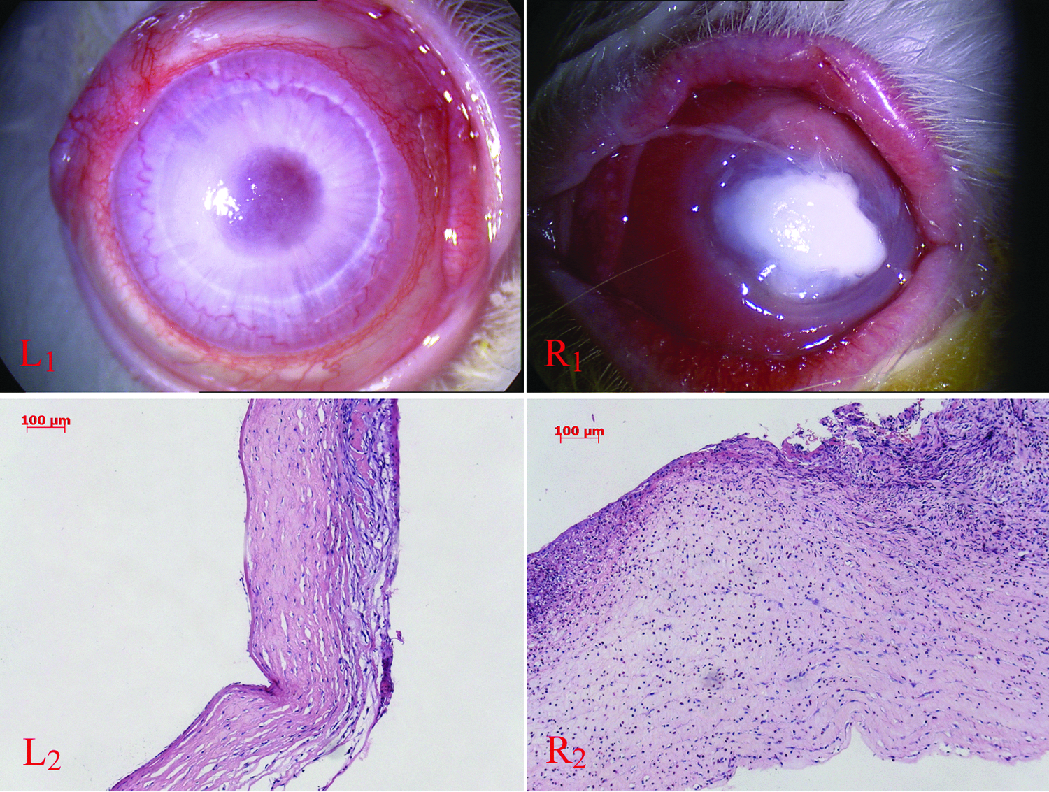
Differences in inflammation and histological examinations of both corneas. Corneal edema, epithelium erosion, and ciliary hyperemia were slight in the left eye (L). The right eye was seriously infected, and the inflammation of the corneal tissue was too intensive to identify the pupil. (L1, R1: Photographs taken under a slit lamp, magnification × 25; L2, R2: histopathology of the cornea, magnification × 100).
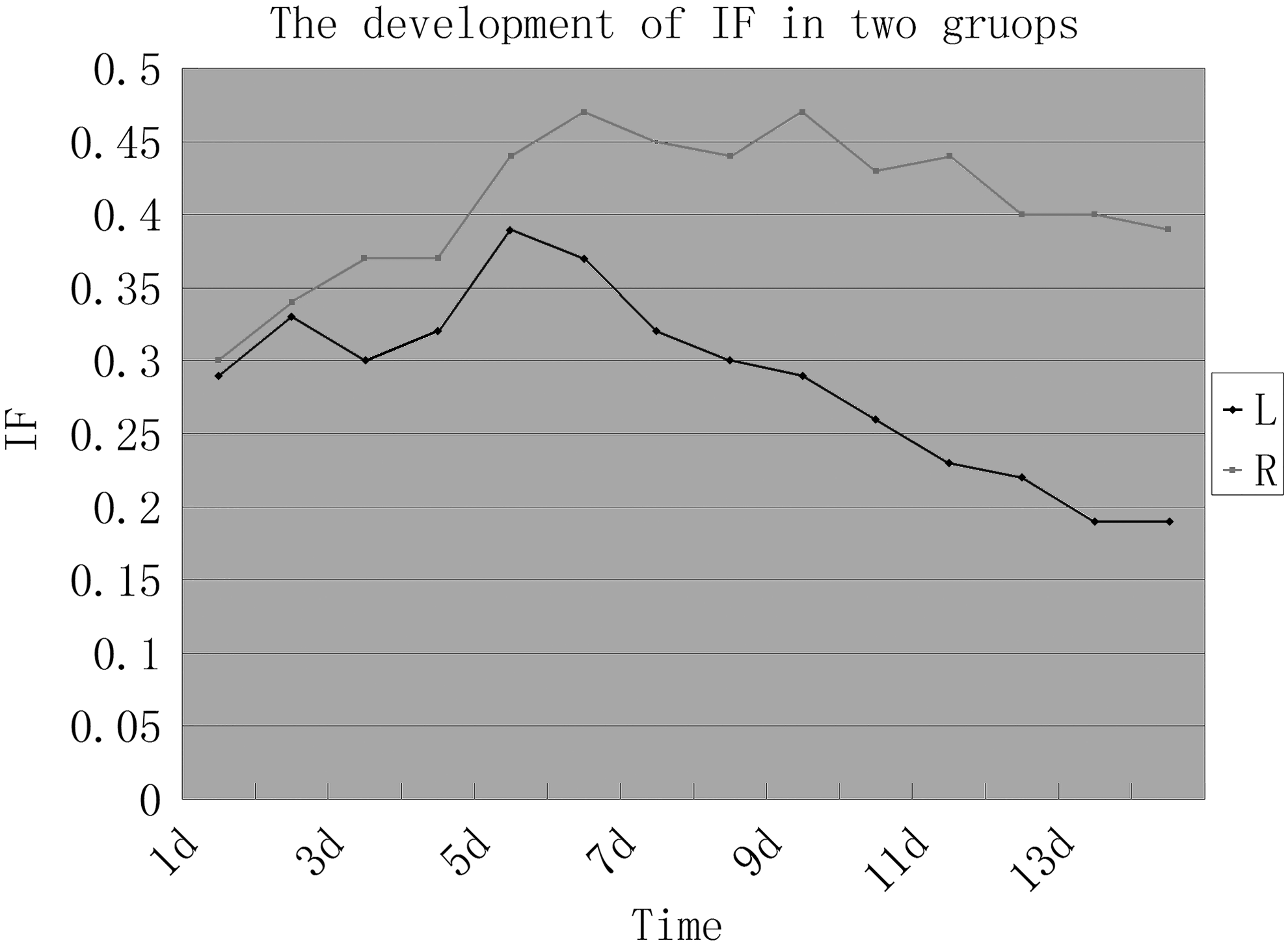
Differences in IF between the 2 treatments. The IF scores were lower in the left eyes (group A) than in the right eyes (group B) in general. The difference was small during the first 5 days. However, it later increased dramatically and reached a peak on day 14 after injury.
Evaluation of qRT-PCR
Compared with other factors (MMP-2, TNF-α, VEGF, and NF-kβ), the expression of MMP-9 was dramatically upregulated in wounded corneas. The relative mRNA expression of MMP-9 was >20-fold higher than other factors on the 4th and 9th days after corneal injury. Although the expression of MMP-9 was significantly decreased on the 14th day, it was nearly 3-fold higher than that of MMP-2 and VEGF and 4-fold higher than that of NF-kβ (Fig. 6A). These results suggest that MMP-9 might play a key role in the processes underlying corneal wound healing. Compared with the right corneas (treated with saline), in the left corneas (treated by Biohcly), the expression levels of MMP-2, MMP-9, VEGF, and NF-kβ were significantly lower on the 4th day after corneal injury, suggesting that Biohcly played a role in suppressing inflammatory factors during the early phase of corneal wound healing. On the 9th day after corneal injury, both MMP-9 and NF-kβ mRNA expression levels were significantly lower in the left corneas than in the right corneas. On the 14th day after injury, the expression levels of TNF-α and VEGF were lower in the left corneas than in the right corneas, and these differences were significant (Fig. 6B–F).
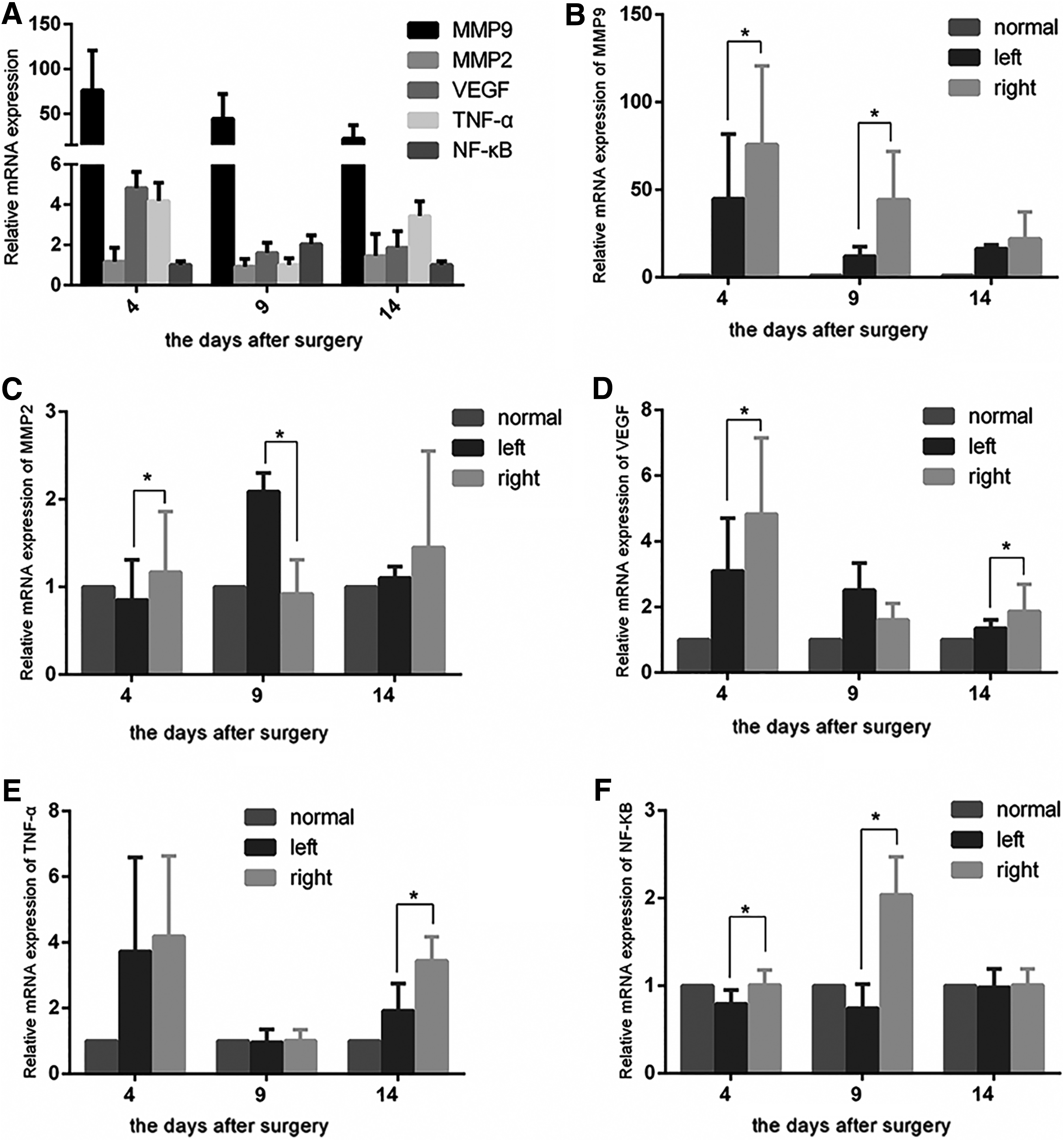
Quantitative RT-PCR analysis of the results of Biohcly (left eye) or sterile saline (right eye) in the cornea “acute” mechanical wound rabbit ocular surface. On the 4th, 9th, and 14th days after surgery, the corneas of the left and right eyes from each rabbit were secured and prepared separately for qRT-PCR. The mRNA levels of MMP-9, MMP-2, VEGF, TNF-α, and NF-κB were determined. The mRNA levels of 5 indexes in right eye corneas were compared across the 4th, 9th, and 14th days after surgery
Evaluation of the MMP-9 protein
Because the MMP-9 mRNA levels were dramatically increased compared with those of the other factors, we focused on the expression of the MMP-9 protein and examined the changes in MMP-9 labeling after corneal injury using ELISA and immunohistochemical staining. In the immunohistochemistry examination, we found that there were no MMP-9-positive cells in the normal corneas. Labeling for MMP-9 was dramatically increased on the 4th day, moderately increased on the 9th day, and mildly increased on the 14th day after injury. MMP-9-positive cells emerged mainly in the corneal epithelium and superficial stroma (Fig. 7A–G). Based on the ELISA results, the expression level in the corneas treated with saline was 1.85 times higher than that in the corneas treated with Biohcly during the early stage of injury (4 days after injury). The difference between the 2 groups was statistically significant (P < 0.05). Subsequently, the expression of MMP-9 gradually decreased, but on the 9th day after injury, the right corneas still exhibited significantly higher expression than the left corneas (P < 0.05) (Fig. 7H). This finding suggested that Biohcly exerted an inhibitory effect on MMP-9 protein expression during the course of corneal wound healing.

Immunohistochemistry for MMP-9 in injured rabbit corneas. There were no MMP-9-positive cells in the normal cornea
Discussion
In this study, Biohcly, an ANW that was previously demonstrated to play a role in wound healing and tissue repair without any systemic or local side effects and to display extraordinary biocompatibility, was verified to exert a positive effect on corneal wound healing after mechanical injury. First, the WHR and wound depth were compared across the 2 treatment groups on days 4, 9, and 14 after injury, and the results showed that the topical use of Biohcly drops significantly promoted corneal wound healing after injury. Second, the WHR was compared across the unhealed corneas with the 2 treatments, and the WHR was much higher in the cases treated with Biohcly than in those treated with saline. Third, among the cases in which ulceration had been healed, the recovery was not as ideal in the corneas treated with saline as in those treated with Biohcly. On the basis of the above findings, we propose that Biohcly performed better than saline in promoting recovery after corneal trauma by accelerating the course of wound healing.
We also found that the WHR of cases treated with saline gradually declined from day 4 to day 14 after injury but continued to increase during the same period in the cases treated with Biohcly. However, the velocity of wound healing was much slower during this period than in the 4 days after injury. Inflammation of the cornea was compared, and we found that changes in IF were significantly and inversely correlated with WHR, indicating that less corneal inflammation occurred, and a higher corneal healing rate was observed. The IF scores of all cases treated with either treatment increased dramatically on day 5 after injury, when corneal inflammation was only slowly resolving in the wounds treated with Biohcly. That of saline remained high, and the IF scores were nearly parallel for both treatments during the last 9 days after the injury. Biohcly played a role in inhibiting corneal inflammation in that the cases treated with Biohcly had much lower IF scores than those treated with saline. In addition, we examined the expression of NF-kβ and TNF-α, 2 important inflammatory factors, in the wounded corneas. Our results show that on the 9th day after injury, the level of the NF-kβ mRNA in the right corneas was almost double that observed on the 4th day after injury, whereas in the left corneas, the expression of NF-kβ remained at nearly the same level between the 4th and 9th days after injury. Moreover, the expression of TNF-α was significantly lower in the left corneas than in the right corneas. The variation in IF and the significant upregulation of NF-kβ on the 9th day and TNF-α on the 14th day observed in the right corneas could partially explain the expansion in corneal ulceration that occurred beginning on day 4 after injury in the cases treated with saline. Considering that the WHR of Biohcly was much faster than that of saline in the later phase of corneal wound healing in addition to the clearly anti-infection characteristics of ANW described in a previous study, the anti-inflammatory feature of Biohcly might play a helpful and important role in corneal ulceration healing.
In addition, our study shows that Biohcly plays a role in reducing corneal scarring and neovascularization during the course of wound healing. To determine the possible mechanisms underlying this effect, we examined the expression levels of MMP-2 and MMP-9 in injured corneas treated with Biohcly or saline. MMP-2 and MMP-9 (also called type IV and V collagenase or gelatinase A and gelatinase B, respectively) exhibit large-scale catalytic activity and are considered 2 enzymes that are pivotal during the period of wound healing and tissue remodeling. 23 The prolonged and sustained expression of MMP-2 during dermal wound repair suggests that MMP-2 plays a role in angiogenesis and matrix remodeling; however, the sudden MMP-9 upregulation observed during the primary stage of healing suggests that this enzyme is involved in reepithelialization.24,25 Surprisingly, compared with the MMP-2 expression, the expression of MMP-9 was dramatically increased at both the mRNA and protein levels during the study. In the left corneas, the mRNA levels of MMP-9 were 51.7-fold higher on the 4th day, 4.7-fold higher on the 9th day, and 13.8-fold higher on the 14th day than the levels of MMP-2 in the left corneas. They were 63.8-fold higher on the 4th day, 47.2-fold higher on the 9th day, and 14.1-fold higher on the 14th day in the right corneas. A similar result was described in a study by Kim et al., 26 who also used a trephine to make a corneal wound model. We believed that there were 2 reasons that could explain the significant differences between the expression levels of MMP-2 and MMP-9 in the injured corneas. First, MMP-9, which is the primary MMP synthesized, is secreted by basal corneal epithelial cells that migrate to the defective epithelium.
MMP-9 is the primary MMP synthesized and secreted by basal corneal epithelial cells migrating to resurface a wound, whereas MMP-2 is mainly secreted by the corneal stroma.27,28 In this study, the corneal epithelium was entirely removed, and this induced a dramatic upregulation of MMP-9. Second, the pattern of MMP-9 synthesis is consistent with the timing of basement membrane degradation in that there was a rapid increase in expression within several days of wounding, followed by a loss of expression after a few weeks. 11 This time course differs from that of MMP-2, which gradually increases over many months of matrix remodeling. 27
Recently, MMP-9 was shown to be correlated with the formation of scars. Manuel and Gawronska-Kozak examined the expression of MMP-9 during the remodeling phase of wound healing in nude mice and found that MMP-9 was upregulated during scarless wound healing, suggesting that MMP-9 expression might be a key factor in the ability of tissue to achieve scar-free healing. 29 Studies of MMP-9 knockout mice have shown that these mice exhibit reduced glial scar formation after spinal cord injury and impaired migratory activities. 30 Moreover, the production of cytokines, such as interferon-γ and TGF-β, which are considered to induce glial scar formation, may be reinforced by MMP-9. 31 Therefore, the improved locomotor function observed in the injured MMP-9 null mice may have been caused not only by a blockade of early inflammatory cells but also by a reduction in glial scarring. 32 In addition, it has also been shown that mice lacking MMP-2 produce more extensive glial scarring during the period of spinal cord injury. 33 However, these mice also exhibit a compensatory increase in MMP-9, which suggests that the compensatory upregulation of MMP-9 induces more severe glial scarring. Recent studies investigating trachoma and glaucoma have shown that the overexpression of MMP-9 plays a role in the development of scars by causing blinding trachoma or filtering surgery failure.34,35 In combination with our results showing that Biohcly had an inhibitory effect on the formation and hyperplasia of corneal scarring, the downregulation of MMP-9 by Biohcly treatment might be responsible for this effect.
In addition to its association with corneal ulcers and scar formation, MMP-9 may also be involved in mediating corneal angiogenesis. Mohan et al. implanted fibroblast growth factor 2 pellets into mouse corneas to create an angiogenic response and found that gelatinase B (MMP-9) expression was also induced. The increase in gelatinase B in this model was coincident with an increase in angiogenic activity and was mediated by the actions of increased AP-1 transcription factor binding. Furthermore, the inhibition of gelatinase B activity in these corneas was associated with a reduction in the angiogenic response. 36 In this study, we observed not only a dramatic reduction in MMP-9 but also a significant reduction in VEGF expression in the Biohcly-treated corneas after injury. Therefore, the downregulation of MMP-9 and VEGF after treatment with Biohcly might contribute to corneal avascularity.
In summary, Biohcly not only accelerated the WHR but also reduced the formation of corneal scarring after corneal mechanical trauma. Moreover, its obvious anti-infection and anti-angiogenesis characteristics are critical to retaining corneal transparency. Considering its safety and small side effects, Biohcly shows bright promise for applications aimed at promoting corneal recovery after injury and related fields.
Footnotes
Acknowledgments
The study was supported by the Guangdong Natural Science Foundation (No. 2015B020226003 and No. 2016A030313208), the Guangdong Pharmaceutical Society project (No. 2015RL13), and the Guangdong Science and Technology Project (No. 2014A020212393). We thank Dr. Chaoyang Li, Dr. Aihua Jiang and Dr. Hui Zhang for their invaluable technical support.
Author Disclosure Statement
No competing financial interests exist.