
Abstract
Purpose:
The intraocular lens (IOL) is a common, yet important, implantable device used in treatment of cataract in clinics. However, the unexpected adhesion of postoperative residual lens epithelial cells (LECs) often causes serious complications, such as posterior capsular opacification (PCO), which lead to vision loss again. In this investigation, a poly(sulfobetaine methacrylate) (PSBMA) brush coating was fabricated on an IOL to generate a hydrophilic surface coating on the IOL for enhanced cell adhesion resistance so as to decrease PCO incidence.
Methods:
The PSBMA brush coating on the IOL surface was fabricated using surface-initiated reversible addition–fragmentation chain transfer polymerization. X-ray photoelectron spectroscopy (XPS) was used to demonstrate the surface coating preparation. The water contact angle (WCA) measurement was used to test surface hydrophilicity. In vitro LEC culture was use to evaluate the cell behavior on the IOL material surfaces, with or without PSBMA coating modification. Finally, animal cataract surgeries were carried out to evaluate in vivo biocompatibilities and anti-PCO effects.
Results:
The XPS and WCA measurements illustrate successful surface modification and good surface hydrophilicity. The in vitro cell culture results show that the hydrophilic PSBMA polymer brush coating evidently decreases adhesion and proliferation of LECs. Results of the in vivo cataract surgery with intraocular implantation show that PSBMA modification on the IOL surface does not induce side effects in nearby tissues, whereas posterior capsular hyperplasia can be evidently reduced.
Conclusion:
The PSBMA brush surface-modified IOL has good in vivo biocompatibility and it can effectively reduce the incidence of postoperative PCO.
Introduction
Cataract continues to be the leading cause of blindness worldwide, both in underdeveloped countries and developed countries with the availability of effective surgery. 1 It is an opacification of the natural crystal lens, thus obstructing light and causing gradual vision loss. Primary cataract surgery, including opaque lens removal and intraocular lens (IOL) implantation, is the most frequently performed and the only effective method for clinical treatment of this ocular disease. This surgical intervention obviously improved patients' vision, but it is not perfect. A number of complications may occur after this surgery. One of the most common complications is posterior capsular opacification (PCO), also called secondary cataract, which is the result of proliferation and trans-differentiation of residual lens epithelial cells (LECs) within the lens capsule after cataract surgery.2,3
With advances in IOL design and surgical techniques over the past decades, the incidence rates of PCO have been reduced from 50% to current levels of 14%–18%. Yet, it remains a major medical problem with profound consequences for the patient's well-being and also is a significant financial burden due to the costs of follow-up treatment.
Surface modification to fabricate a functional organic coating on biomaterials is a feasible, yet effective, method for biocompatibility improvements.4–8 As described above, PCO is the result of LEC proliferation and trans-differentiation on the IOL surface, and the initial cell adhesion is of vital importance. Therefore, surface modification has been carried out to generate hydrophilic coatings on the hydrophobic IOL to decrease the initial cell adhesion observed in previous studies. Several organic coating technologies have been introduced onto the IOL surface. For example, IOL surfaces were treated with O2 plasma and chemically grafted with hydrophilic moieties, such as poly(ethylene glycol), phosphorylcholine, or heparin.9–14
These surface treatments improve their hydrophilicity and thus decrease the initial cell adhesion. Among them, the surface-heparinized IOL is a clinically approved, implantable medical device and widely used in cataract patients. 14 After heparinization, the surface hydrophilicity of the IOL increases to some extent. However, such surface modifications just decrease cell adhesion to a certain degree. The remaining cell adhesion on these surfaces still cannot be neglected. Long-term clinical observation also indicates that there is no significant difference between cataract patients who are implanted with a heparinized IOL and pristine IOL. This may due to the sparse immobilization density of hydrophilic molecules by the random grafting methods. 15 Thus, the enhanced hydrophilic surface coating is in great demand.
Reversible addition–fragmentation chain transfer (RAFT) polymerization is a type of well-controlled free radical polymerization. In our previous investigations, surface-initiated RAFT (SI-RAFT) polymerization was carried out on the IOL surface, generating a highly hydrophilic surface coating and thus enhanced cell adhesion resistance.16–18 The comb-like, enhanced, hydrophilic polymer brush such as the poly(polyethylene glycol methacrylate) (PPEGMA) or poly(2-methacryloyloxyethyl phosphorylcholine) brush was obtained on the IOL surface through the SI-RAFT polymerization method. However, the PPEGMA brush coating does not eliminate the PCO occurring in in vivo trails as usual.
Zwitterionic betaines have been developed recently for low-cost and easy preparation. Polymers with betaine groups have unique characteristics such as biocompatibility, lubrication, and antifouling properties in the hydrated state.19,20 In the present investigation, a hydrophilic, sulfobetaine methacrylate (SBMA) polymer brush was fabricated on the IOL surface through SI-RAFT polymerization to further verify the effect of the hydrophilic coating on PCO prevention. The hydrophilicity of the coating was investigated and the LEC-resistant property was tested. The sulfobetaine polymer brush-modified IOL was then implanted into rabbit eyes by standard cataract surgery to evaluate in vivo biocompatibility and effects on postoperative PCO prevention.
Methods
Materials
The surface activation agent, (3-aminopropyl) triethoxysilane (APTES), the cell-staining dye, fluorescein diacetate (FDA), and agents involved in SI-RAFT polymerization, including SBMA, 4-cyano-4-(phenylcarbonothioylthio) pentanoic acid (CPCTTPA), 4, 4′-azobis-(4-cyanovaleric acid) (V501), 2-morpholino-ethanesulfonic acid (MES), N-(3-dimethylaminopropyl)-N′-ethylcarbodiimide hydrochloride (EDC), and N-hydroxysulfosuccinimide sodium salt (NHSS), were supplied by Sigma-Aldrich. Masson's trichrome staining kits were purchased from Leagene Biotech Co., Ltd.
Silicone (polydimethylsiloxane) used as the hydrophobic IOL material substrate for characterization was prepared using Sylgard® 184 of Dow Corning.21–23 The hydrophobic, acrylic foldable IOL was supplied by 66Vision Tech Co., Ltd. The human lens epithelial cell line (HLE B3, CRL-11421™) was originally obtained from American type culture collection (ATCC). The postoperative administration eye drops and ointments such as dexamethasone, tobramycin, levofloxacin, triamcinolone acetonide, and prednisolone acetate were obtained from the Eye Hospital of Wenzhou Medical University and used according to the instruction manual.
IOL surface modification and characterization
The SI-RAFT polymerization procedure was similar to our previous investigations.17,18 As illustrated in schematic Fig. 1, each experiment was initiated by 2% (v/v) APTES treatment, which generates amino groups on the silicone materials or hydrophobic acrylic IOL surfaces. The RAFT agent (CPCTTPA) was then grafted onto the aminated substrates through carbodiimide chemistry. After washing, the substrates were put into glass vials with 1 mg/mL V501 and 100 mg/mL SBMA in MES buffer (pH 5.2). After sealing and degassing, SI-RAFT polymerization was carried out in a microwave reactor (Initiator 60, Biotage) at 60°C for 6 h. Finally, the poly(sulfobetaine methacrylate) (PSBMA)-modified IOL materials were washed with pure water and dried for further use.
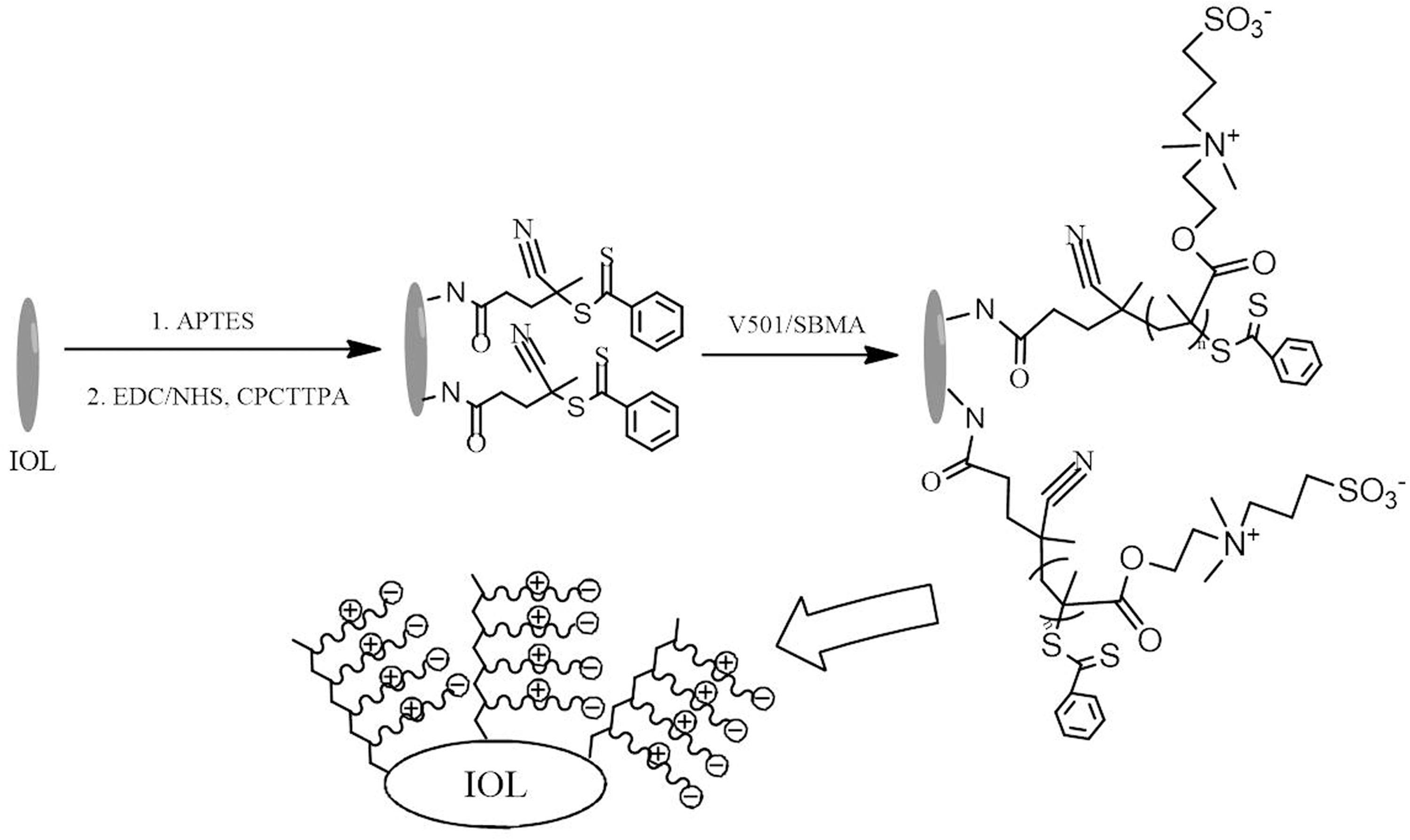
Schematic illustration of PSBMA brush modification on the IOL surface through SI-RAFT polymerization. APTES, (3-aminopropyl) triethoxysilane; CPCTTPA, 4-cyano-4-(phenylcarbonothioylthio) pentanoic acid; EDC, N-(3-dimethylaminopropyl)-N′-ethylcarbodiimide hydrochloride; IOL, intraocular lens; NHS, N-hydroxysulfosuccinimide sodium; PSBMA, poly(sulfobetaine methacrylate); SI-RAFT, surface-initiated reversible addition–fragmentation chain transfer.
X-ray photoelectron spectroscopy (XPS) (PerkinElmer Co., USA) was used to characterize the chemical composition of the material surface. The water contact angle (WCA) analysis (OCA20; DataPhysics Co., Germany) was used to measure the hydrophilicity of the coating. American type culture collection (Ultraviolet-Visible [UV-Vis]) spectroscopy (Cary 300; Agilent Technologies, USA) was used to characterize the IOL transmittance before and after modification of the PSBMA brush coating.
In vitro cell viability assay
Human lens epithelial cells were used to evaluate initial cell adhesion and proliferation on the surface-modified IOL materials. The cell culture and cell seeding onto the material surfaces were the same as in our previous studies.18,21 The cells were seeded with a density of 5.0 × 10³ cells per well. After incubation for 1 day or 3 days, cells were stained with FDA and fluorescent images were obtained by fluorescence microscopy (Nikon, Japan) with a fluorescein filter (488 nm/excitation).
Intraocular implantation and evaluation
In vivo intraocular implantation of the PSBMA-modified IOL and postoperative evaluation were carried out according to our previous publications.17,18,21–23 Briefly, 2-month-old New Zealand White rabbits were used to evaluate the intraocular tissue biocompatibility of PSBMA-modified IOLs. After general anesthesia, standard phacoemulsification surgeries were performed on the left eyes of rabbits using a phacoemulsification instrument (Alcon, USA). Postoperative topical therapy included a combination of levofloxacin eye drops and tobramycin–dexamethasone ointment during the first postoperative week and prednisolone acetate drops, which were tapered during the 1st and 2nd weeks. After IOL implantation, the eyes were dilated and observed by slit-lamp microscopy at intervals. The animals were anesthetized and humanely sacrificed with air embolism after 6 months. The ocular tissue sections were prepared according to the standard procedure, followed by Masson's staining and microscopic observation.
Ethical approval
New Zealand White rabbits, weighing 2–2.5 kg, were obtained from the Wenzhou Medical University Animal Laboratory. The rabbit experiment protocol was approved by the Laboratory Animal Ethics Committee of Wenzhou Medical University. All in vivo experiments that were performed were consistent with the Animal Experimentation Guidelines of Wenzhou Medical University and adhered to the Association for Research in Vision and Ophthalmology statement for the Use of Animals in Ophthalmic and Vision Research.
Results and Discussion
Surface modification of the IOL
The IOL surface modification with the PSBMA brush through SI-RAFT polymerization is schematically shown in Fig. 1. The material surface was first activated by APTES silanization, which generated amino groups on the material surface. The carboxyl group-ended RAFT agent was then covalently grafted onto the surface through EDC/NHS chemistry. Then, SI-RAFT polymerization of SBMA was carried out to fabricate the PSBMA polymer brush on the IOL surface.17,18
As an effective surface characterization method, XPS was performed to analyze the changes in elemental composition on the surface after each step. As shown in Fig. 2a, the pristine silicone surface consisted mainly of C, O, and Si elements.17,18 Characteristic peaks of O1s, C1s, Si2s, and Si2p were found at 530.9, 283.9, 149.9, and 98.9 eV in the spectrum of the bare silicone IOL material surface. 17 After APTES activation and RAFT agent immobilization, minor characteristic peaks at 397.8 and 399.8 eV arise (N1s signal), which are due to introduction of the N element onto the surface. Moreover, the characteristic peak of the S1s signal appears at 167.8 eV after CPCTTPA grafting, which further confirmed the immobilization of the CPCTTPA molecule (Fig. 2b).
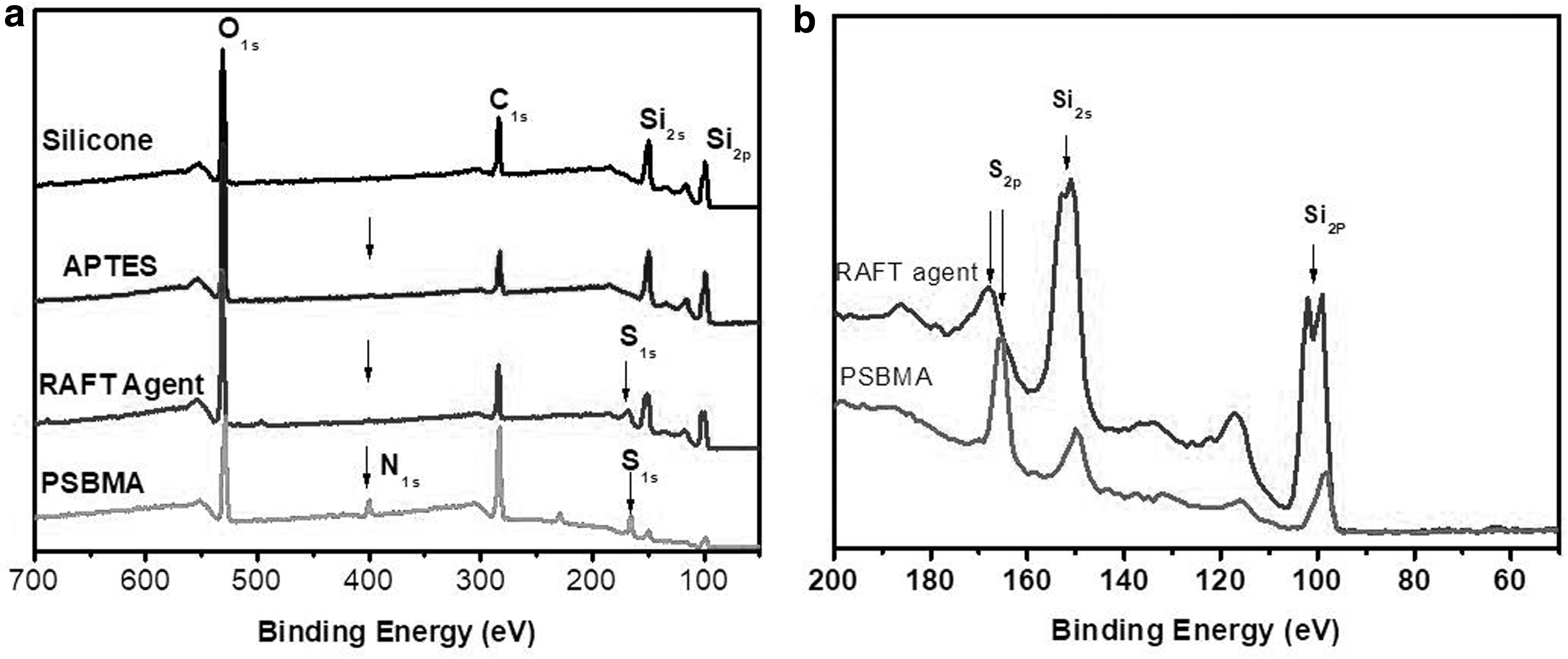
Nitrogen and sulfur elements are also characteristic in SBMA molecules. Generation of the PSBMA brush on the surface evidently increases the intensity of N1s and S1s signals (Fig. 2). On the other hand, it can be also observed from Fig. 2a that the signal intensity of Si2s and Si2p is gradually reduced with the surface modification. As the XPS detection limitation is less than 10 nm in surface thickness, the surface modification gradually increases the coating thickness, which resulted in the decrease of signals of the substrate elements. 24 These results clearly verified the PSBMA fabrication on the surface through SI-RAFT polymerization.
The surface wettability is also evaluated after PSBMA brush modification. As indicated in Fig. 3a, the surface WCA of the model hydrophobic IOL materials (silicone) was 114°. The surface activation through APTES silanization, as well as the subsequent RAFT agent grafting, does not significantly change the surface wettability. However, after modification of the zwitterionic PSBMA brush coating, the surface hydrophilicity was greatly enhanced. The WCA on the PSBMA brush coating surface was about 26.9 ± 2.5° (Fig. 3a). The SI-RAFT polymerization time also affects the hydrophilicity on the PSBMA brush coating surface. As shown in Fig. 3b, the surface hydrophilicity is effectively enhanced with increasing reaction time in the first reaction hour. The WCA is further (slightly) decreased when the reaction time is extended and reaches a plateau during a period of 4–9 h. However, further increase of the reaction time does not increase hydrophilicity, but has negative effects on the surface. As shown in Fig. 3b, the WCA increased to 45.5 ± 10.1° when the polymerization time was 12 h.
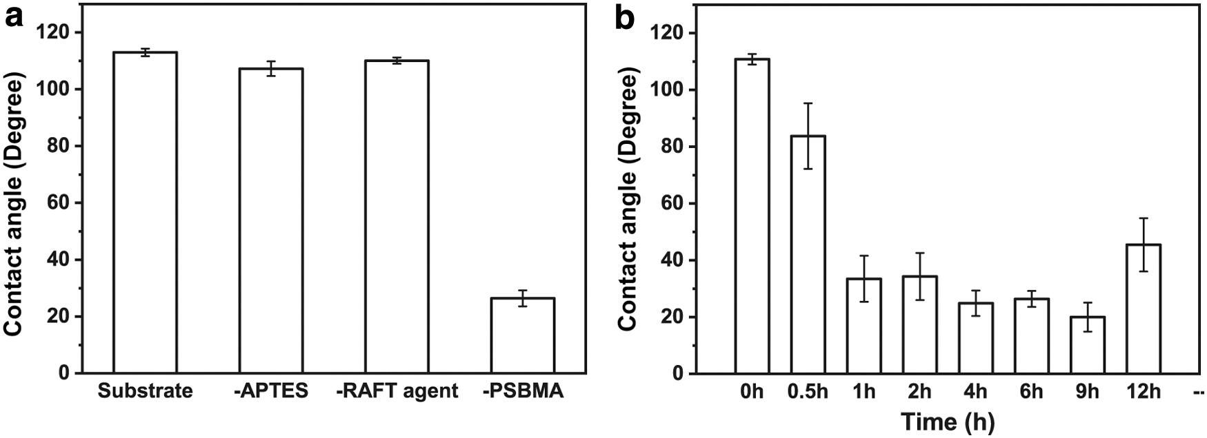
Water contact angle of silicone IOL materials during different steps of surface modification
The WCA changes can be visualized by the images of water drops on the surface after different reaction times of the modification (Fig. 4). This phenomenon also exists in surface grafting of polyethylene glycol (PEG), which may be due to the loop effect when the polymer chain is longer than a critical value and exposes the carbon backbone. 18 For balance of the reaction time and hydrophilicity, a 6-h time period is adopted for the surface modification. The evidence of decrease in the WCA not only proves PSBMA grafting through SI-RAFT polymerization but also shows the excellent hydrophilicity of the modified surface.
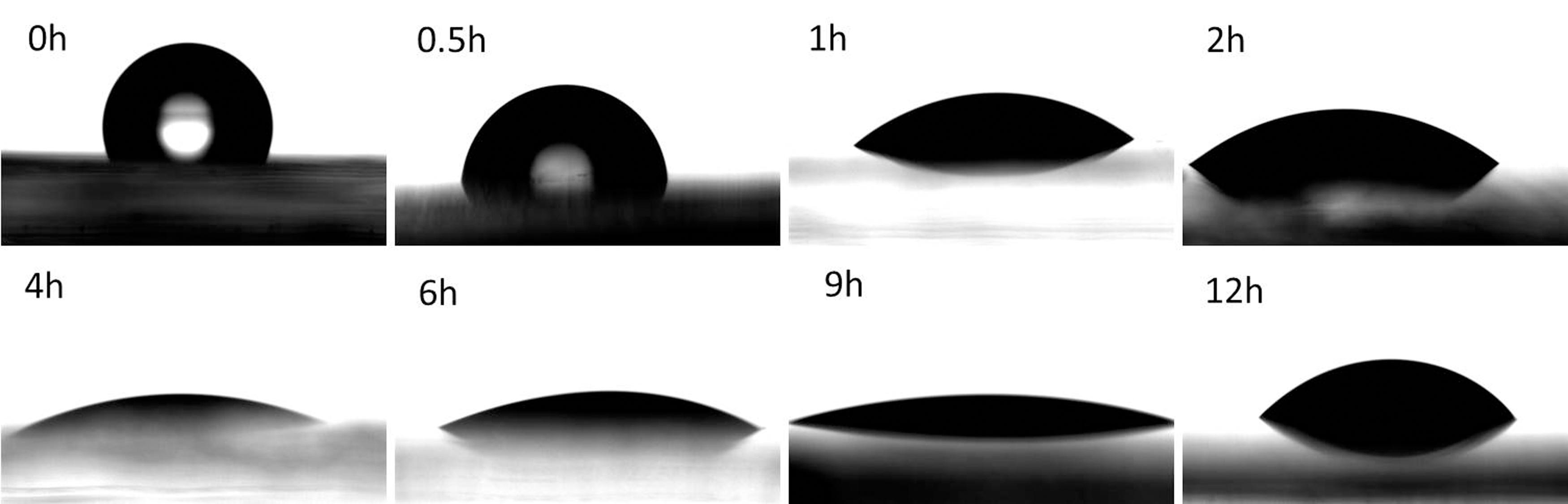
Visualization of images of water drops on the surface after different SI-RAFT reaction times of the PSBMA brush modification.
Besides surface hydrophilicity, transmittance is another key issue of IOL materials. Herein, the transmittance levels of the materials before and after PSBMA brush surface modification were tested. As shown in Fig. 5, the PSBMA brush surface modification does not evidently influence light transmission of the bulk materials. The total light transmittance of both types of materials is around 90%. The modified PSBMA brush coating possesses excellent transparency, so it is sufficient for IOL applications.
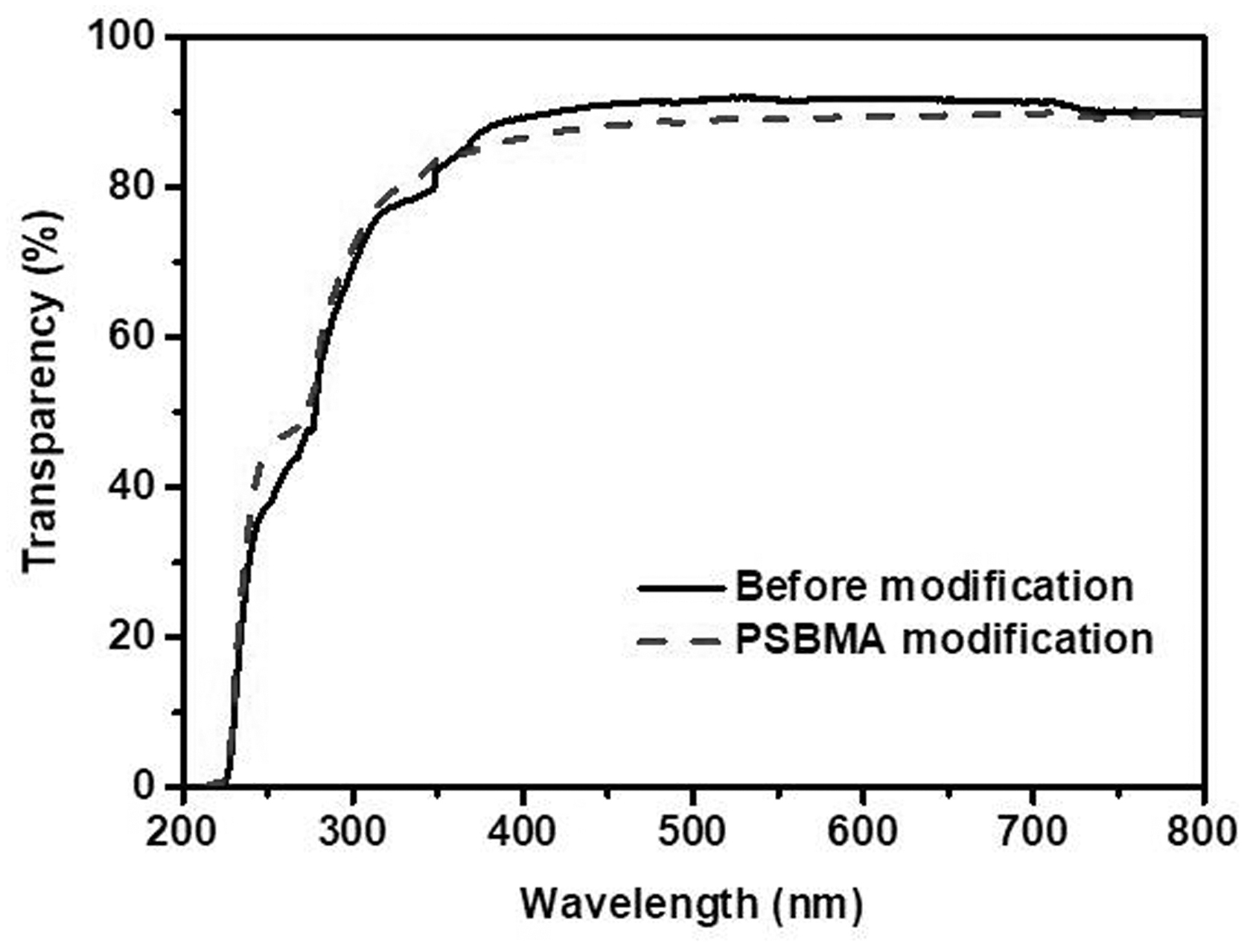
Light transmittance of the IOL material before and after PSBMA modification.
In vitro cell resistance evaluation
As mentioned above, PCO is the result of the residual LEC adhesion and proliferation after the cataract surgery, which decreases vision again after IOL implantation. As a result, the reduction in the initial cell adhesion plays an important role in IOL biocompatibility. In this investigation, the zwitterionic PSBMA brush coating was fabricated on the IOL material surface through SI-RAFT polymerization to reduce LEC adhesion. In vitro LEC adhesion on the PSBMA-modified IOL surface is shown in Fig. 6. Moderate cell adhesion was found on the pristine IOL surface in the initial stage, whereas it was seldom found on the PSBMA-modified surface. What is more, the adherent cells spread well on the pristine surface. On the contrary, only few cells were found on the PSBMA coated surface. These adherent cells were rendering round and unspreading morphologies which indicate the unfavorable condition to the cells. After culturing for 3 days, high cell proliferation was obtained on the pristine IOL surface. However, introduction of the PSBMA coating on the surface greatly reduces not only LEC adhesion but also cell proliferation. Almost no cells were detected on such surface after the 3-day culture.
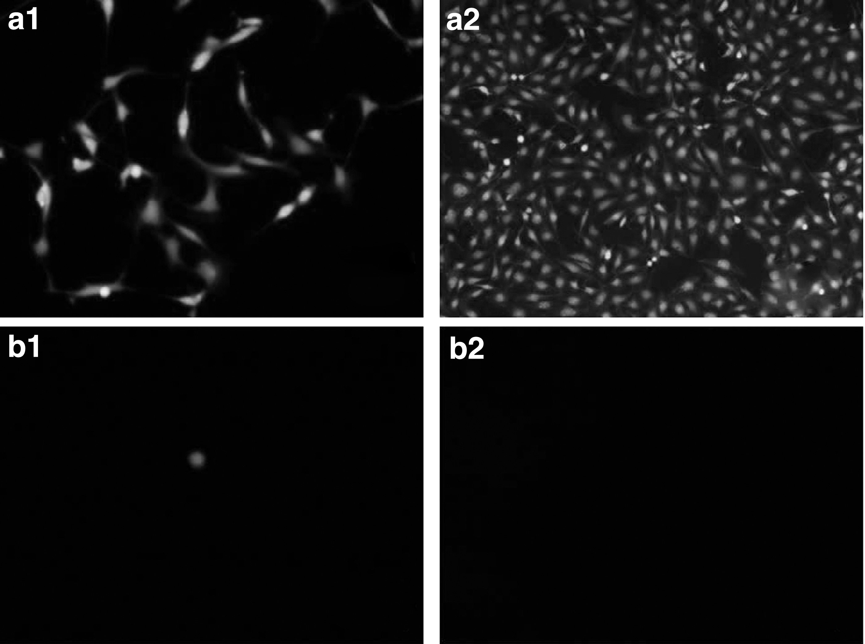
The representative fluorescent images of LECs on IOL materials before
With their zwitterionic nature, betaines, such as SBMA, have recently been widely used for surface modification of biomaterials. The zwitterionic surface shows great promise for minimizing cell adhesion.25–28 Herein, the excellent LEC-resistant property is also attributed to the hydrophilicity of the zwitterionic surface. It is demonstrated that residual LEC adhesion to the IOL may be the reason for PCO development.21,29,30 As a result, the adhesion-resistant surface coating can restrain the formation of a multicellular secondary membrane resulting from proliferation, migration, and fibrosis of residual LECs on the posterior capsule and can eventually reduce the incidence of PCO formation. 21
In vivo ocular implantation
To evaluate in vivo biocompatibility and the effects of the surface coating on PCO prevention, the PSBMA brush-modified IOL was consequently implanted into the rabbit eye during clinical cataract surgery. Regular slit-lamp observation was carried out postoperatively and images were obtained on days 1, 3, 7, 30, and 45. Figure 7 shows a group of representative slit-lamp images of IOLs in rabbit eyes before and after PSBMA brush modification. Some acute anterior chamber inflammation occurred in the pristine IOL group after surgery (day 1; Fig. 7a1), which was quickly eliminated by organism absorption. The anterior chamber becomes clear after 3 days postoperatively (Fig. 7a2, a3). No acute anterior chamber inflammation was found in the PSBMA modification group (Fig. 7b1–b3). The ocular surface and anterior chamber are clear and no apparent toxic damage was found in the cornea and iris in the observation period, which indicates that there is no evidence of acute tissue incompatibility of the PSBMA-modified IOLs.
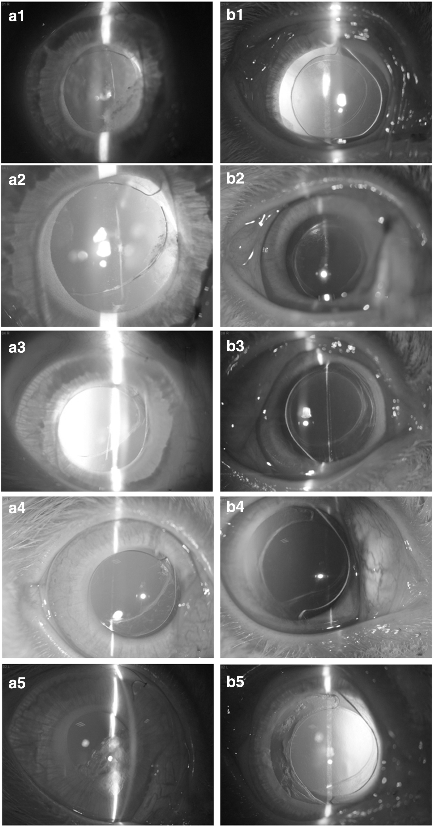
Representative slit-lamp images of the animal eyes with pristine hydrophobic IOL
Long-term, in vivo tissue compatibility was further demonstrated by histological observation study. The ocular tissues were cross cut, stained with Masson staining, and examined. The cornea and iris are important ocular tissues that exist near the lens. They serve as the refractive medium and ocular blood supply medium in the eye, respectively. Figure 8 shows the ocular tissue morphologies in the eyes with either pristine or PSBMA-modified IOL implantation. It can be observed that all ocular tissues neighboring the lens, including the cornea and iris (Fig. 8a, b), were visible with their normal morphologies and intact structures and no morphological change was found between common and PSBMA brush coating-modified IOL-implanted eyes, which indicate the excellent long-term in vivo biocompatibility and safety of PSBMA-modified IOLs. The five-layered corneal structure remains intact in both the common and surface-modified IOL-implanted groups, including the epithelium, Bowman's membrane, stroma, Descemet's membrane, and endothelium (Fig. 8a1, a2).21,31 Plenty of blood vessels and chorionic villi are found in the iris in these two groups (Fig. 8b1, b2).
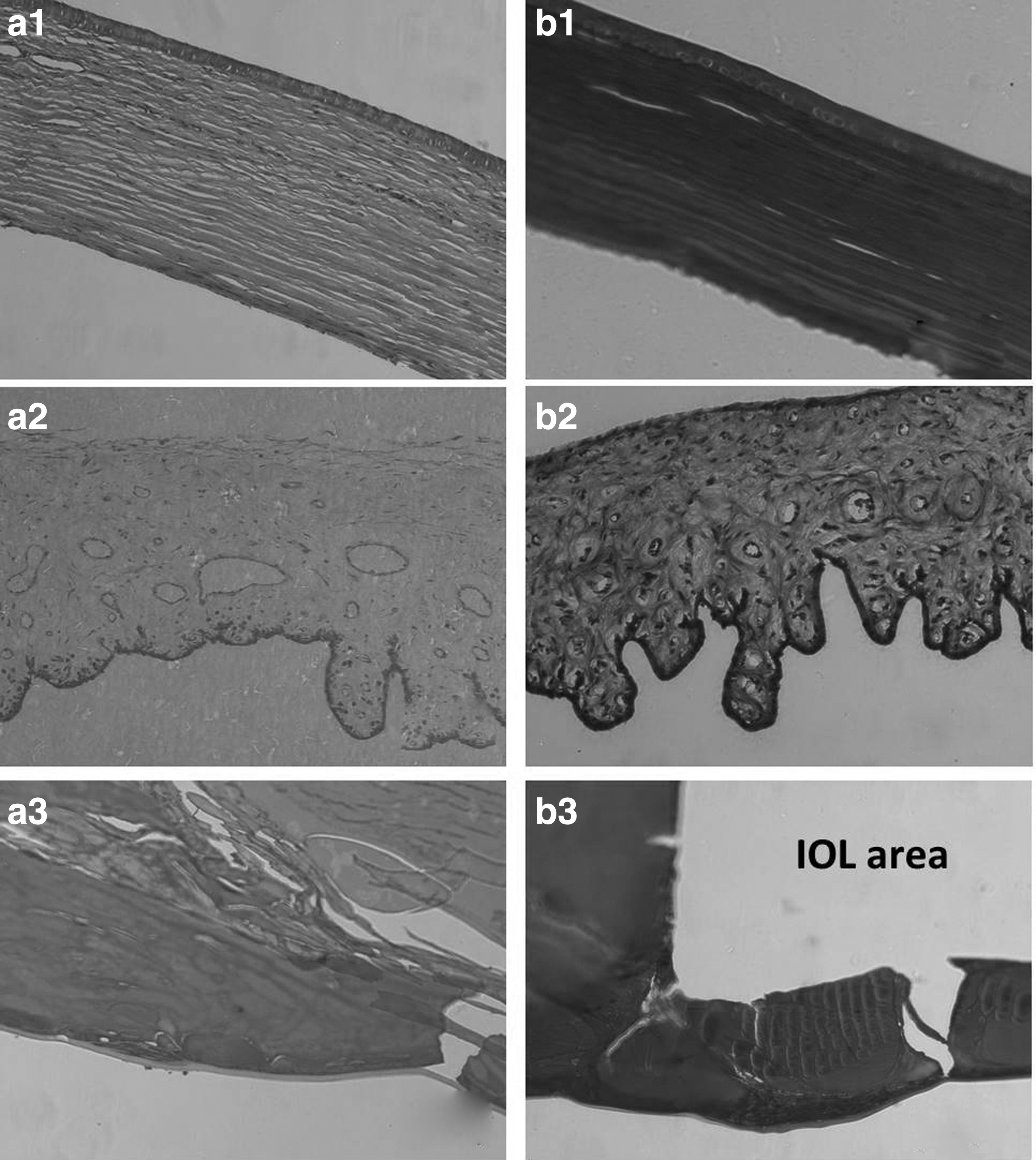
Masson staining of histological sections of IOL implantation.
Some cell adhesion onto the pristine IOLs was found after 1-month implantation, as indicated by the white scatter points and fibrosis (Fig. 7a4). Once it appears, PCO may develop rapidly. Heavy synechia and PCO were found in the pristine IOL 45 days postoperatively (Fig. 7a5), whereas neither in vivo cell adhesion nor PCO development was found on the PSBMA-modified IOL in these conditions (Fig. 7b4, b5), which is confirmed by the histological observation of the lens capsule. Serious hyperplasia was found in the lens capsule in the pristine IOL after 6 months (Fig. 8a3), whereas hyperplasia was less in the PSBMA-modified IOL case (Fig. 8b3). Posterior capsule hyperplasia is a typical symptom of PCO. These results indicate that the hydrophilic PSBMA brush modification enhances resistance to cell adhesion and thus delays and decreases the PCO development.
Bioadhesion is much more likely to occur on the hydrophobic IOL material surface. The residual LECs after surgery may adhere to the IOL material surface and proliferate between the surface and capsule, which is the leading cause of secondary cataract. 32 The bacterial adhesion in IOL implantation may cause endophthalmitis. Researchers and clinicians have tried several methods to improve the biocompatibility of IOLs, such as improving surgical techniques, optimizing the IOL design, developing new IOL materials, or injecting drugs into surgical sites.33–38 On the other hand, biomaterial surface modification provides an alternative approach to improve their biocompatibility.4–6,39,40 A surface with excellent cell-resistant property can be obtained through hydrophilic macromolecule immobilization or hydrogel-like surface coating.41–43
In the present investigation, an excellent, hydrophilic, zwitterionic PSBMA brush coating was generated on the hydrophobic IOL surface through SI-RAFT polymerization. Such zwitterionic PSBMA brush modification can effectively reduce residual LECs or the initial bacterial adhesion and can prospectively decrease the complication incidence in vivo.
Conclusions
Zwitterionic PSBMA brush can be fabricated on the hydrophobic IOL surface through SI-RAFT polymerization and greatly improves surface hydrophilicity. As a result, the initial LEC adhesion is distinctly reduced after the PSBMA brush modification. Intraocular IOL implantation results reveal excellent ocular tissue compatibility as the IOL implantation does not induce pathological changes in the neighboring ocular tissues, such as the cornea and iris. Although such modification cannot thoroughly eliminate PCO, the PCO incidence can be postponed and PCO severity can also be greatly alleviated when comparing with the pristine IOL. These results demonstrate that hydrophilic modification of the zwitterionic PSBMA brush can be taken into consideration to improve the biocompatibility of IOLs after implantation.
Footnotes
Author Disclosure Statement
No competing financial interests exist.
Funding Information
The authors acknowledge the funding from the National Key R&D Program (2017YFC1104602) and National Natural Science Foundation of China (81771984).