
Abstract
Purpose:
Particulate matter (PM) is a primary cause for the development of acute and chronic dry eye disease, especially irritant-induced conjunctivitis. The purpose of the present study was to determine the effects of fine atmospheric PM on the rabbit ocular surface, and determine the protective effects of a synthetic antioxidant, manganese(III) tetrakis(1-methyl-4-pyridyl) porphyrin (Mn-TM-2-PyP), in vitro and in vivo.
Methods:
Rabbit corneal epithelial cells (SIRC) were exposed to increasing concentrations of PM to determine the effects on cell motility and viability. The in vivo effects of topically instilled PM were tested in New Zealand White rabbits. Comprehensive ophthalmic exams and corneal fluorescein staining were performed.
Results:
Exposure to PM resulted in dose-dependent cell death and impaired cellular motility; Mn-TM-2-PyP protected against PM-induced cytotoxicity and significantly increased SIRC cell motility. In vivo, exposure to PM (5 mg/ml, topical, 3 times daily for 7 days) resulted in signs of dry eye, notably hyperemia, increased corneal fluorescein staining, and decreased tear volumes. Mn-TM-2-PyP significantly improved hyperemia and corneal fluorescein readouts but had no effect on tear production. Lifitegrast (Xiidra®) showed similar pharmacologic efficacy to Mn-TM-2-PyP.
Conclusion:
Overall, these data provide evidence that PM induces phenotypes of ocular surface disease responsive to antioxidant and immunosuppressant therapy. To our knowledge this is the first report of a large animal model to study PM-induced ocular surface disease. The present work provides standardized experimental paradigms for the comprehensive in vitro and in vivo testing of novel therapeutic approaches targeting PM-induced conjunctivitis and dry-eye.
Introduction
Air pollution and military environmental exposure from burn pits has emerged as a significant health concern, including an increased risk of developing ocular surface disease. 1 Particulate matter (PM) is a specific air pollutant that represents an urgent environmental concern in areas of dense urban populations, such as India and China. In addition, PM has also been the cause for ocular surface disease in the military population resulting from exposure to burn pits and desert climates.
PM is a heterogeneous mixture of toxic pollutant compounds, such as polycyclic aromatic hydrocarbons, carbon oxides, nitrogen oxides, heavy metals, and chlorinated pesticides.2,3
Multifactorial damage has been noted when the eyes are directly exposed to the toxins of PM. Specifically, experimental conditions mimicking airborne exposure have shown impaired migration and proliferation of human corneal epithelial cells. 4 Clinically, locations like Hangzhou, China have reported a significant association between air pollution and patients presenting with dry eye syndrome. 5
Therefore, it is likely that PM exposure may represent a significant contributor to the exacerbation of ocular surface disease, especially irritant-induced conjunctivitis following acute exposure to PM. This common ocular surface disease is the consequence of mechanical and chemical injury to the ocular surface that may also trigger a type I hypersensitivity response. It can present with mild-to-moderate symptoms and develop into chronic forms with symptoms resembling dry eye disease. In irritant-induced conjunctivitis, the conjunctiva, that is, the mucous membrane tissue that lines the front and inner surface of the eyelids, becomes inflamed resulting in hyperemia and other signs of ocular surface disease. While different pollutants can cause conjunctivitis, exogenous PM is the primary cause for the special populations described earlier.
Previous experimental studies have primarily used rodents to study PM-induced ocular surface disease. For example, administration of PM in male specific pathogen-free BALB/c mice resulted in damage to tear film function and destruction of the structural organization of the ocular surface. 6 Administration in 6-week-old female Sprague-Dawley rats induced dry eye syndrome by reduction in tear volume, damage to the corneal epithelium, and loss of conjunctival goblet cells. 7 These studies provide strong preclinical evidence that PM can induce phenotypes of ocular surface disease in mouse and rat models.
However, no validated preclinical large animal models for PM-induced ocular surface disease exist. Rabbit models are highly desirable to investigate the effects of PM, especially as the rabbit's eye can mount an immunogenic response much more easily than the rat immune system. For this reason, rabbits are often used as reliable disease models for both mechanistic and efficacy studies involving novel therapeutics and drug candidates. However, the lack of a rabbit preclinical screening tool has significantly stalled both novel studies into the pathophysiology of this unique conjunctivitis subtype and the preclinical development of novel targeted therapeutics.
Herein, we developed a novel PM-induced conjunctivitis model in New Zealand White (NZW) rabbits by topical instillation of PM and tested the pharmacologic efficacy of an experimental antioxidant when compared with ophthalmic lifitegrast solution (formulated as Xiidra®). Furthermore, we standardized in vitro experimental paradigms in rabbit corneal epithelial cells assessing cytotoxicity and cell motility in response to PM exposure.
Ocular surface disease by exposure of PM presents an urgent and unmet clinical need in areas of dense population and poor air pollution as well as individuals in the veteran population exposed to burn pits and desert climates. The research presented herein offers new experimental tools for the comprehensive in vitro and in vivo testing of novel therapeutic approaches targeting PM-induced conjunctivitis and pollutant-induced dry eye disease.
Methods
Culture of rabbit corneal epithelial cells
Rabbit corneal epithelial cells [Statens Serum Institute Rabbit Cornea (SIRC)] were obtained under Material Transfer Agreement from American Tissue Type Collection (CCL-60®; ATTC, Manassas, VA) and cultured according to the supplier's instructions. Briefly, SIRC cells were maintained in complete media consisting of Eagle's Minimum Essential Medium (ATCC) supplemented with 10% fetal bovine serum (Corning, Inc., Corning, NY) and 1% penicillin/streptomycin. Passages 1–8 were used for experiments described herein.
Test articles
Fine atmospheric PM of 4 μm diameter and smaller was obtained from National Institute of Standards and Technology (SRM® 2786; Gaithersburg, MD). For in vitro studies, PM was dissolved directly into complete growth medium at a concentration of 1 mg/mL, then diluted to desired doses for treatments. For in vivo administration, PM was dissolved at a concentration of 5 mg/mL into physiological saline (B. Braun Medical, Inc., Bethlehem, PA).
Manganese(III) tetrakis(1-methyl-4-pyridyl) porphyrin (Mn-TM-2-PyP) was obtained from Cayman Chemicals (Ann Arbor, MI) and diluted to a 1% stock solution in physiological saline (B. Braun Medical, Inc.). For in vitro studies, the stock solution was diluted in tissue culture media. Mn-TM-2-PyP was diluted to 0.05% w/v in saline for in vivo studies.
Ophthalmic lifitegrast solution was obtained as single-use aliquots in the FDA-approved formulation (5%; Xiidra®) through a local pharmacy.
Cell viability and proliferation assays
Cell viability was assessed using 3-(4,5-dimethylthiazol-2-yl)-2,5-diphenyltetrazolium bromide (MTT) uptake and lactate dehydrogenase (LDH) release assays, performed in 96-well plates (Techno Plastic Products, Midwest Scientific, Inc., St. Louis, MO) essentially as previously described in detail for human corneal epithelial cells.8,9 Response of SIRC cells to exogenously applied chemically induced oxidative stress was tested using tert-butyl hydroperoxide (tBHP). For tBHP treatments, cells were treated with indicated doses up to 1 mM for 6 h, as previously reported.8,9
Briefly, 50 μL of media was taken from the cells into a new 96-well plate and incubated for 1 h in the dark with 50 μL LDH assay buffer containing 2 mM iodonitrotetrazolium chloride, 3.2 mM β-nicotinamide adenine dinucleotide sodium salt, 160 mM lithium lactate, and 7.5 μM 1-methoxyphenazine methosulfate in 0.2 M Tris-HCl buffer (pH 8.2). After incubation, 50 μL of 1 M acetic acid was added to stop the reaction, and absorbance was measured at 490 nm using a Cytation 5 imaging plate reader (Biotek, Winooski, VT).
The remaining media were aspirated and cells were incubated with 100 μL of 1.2 mM MTT in Hank's Balanced Salt Solution with calcium and magnesium (Corning, Inc.) and 10 mM 4-(2-hydroxyethyl) piperazine-1-ethanesulfonic acid (pH 7.3) for 2 h in a 37°C oven. After incubation, the dye was removed and cells were lysed with 100 μL dimethyl sulfoxide while shaking gently. Absorbance was measured at 570 nm using a Cytation 5 imaging plate reader (Biotek).
Separate biological replicates (n) consisted of 4–8 technical replicates. Data were normalized to the control condition, expressed as a fold change, and analyzed in Prism software (GraphPad, Inc., La Jolla, CA).
Scratch wound healing and motility assay
Cells were seeded at a density of 300,000 per well in 6-well plates, and grown to confluency in complete media over a period of 48 h. A scratch was made in each well using a sterile 20 μL pipette tip, and 3 baseline brightfield images were taken of each scratch. Media were removed, cells were rinsed 3 times with Hank's Balanced Salt Solution with calcium and magnesium, and incubated with either serum-free media, serum-free media supplemented with 200 μg/mL PM, or serum-free media supplemented with 200 μg/mL PM containing either 0.005% or 0.05% Mn-TM-2-PyP for 24 h. After the incubation, imaging was repeated. The width of scratch was quantified in Fiji (ImageJ; NIH, Bethesda, MD) and the treatment effect presented as the width of the scratch expressed as percentage from baseline.
Animals
All animals were treated in accordance with the ARVO Statement for the Use of Animals in Ophthalmic and Vision Research using protocols approved and monitored by the Institutional Animal Care and Use Committee of the University of North Texas–Health Science Center at Fort Worth (Protocol No. 2021-0009, approved April 1, 2021). NZW rabbits (3–4 months of age) were purchased from Robinson Services Incorporated (Mocksville, NC) and singly housed at a constant temperature (22°C ± 1°C) and in a light-controlled environment (lights on from 7 a.m. to 7 p.m.) with ad libitum access to food and water.
Treatments
Rabbits were fully randomized and received 3 times daily 35 μL topical instillations (TID) of 5 mg/mL of PM for a period of 10 days. To assess treatment effects of pharmacological agents, rabbit eyes were treated with physiological saline, 0.05% Mn-TM-2-PyP diluted in physiological saline, 5% ophthalmic lifitegrast solution (Xiidra; Novartis, East Hanover, NJ), or remained untreated. Treatments were administered topically (35 μL BID) for 10 days immediately after discontinuation of PM dosing. At the end of the study, animals were sacrificed by phenytoin/pentobarbital overdose (Euthasol®; Virbac Animal Health, Carros, France).
Corneal fluorescein staining
Rabbits were anesthetized by 4% isoflurane in oxygen (Pivetal, Loveland, CO) during the procedure. After the animal was positioned for imaging, a drop of sterile saline was applied to a fluorescein strip (BioGlo™ ophthalmic strips; Hub Pharmaceuticals, Scottsdale, AZ) and the strip was applied on the superior sclera of the animal. The eyelid was closed mimicking the “blinking” movement for a couple of times. After 30 s, the eyelid was opened and the eye washed with 5–10 mL physiological saline (Advanced Eye Relief Eye Wash; Bausch + Lomb, Rochester, NY). Images were recorded with a Spectralis HRT system (Heidelberg Engineering, Heidelberg, Germany) in fluorescein angiography mode. Images were scored using 2 different scoring systems: the Oxford scoring system (0–5 scale 10 ) and the NEI scoring system (0–4 scale per quadrant with a maximum score of 1611), both of which measure the amount of fluorescein present on the cornea.
Schirmer tear test
Schirmer tear test was collected using TearFlo™ strips (Hub Pharmaceuticals) that were inserted in the medial lower lid for 1 min. Readings were then collected and documented.
Ophthalmic exams
Ophthalmic exams were performed throughout the entire study period by a veterinary ophthalmologist. The anterior segment of the eye was assessed using a handheld slit lamp (SL-17; Kowa Company Ltd., Tokyo, Japan) followed by a drop of 1% tropicamide (Bausch + Lomb). After 10 min, an indirect ophthalmoscope (All Pupil II; Keeler, Malvern, PA) and a 28D lens were used to assess the posterior part of the eye. Exams were scored using the SPOTS system. 12 The hyperemia scoring presented herein is part of the SPOTS scoring system and classifies hyperemia from 0 (normal bulbar conjunctiva) to 3 (red-to-dark red, engorged bulbar conjunctival vessels with extensive branching and/or tortuosity). 12
Ophthalmic exams were performed by a veterinary ophthalmologist by visual assessment and recorded using a standardized ophthalmic exam form and subsequently transferred to digital format. All ophthalmic exams were performed in awake animals.
Data acquisition and statistical analysis
All data were acquired and analyzed using a masked design, with investigators blinded for treatment group. Ophthalmic exams were performed by a veterinary ophthalmologist blinded for treatment group. Corneal fluorescein images were acquired by 1 investigator blinded for treatment group, and subsequently scored by 3 separate masked investigators, before the treatment code was unveiled.
Data were graphed and analyzed using Prism version 9 (GraphPad, Inc.). Data were analyzed using Student's t-test or 2-way ANOVA with Holm–Šídák multiple comparisons test, as appropriate. Data are shown as mean ± standard deviation or mean ± standard error of the mean for parametric data or median ± interquartile range for score-based data. The significance threshold was set as P < 0.05. The number of replicates (n) represent biological and/or technical replicates as indicated. Cell viability data were fitted in Prism 9.0 software (GraphPad, Inc.) by nonlinear regression using a 4-parameter logistic equation with variable Hill slope.
Results
SIRC cells exhibit dose-dependent cell death to exogenously applied oxidative stress and PM
SIRC cells were exposed to increasing doses of tBHP (0–1 mM) for 6 h to induce oxidative stress. Two separate MTT and LDH assay experiments, each with 12 technical replicates were performed to assess oxidative stress-induced cytotoxicity. MTT assay revealed dose-dependent cell death with an IC50 of 28.9 (CI: 22.3–41.8) μM tBHP (Fig. 1A). Concomitantly, LDH release increased dose dependently with an EC50 of 87.4 (CI: 65.1–138.5) μM tBHP (Fig. 1B). The response of SIRC cells to oxidative stress is similar to previous reports for corneal epithelial cells using the HCE-T cell line.8,9
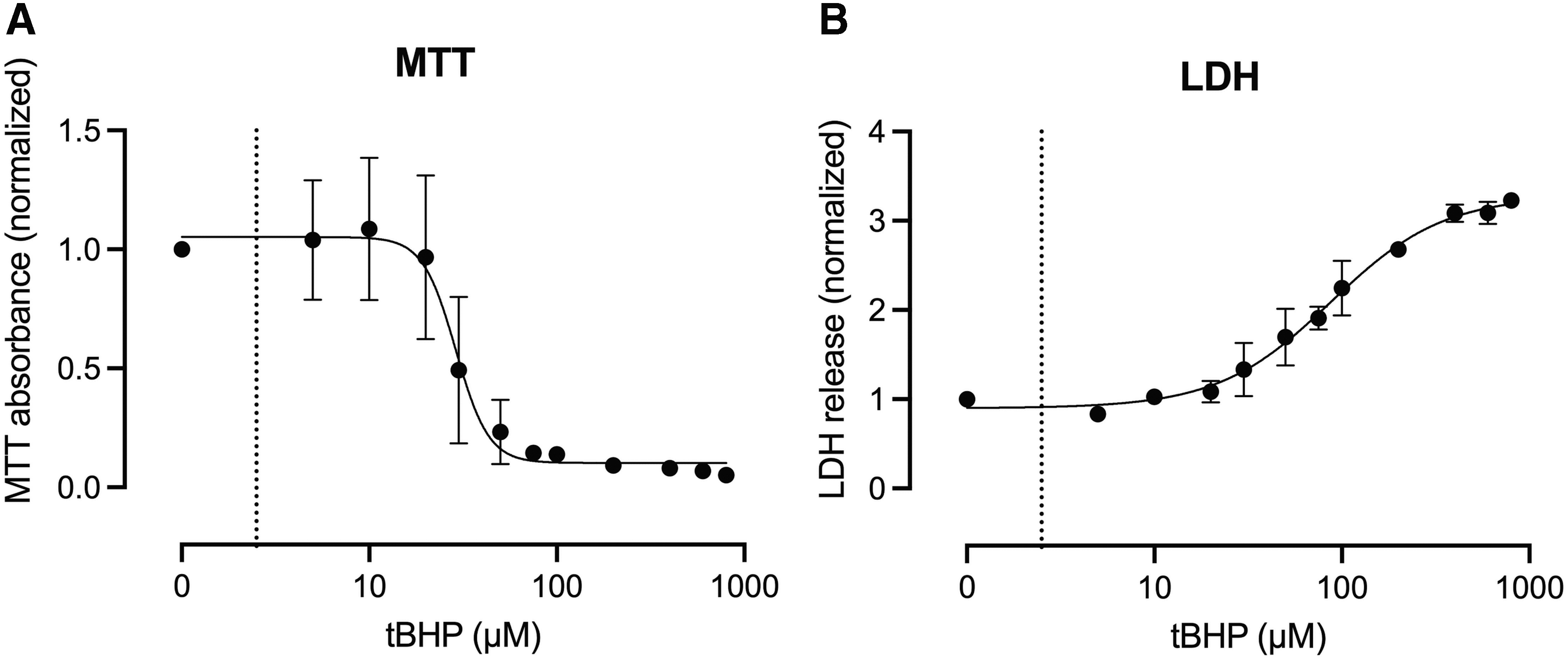
Exogenously applied chemically induced oxidative stress results in dose-dependent cytotoxicity in SIRC cells.
In a separate experiment, SIRC cells were exposed to a dose range of PM (1–300 μg/mL) for 24 h. MTT assay revealed dose-dependent loss of cell viability and proliferation with an IC50 of 13.6 μg/mL PM (n = 3–6; Fig. 2A), while LDH release was dose dependently increased with an EC50 of 149.1 μg/mL PM (n = 3–6; Fig. 2B). To our knowledge, this is the first report evaluating the effects of PM on SIRC cells.
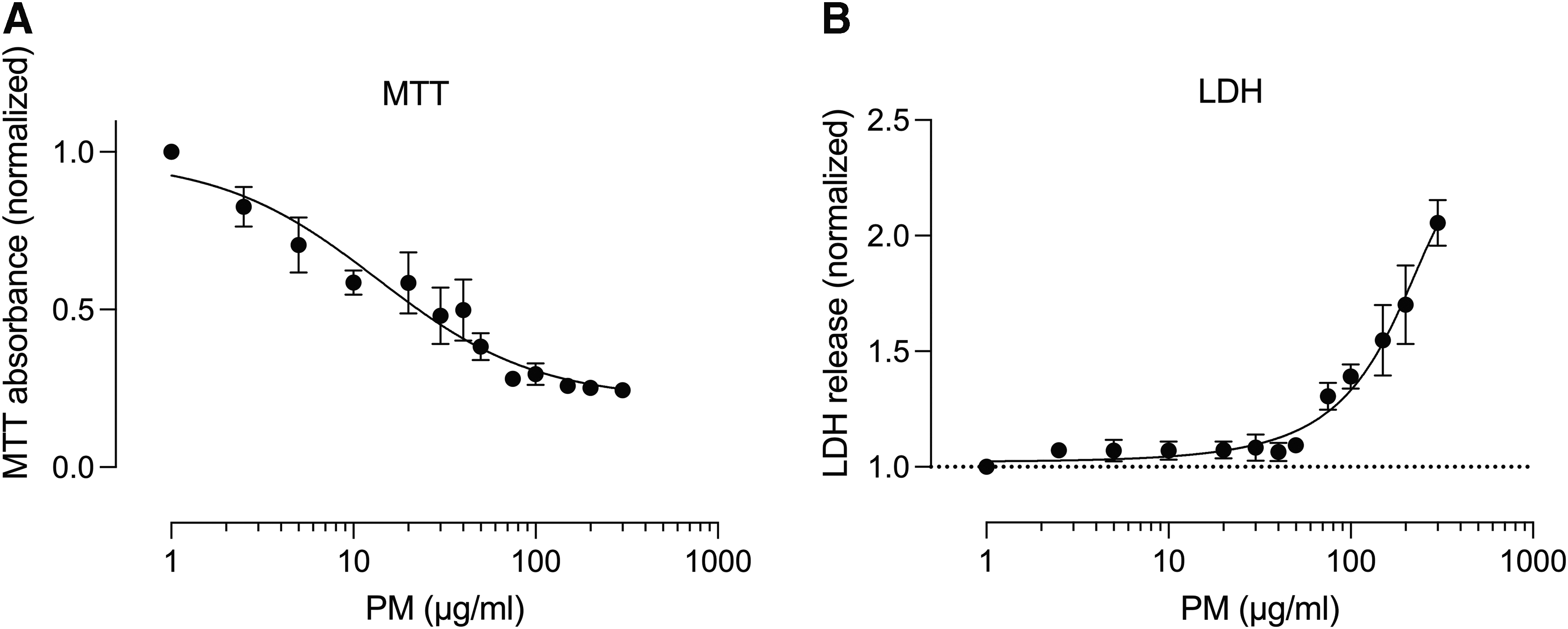
PM induces dose-dependent cell death of SIRC cells.
Mn-TM-2-PyP protects against PM-induced SIRC cell death
Two concentrations of PM (100 and 200 μg/mL) were selected to investigate the effect of the superoxide dismutase mimetic, Mn-TM-2-PyP, on PM-induced cytotoxicity. SIRC cells were pretreated for 30 min with Mn-TM-2-PyP (0.005% or 0.05%, based on previous studies in HCE-T cells 9 ) or left untreated (control), then exposed to PM at the indicated doses for a period of 24 h. Cells without PM were included as additional control group.
Both concentrations of Mn-TM-2-PyP reduced LDH release and prevented PM-induced (100 μg/mL) increases in LDH release (n = 3; Fig. 3A). A similar effect was noted when SIRC cells were treated with 200 μg/mL PM, however, this effect did not reach statistical significance, likely due to the higher level of LDH release and associated biological variability at this PM concentration (n = 3; Fig. 3B).
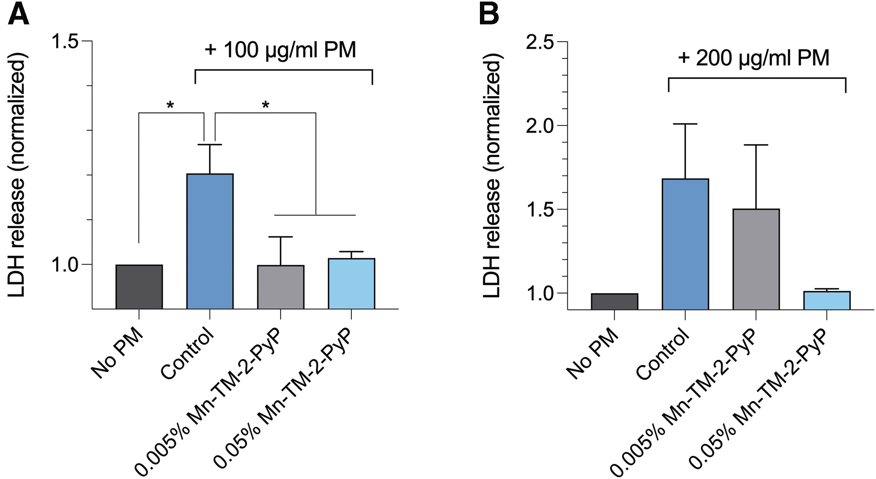
Mn-TM-2-PyP protects against PM-induced cytotoxicity in SIRC cells.
Mn-TM-2-PyP protects against PM-induced impairment of motility of SIRC cells
Scratch assays were performed on SIRC cells to assess the ability of Mn-TM-2-PyP to protect against PM-induced impairment of epithelial cell motility. Scratches were made using a 20-μL pipet tip and baseline images were taken immediately thereafter to measure the initial scratch width. Motility was assessed over a 24-h period in serum-free media, or serum-free media supplemented with 200 μg/mL PM in the presence or absence of 0.05% Mn-TM-2-PyP. Images were acquired after 24 h.
Two separate experiments were performed in different SIRC cell passages (P94 and P95). PM resulted in significantly impaired SIRC cell motility (1-way ANOVA, P < 0.001). Specifically, the widths of the scratch after 24 h were 54.4% ± 2.4% and 52.0% ± 0.8% of the original width in untreated cells. In the presence of PM, the scratch remained at 79.8% ± 2.6% and 69% ± 2.1% of the original scratch width after 24 h (P < 0.001; Fig. 4A, B). In contrast, Mn-TM-2-PyP improved PM-treated SIRC motility beyond the level in untreated cells; scratch widths were 42.0% ± 1.9% and 38.9% ± 0.8% (P < 0.001; Fig. 4A, B).
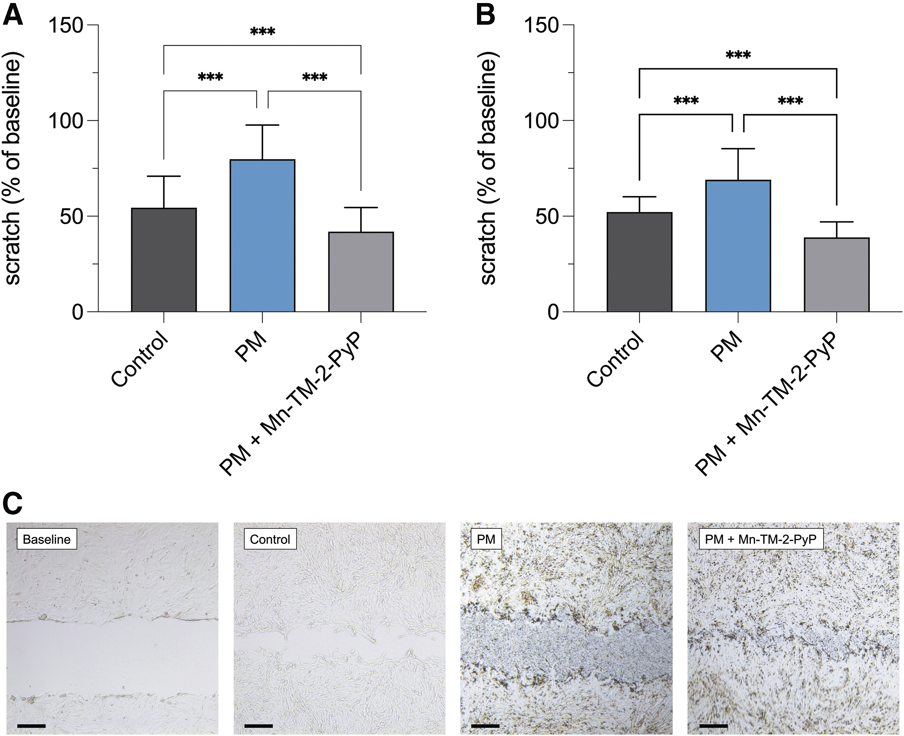
Mn-TM-2-PyP protects against PM-induced impairment of motility of SIRC cells. PM exposure significantly reduced motility of SIRC cells after 24 h in serum-free media, compared with control (no treatment). Mn-TM-2-PyP significantly enhanced SIRC cell motility in the presence of PM. Data were generated in 2 separate passages of cells:
Topical instillation of PM resulted in signs of irritant-induced conjunctivitis in rabbits
To determine the effects of PM exposure in vivo, a study in NZW rabbits was conducted. Baseline ophthalmic exams were performed to exclude any underlying ocular abnormalities. All rabbits showed absence of hyperemia (score 0) or any other ocular abnormality. Eyes were subsequently treated for a period of 7 days by 3 times-daily topical instillation with PM (5 mg/mL), which resulted in the presence of severe hyperemia (n = 16 eyes, median score 2, P < 0.001; Fig. 5A).
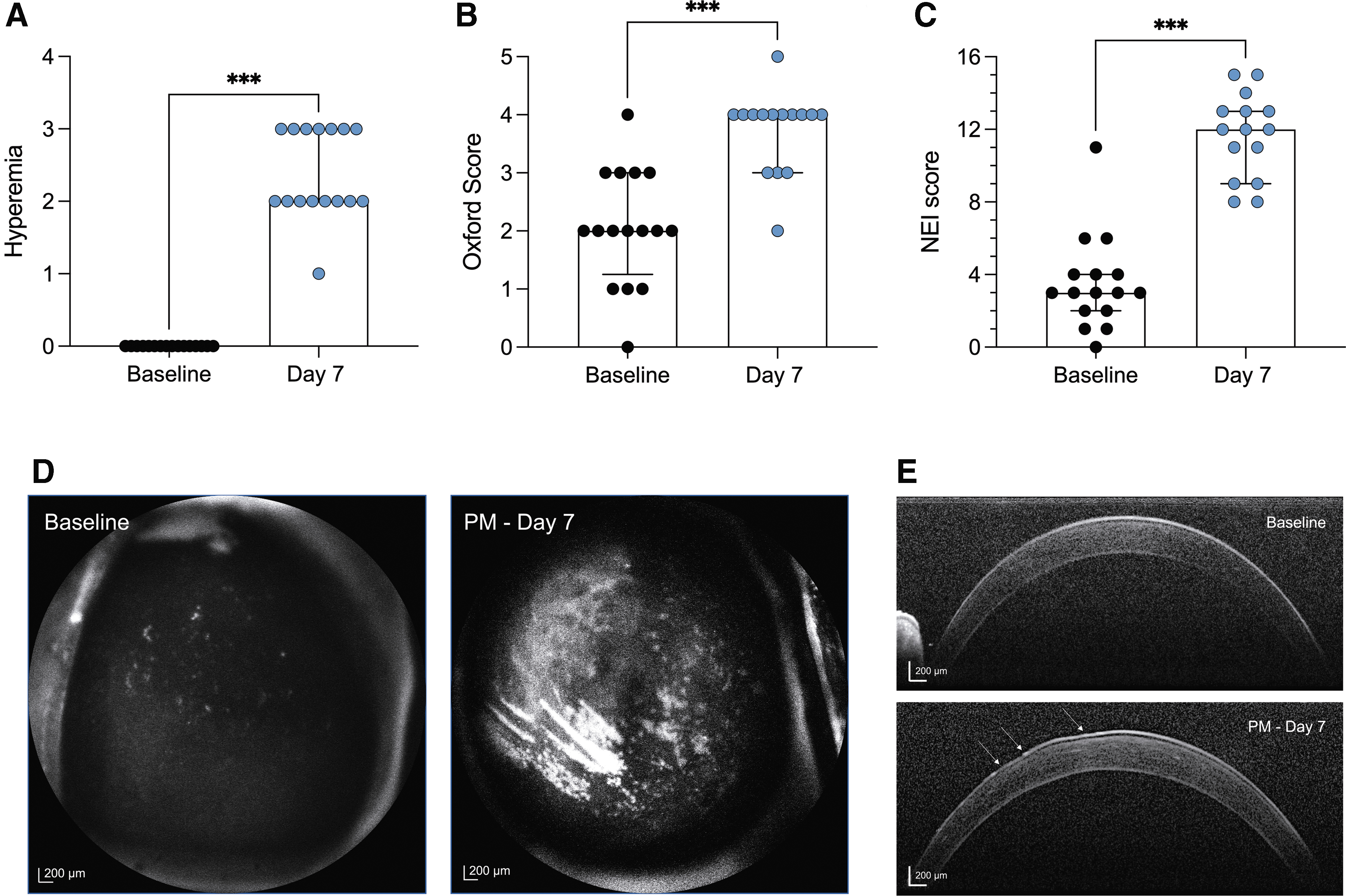
Topical instillation of PM elicits ocular surface damage, conjunctivitis, and signs of irritant-induced dry eye.
Tear volumes, assessed by Schirmer's test, showed a small but significant reduction (10 ± 1 mm vs. 7.7 ± 0.8 mm, n = 16 eyes, P < 0.05). Corneal fluorescein staining as clinically relevant assessment of corneal damage was significantly increased following PM exposure compared with baseline. Specifically, PM treatment increased median corneal fluorescein staining by 2 scores when analyzed using the Oxford grading (n = 15–16 eyes, P < 0.001; Fig. 5B, D) and by 9 scores when analyzed using the NEI grading (n = 15–16 eyes, P < 0.001; Fig. 5C, D). Changes to the tear film and cornea were visible by anterior segment spectral-domain optical coherence tomography (SD-OCT) imaging (Fig. 5E).
Mn-TM-2-PyP and ophthalmic lifitegrast reduce signs of PM-induced conjunctivitis
To evaluate the ability of lifitegrast and the antioxidant, Mn-TM-2-PyP, to attenuate conjunctivitis phenotypes induced by 10 days' exposure to PM, rabbits were randomized into 2 cohorts, each cohort comprising 2 male and 2 female rabbits. Cohort I was treated with lifitegrast (OS), whereas the contralateral remained untreated; cohort II was treated with 0.05% Mn-TM-2-PyP (OS), whereas the contralateral eye (OD) received saline.
Discontinuation of PM treatment did not improve hyperemia (Fig. 6A) or corneal fluorescein staining (Fig. 6B–D) in untreated and saline-treated eyes after 10 days. In contrast, topical instillation of lifitegrast attenuated both hyperemia (n = 4 eyes, P < 0.05; Fig. 6A) and attenuated corneal fluorescein staining when scored using both the Oxford (n = 4, P < 0.01; Fig. 6B) and NEI (n = 4, P < 0.001; Fig. 6C) scale. Similarly, Mn-TM-2-PyP significantly reduced ocular surface pathology. Specifically, hyperemia resolved after 10 days of treatment (n = 4, P < 0.001, Fig. 6A) with Mn-TM-2-PyP. Concomitantly, corneal fluorescein staining was reduced significantly when scored using both the Oxford (n = 4, P < 0.001; Fig. 6B) and NEI (n = 4, P < 0.001; Fig. 6C) scale. Treatments had no effect on tear volume (Table 1).
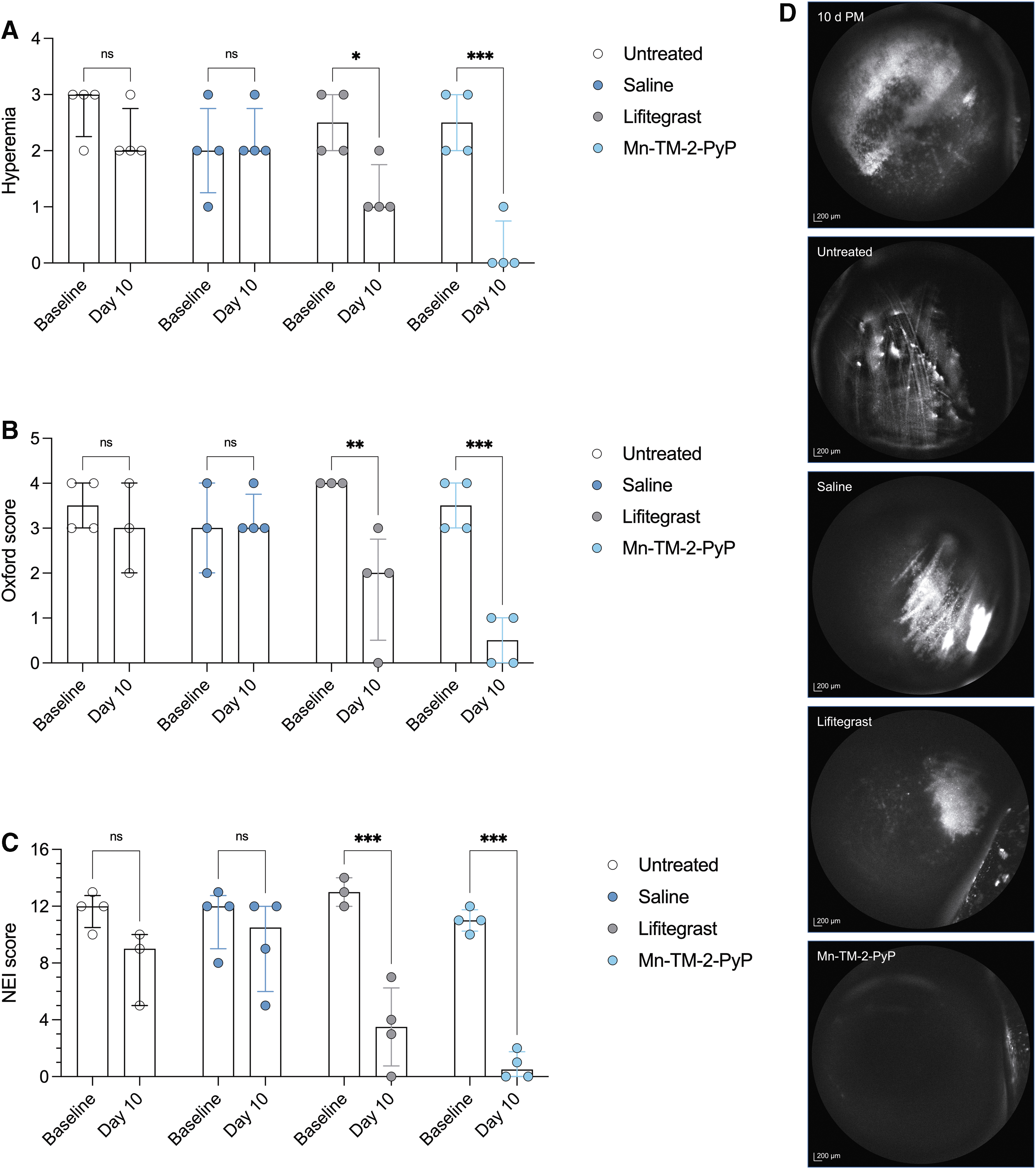
Mn-TM-2PyP significantly improves signs of PM-induced dry eye.
Tear Volumes at Baseline and Day 10
STT were sampled at baseline (following 10 days induction of allergic conjunctivitis by PM) and again following 10 days of topical treatments. There was no statistically significant effect of treatments on tear volume (2-way ANOVA; treatment, P = 0.26; time, P = 0.26; treatment × time, P = 0.96). Data are presented as mean ± SEM from n = 4 eyes per treatment group.
Mn-TM-2-PyP, manganese(III) tetrakis(1-methyl-4-pyridyl) porphyrin; PM, particulate matter; SEM, standard error of the mean; STT, Schirmer tear test.
Discussion
Exposure to pollution, smoke, and other allergens represents a significant health concern in both the civilian and military populations, given a strong clinical correlation between PM exposure and acute and chronic changes to the ocular surface.1,13 Especially individuals in densely populated areas exposed to urban air pollution are at risk for developing chronic signs and symptoms of ocular surface disease, including conjunctivitis and dry eye disease. However, a significant knowledge gap exists pertaining to the mechanisms of PM-induced ocular surface disease; standardized and validated experimental tools for drug discovery targeting the etiology of urban pollution are scarce.
The research presented herein offers new experimental tools for the comprehensive in vitro and in vivo testing of novel therapeutic approaches targeting PM-induced conjunctivitis and dry eye. Specifically, we herein present the first report of a rabbit model of PM-induced ocular surface disease that presents with clinical phenotypes of irritant-induced conjunctivitis.
Irritant-induced conjunctivitis is a common ocular surface disease that, when chronic, is often described under the umbrella term of dry eye disease or, more specifically, irritant- or pollutant-induced dry eye disease. In addition, we used rabbit corneal epithelial (SIRC) cells for the in vitro studies presented herein. While human corneal epithelial cells remain the cellular model of choice for permeability assays due to their ability to form stratified corneal epithelia, 14 SIRC cells provide an important drug discovery tool to establish target engagement and in vitro efficacy in rabbit cornea cells before performing in vivo efficacy studies.
In the past few years, PM-induced models of ocular surface disease have become more widely used, particularly in rodents. The first report published in 2017 exposed male BALB/c mice to topical treatments of PM up to 10 μm in size (PM10). Exposure (5 mg/mL, 4 × daily for 14 days) resulted in typical signs of ocular surface disease, including increased corneal fluorescein staining, increased TNFα, as well as concomitantly decreased tear breakup time, tear volumes, and conjunctival goblet cell number. 6 A follow-up study by the same group using the same experimental paradigm, but PM of smaller size (<2.5 μm; PM2.5), yielding overall similar results. 15
The first report of testing the effects of PM in Sprague-Dawley rats was published in 2019. In that study, topical PM instillation (20 mg/mL, 3 × daily PM10 for 5 days) resulted in increased corneal fluorescein, TNFα levels and matrix metalloproteinase 9 activity, all clinically relevant biomarkers for the diagnosis of ocular surface disease. Similar to published studies in mice, tear volumes were reduced in rats exposed to PM. Subsequently, several studies have demonstrated the pharmacologic efficacy of natural plant extract and phytochemicals in the rat model.16–18
Some studies since have expanded the experimental paradigm to testing dose ranges of PM, 19 utilizing different animal strains,19,20 formulating PM as aerosol, 7 and evaluating different types of PM, including titanium oxide. 20 The data from rabbits presented herein are consistent with these observations in rodents, suggesting conserved mechanisms of PM-induced ocular surface disease across species.
We have previously demonstrated efficacy of the synthetic antioxidant, Mn-TM-2-PyP, in the SiccaSystem® desiccating stress/scopolamine-induced murine model for dry eye disease. Mn-TM-2-PyP is a synthetic antioxidant of the porphyrin class that acts as catalytic antioxidant and superoxide dismutase mimetic, thereby scavenging reactive oxygen species. 21 Specifically, Mn-TM-2-PyP prevented against corneal damage to a greater extent than clinical standard of care, ophthalmic cyclosporine emulsion (Restasis®), and significantly reduced oxidative DNA damage in the cornea. 9
These data provided the first evidence of specific oxidative damage in the cornea using the desiccating stress/scopolamine model, confirming the etiological role of oxidative stress in dry eye disease that had been postulated based on genetic studies in rodents 22 and observations from clinical studies. Given in vitro studies implicating oxidative stress in PM-induced cytotoxicity in vitro, as well as our prior in vivo data for Mn-TM-2-PyP in human corneal epithelial cells and the desiccating stress/scopolamine mouse model, 9 Mn-TM-2-PyP was selected as a strong candidate for PM-induced dry eye.
PM has been extensively characterized for their chemical composition and contain several classes of chemicals well known to cause toxicity upon exposure, including (nitrated) polycyclic aromatic hydrocarbons. 3 Polycyclic aromatic hydrocarbons have been shown to elicit cytotoxicity and carcinogenicity through oxidative DNA damage.23,24
In vitro, PM exposure has been linked to the generation of reactive oxygen species and elevated levels of cellular oxidative stress.4,25,26 However, while several in vivo studies tested phytochemicals with antioxidative and anti-inflammatory properties,16–18 no study so far has evaluated the preclinical efficacy of synthetic antioxidants in models for PM-induced ocular surface disease. Given the mechanism of action of polycyclic aromatic hydrocarbons in inducing oxidative DNA damage, 24 and our previous work demonstrating that Mn-TM-2-PyP can protect against oxidative DNA damage in corneal epithelial cells in vivo, 9 it is likely that generation of reactive oxygen species and oxidative DNA damage is a contributor to PM-induced ocular surface damage in this model in addition to mechanical damage by PM particles.
Ophthalmic therapeutic development relies heavily on the use of rabbits as the large animal species of choice to test pharmacologic efficacy, especially given the size of the rabbit eye and the thickness of rabbit cornea that more closely resembles the human condition. 27
Rabbit corneal epithelial (SIRC) cells were selected for the in vitro studies presented herein based on the rationale that demonstrating target engagement in the selected species is needed before engaging in efficacy studies. While SIRC cells possess a more fibroblast phenotype and do not form strong tight junctions or stratified epithelia, 14 SIRC cells can respond potently to exogenous triggers with activation of the endogenous antioxidant system and secretion of proinflammatory cytokines.28–30 This makes SIRC cells suitable for target engagement and proof-of-concept studies in preparation for subsequent in vivo studies in rabbits.
Mn-TM-2-PyP protected against both PM-induced cytotoxicity and impairment in cellular motility in SIRC cells. It has previously been shown that SIRC cells respond by generation of intracellular oxidative stress, activation of the endogenous antioxidant system, and secretion of proinflammatory cytokines to exogenous stimuli, including ultraviolet radiation, lipopolysaccharide, and chemically induced oxidative stress.28–30 These data are in accordance with previous studies that have suggested generation of cellular oxidative stress as underlying mechanism of the deleterious effects of PM on epithelial cells4,25,26 and data from mouse and human corneal epithelial (HCE-T) cells that showed dose-dependent cytotoxicity and generation of cellular oxidative stress. 31
In vivo, Mn-TM-2-PyP was effective in reducing PM-induced hyperemia and corneal damage, suggesting that oxidative stress is an etiological contributor in PM-induced conjunctivitis in rabbits. Notably, the efficacy of Mn-TM-2-PyP was greater compared with lifitegrast, which was used as a clinical reference standard. Lifitegrast (formulated as Xiidra) is FDA approved for signs and symptoms of dry eye disease through preventing the interaction of lymphocyte function-associated antigen 1 with its ligand, intercellular adhesion molecule 1 (for review, see Haber et al. 32 ).
Overall, data obtained from rabbit corneal epithelial cells and topical instillation of PM onto the cornea in rabbits in vivo suggest the mechanism of PM-induced cytotoxicity and damage, involving impaired motility and generation of oxidative stress. However, the generation of oxidative stress was not evaluated mechanistically, which is one limitation of the present study and subject to ongoing research.
Future studies should also address the chronicity of PM-induced pathology and the effects of steroids and ophthalmic cyclosporine, which remain the clinical standard of care for allergic and irritant-indued conjunctivitis and dry eye disease, respectively. In addition, increasing group sizes must be considered when evaluating novel drug candidates that may exhibit less potent protection against PM-induced pathology. In this study, 1 eye was dosed with either Mn-TM-2-PyP or lifitegrast, while the contralateral received saline or remained untreated, respectively. For future drug discovery studies, it is advisable to treat both eyes with the same test article to eliminate the potential for crosscontamination as a result of the animals licking or scratching.
Conclusions
These data provide evidence that PM exposure in rabbits results in chronic ocular surface disease manifesting with conjunctivitis and corneal damage that is responsive to antioxidant and immunosuppressant therapy. To our knowledge, this is the first report of a large animal model to study PM-induced ocular surface disease. As ocular surface disease by exposure to PM remains an urgent clinical need, the present work provides standardized experimental paradigms for the comprehensive in vitro and in vivo testing of novel therapeutic approaches targeting PM-induced dry eye disease.
Footnotes
Acknowledgments
The authors would like to thank the members of the Department of Laboratory Animal Medicine at the University of North Texas–Health Science Center at Fort Worth, Fort Worth, TX for their dedication to animal welfare. Generous support by the North Texas Eye Research Institute is gratefully acknowledged. Sean Ogle (Experimentica Ltd.) provided excellent Research Operations support.
Authors' Contributions
Conceptualization: A.K.G., M.B.G., and S.K.; methodology: A.K.G., M.B.G., and S.K.; formal analysis: A.K.G., S.I., M.B.G., and S.K.; investigation: A.K.G., M.B.G., S.I., N.E.P., and S.K.; data curation: A.K.G., M.B.G., and S.K.; writing—original draft preparation: A.K.G., M.B.G., S.I., and S.K.; writing—review and editing: A.K.G., M.B.G., S.I., N.E.P., and S.K.; supervision: M.B.G. and S.K.; project administration: M.B.G. and S.K.; funding acquisition: S.K. All authors have read and agreed to the published version of the article.
Author Disclosure Statement
Employment: A.K.G., M.B.G., and N.E.P. (Experimentica Ltd.); Stock/equity ownership: S.K. (Experimentica Ltd.); S.K. (K&P Scientific LLC); A.K.G. (eyeNOS, Inc.). S.K. conducts academic research in areas of interest similar to the business interests of Experimentica Ltd. and K&P Scientific LLC. The terms of this arrangement have been reviewed and approved by Loyola University Chicago in accordance with its conflict-of-interest policy. The funders had no role in the design of the study; in the collection, analyses, or interpretation of data; in the writing of the article, or in the decision to publish the results.
Funding Information
This research was funded, in part, by NIH/R24 grant (EY032440). Additional funding by the Illinois Society for the Prevention of Blindness, the Richard A. Perritt, M.D. Charitable Foundation, and the Dr. John P. and Therese E. Mulcahy Endowed Professorship in Ophthalmology is gratefully acknowledged. Experimentica Ltd. and K&P Scientific LLC have provided in kind resources to conduct this study.