
Abstract
Abstract
Introduction:
The excellent results of harmonic scalpel (HS) for closure of blood vessels encouraged surgeons to use these instruments for cystic ducts. The use of HS on tissues other than blood vessels was started with little data about its efficacy or safety. Therefore, this study was designed to verify the safety and efficacy of HS for achieving safe closure of the cystic ducts after laparoscopic cholecystectomy.
Materials and Methods:
Sixty patients with symptomatic gallstone disease were enrolled in this prospective case control study. The patients were operated with laparoscopic technic and divided into two groups (n = 30). After the dissection of Calot's triangle, proximal cystic ducts on common bile ducts were sealed with single surgical clips (SC) in both groups. In the first group, distal of the cystic ducts was sealed with single SC and the gallbladders were removed with SC. In the second group, distal of the cystic ducts was sealed with HS and the gallbladders were removed as sealed cystic ducts with HS. Twenty-gauge catheters were inserted into the fundus of gallbladders in vitro and connected to the arterial line transducer set. A gradually increasing pressure was applied through a sphygmomanometer. The bursting pressures of the gallbladders were measured, and differences between HS and single SC groups were calculated with independent samples t-test. The value of P < 0.05 was accepted as significant.
Results:
The mean cystic duct bursting pressures in single SC and HS groups were 332.46 ± 4.62 and 343.06 ±4.28 mmHg, respectively. The mean values between the groups were found significant and indicated the superior results of HS.
Conclusions:
HS sealer could be an alternative method for cystic duct closure, especially for avoiding the clip displacement and migration of the clip. Results of this study indicated that HS sealer was as reliable as single SC and it could be accepted as a standard closure technic.
Introduction
We suppose that skepticism might have been a major reason for reluctance to use this technic. The aim of this study was to verify the effectiveness of HS for cystic duct closure in LC.
Materials and Methods
In this prospective study, 60 patients with symptomatic gallstone disease were operated with laparoscopic technic. An informed consent was obtained from all patients. Patients with acute cholecystitis and previous upper abdominal operation were excluded from the study. After preoperative evaluation and preparation for surgery, all cholecystectomies were performed by the same surgical team. Operative procedures were performed under general anesthesia, and the patients were placed in the standard supine Fowler's position with the right shoulder up. A uniform technic of the LC was applied, including the use of four trocar in the “American” position. A pneumoperitoneum was obtained by direct trocar application with a maximum pressure of 14 mmHg. A zero-degree optic scope was used. All cholecystectomies were performed without perforation, as the study design was based on the measuring of bursting pressures of cystic ducts. The patients were divided into SC and HS groups (n = 30). In the first group, dissection of Calot's triangle was performed with an atraumatic monopolar dissecting forceps. Closure of the cystic duct and artery was performed by SC. Mobilization of the gallbladder from the liver bed started from the posterior at the Calot's triangle and continued through the anterior using monopolar scissors. Consequently, gallbladders were removed with SC.
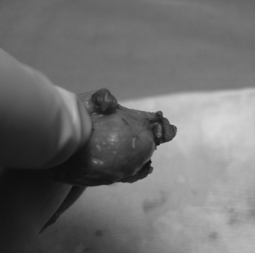
In the second group, the HS was used for dissection in the Calot's triangle, with the power level set at “5” (more cutting-less coagulation) due to avoidance of gallbladder perforation. For the closure and division of both cystic duct and artery, the instrument was set at the power level “2” (less cutting- more coagulation) due to avoidance of bile leakage. Cystic duct closing procedure continued as follows. First, it was confirmed that there were no calculi in the lumen of the cystic duct by moving the jaws of the SC applier up and down. Second, the cystic duct was closed with SC near the CBD, then HS's jaws were closed near the gallbladder-cystic duct junction until a click was heard. Third, the instrument was activated at the power level “2” and just before detaching the sealing was stopped, the jaws were then opened and re-sealed 2 mm away from the previous sealed part of the cystic duct until sealing was completed before detaching the cystic duct (Fig. 1). Finally, the gallbladder was removed from the liver bed via HS and extracted through the subxiphoid port with sealed duct by HS (Fig. 2). A subhepatic tube drain was inserted through the lateral port. Then, twenty-gauge catheters were inserted from the fundus of the gallbladder and fixed with 3/0 silk sutures ex vivo; and catheters were connected to the arterial line transducer set. An increasing pressure was applied through a sphygmomanometer. Bursting pressures were measured with an invasive arterial blood pressure measurement device, and measured systolic pressures were accepted as the bursting pressure (Fig. 3). The maximum level of the pressure that is just before the sudden fall as a result of explosion was recorded as the bursting pressure. Results were written down to Statistical Package for the Social Sciences version 10.0 software (SPSS, Inc., Chicago, IL). At the same time, the operative time was recorded. Patients were discharged on the morning of postoperative day 1 after the removal of the subhepatic drain. Independent samples t-test was used for parametric variables between HS and SC groups. The value of P < 0.05 was accepted as significant.
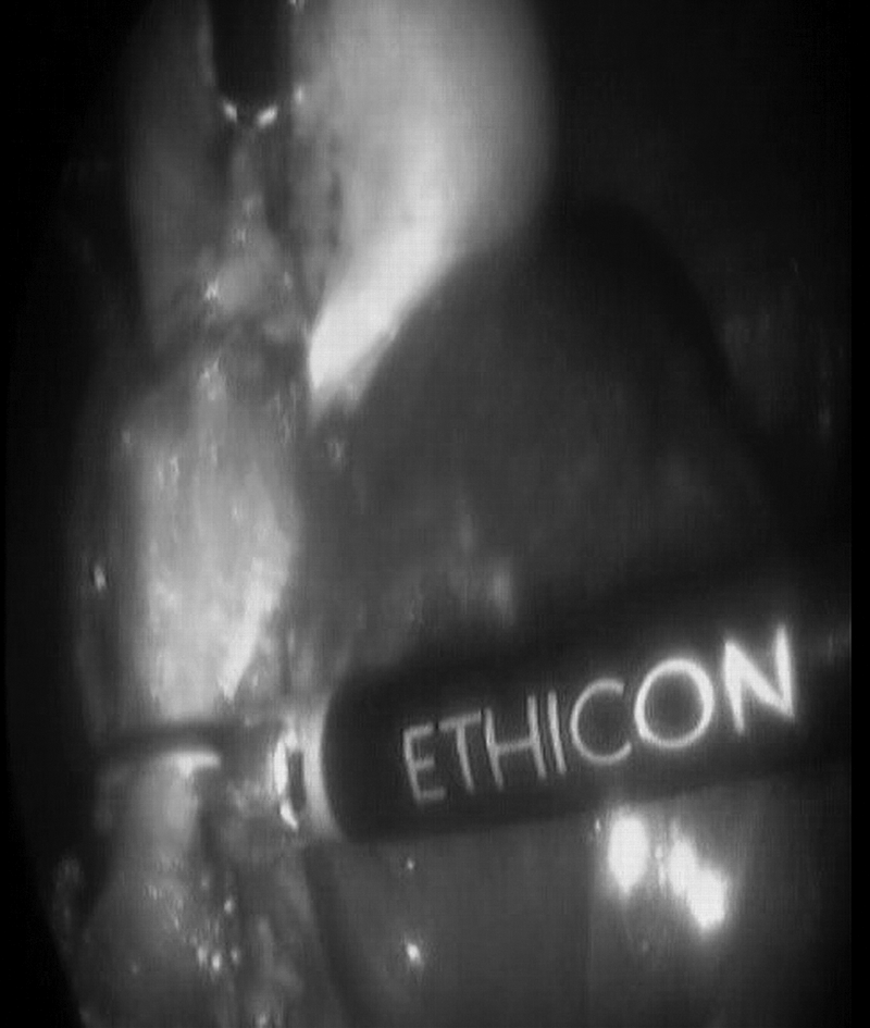
Cystic ducts near the gallbladder-cystic duct junction were sealed by harmonic scalpel in the second group.
Cystic duct sealed by harmonic scalpel.
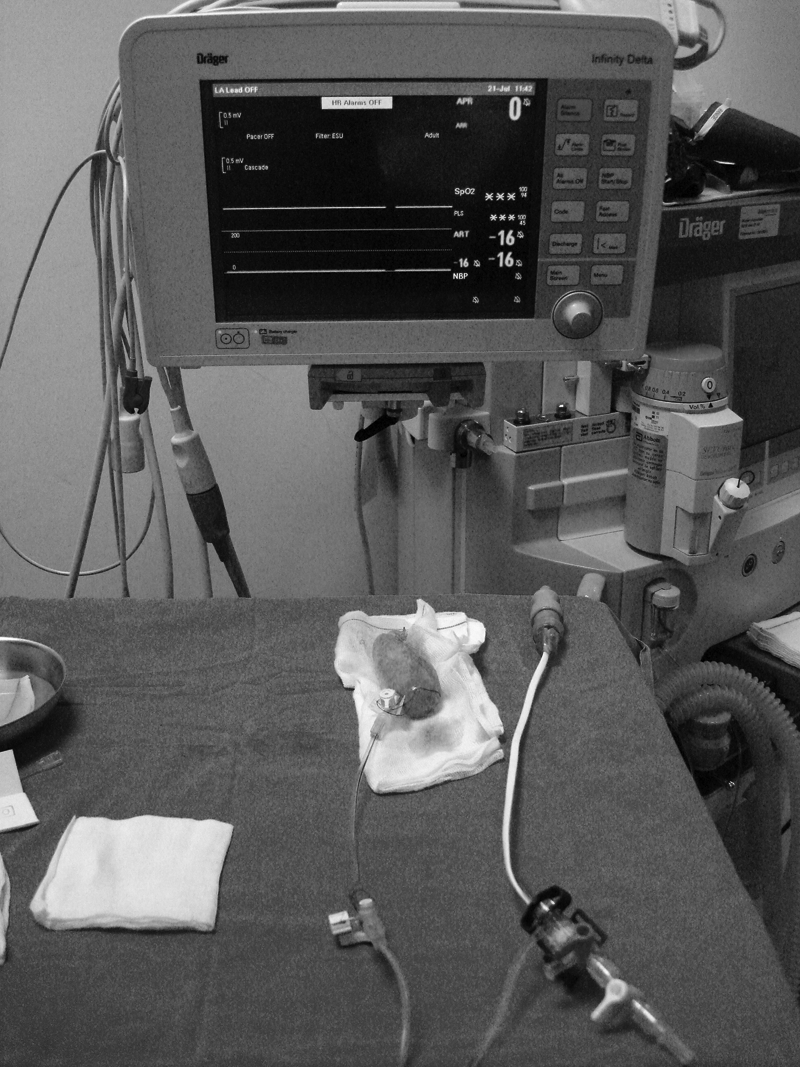
Twenty-gauge catheters were applied to fundus of gallbladder and fixed with 3/0 silk sutures ex vivo, and catheters were connected to the arterial line transducer set.
Results
The present study included 60 patients with symptomatic gallstone disease. There were 39 women (65%) and 21 men (35%). The average age of the patients in HS and SC groups were 47.2 ± 2.36 years (25–80 years) and 46.7 ± 1.43 years (34–63 years), respectively (P = 0.93). The average operation time in HS and SC groups was 33.3 ± 1.43 min and 32.9 ± 1.37 min, respectively (P = 0.85). Neither minor nor major bile leaks were encountered in either group throughout the LC from cystic duct on gallbladder. Ex-vivo measurements of the mean cystic duct bursting pressures were 332.46 ± 4.62 mmHg for SC and 343.06 ± 4.28 mmHg for HS groups. Differences of the mean cystic duct bursting pressures between HS and SC groups indicated the superiority of HS (P = 0.04). All the results were shown in Table 1.
HS, harmonic scalpel; SC, surgical clip; SD, standard deviation.
Discussion
LC was accepted as the gold standard within 5 years of its introduction by consensus.1,2 Closure of cystic duct during LC by SC was the most frequently used technic.3,4 The lumen of the cystic duct must be completely sealed, and there should not be any leakage from the CBD. Although SC was known as a safe closure method, bile leakage due to clip displacement from the cystic duct stump is a potential pitfall of LC. 22 The slippage of the clips and migration into the biliary tract or duodenum varies between the clip types. 23 Further, in the time of application, the metallic clips can fall from the applicator. 24 This can cause recurrent abdominal abscess. 25 Therefore, some alternative technics for the closure of cystic duct such as securing sutures and absorbable clips have been introduced till now.5–7
Some electrothermal energy sources such as monopolar diathermy have been used for the dissection of Calot's triangle and gallbladder bed since the first LC; but this electrothermal energy can result in unrecognized transfer of energy to the biliary tract, which causes bile leak, external biliary fistula, or biliary stricture and it cannot be used for the closure of cystic duct.14,15 After the beginning of the use of HS for sealing of the cystic artery, some surgeons also started to investigate the role of HS for sealing of the cystic duct. According to the results of these studies, the use of HS was accepted as a reasonable alternative for closure of cystic duct smaller than 6 mm in diameter.8–13 According to Westervalt, 8 there were no bile leaks from the cystic-duct stump in his 100 patients in whom the closure and division of the cystic duct were solely achieved by the HS. Similar findings were reported by Tebala. 9 In the study by Huscher et al., 10 bile leaks were encountered in 7 of the 331 patients (2.1%), in whom closure and division of the cystic duct were solely obtained by the HS group, compared with 3 of the 130 patients (2.3%), in whom closure and division of the cystic duct were obtained by HS plus endo-loop of absorbable suture material group. The results of the bile leaks in this study could be explained by the application method of the HS. Huscher stated that the blades were first applied more proximally for a few seconds and then they were applied a few millimeters distal to the previous application site, holding the grasp until the division of the duct was completed. First application of HS to cystic duct can only be made on a visual basis. The instrument has no feedback sensors capable of simple sealing of the cystic duct. We supposed that it would be rather difficult to determine the amount and type of damage under the cystic duct by applying the HS for a few seconds to the proximal site as the first application. At the same time, “a few seconds” varies from surgeon to surgeon. Whether the sites of proximal application were the source of some bile leaks in their study remains uncertain, although the possibility theoretically exists. In our study, as well as in Huscher's study, the HS were applied more proximally for a few seconds and then applied a few millimeters distal to the previous application site; but there was no bile leaks from the cystic-duct stump on the gallbladder during the operation and on the ex vivo experiment until the bursting time. In another study, Bessa et al. 11 reported that the HS was as safe and effective as the commonly used clip and cautery technic in achieving safe closure and division of the cystic duct in the LC. Further, it provides a superior alternative to the currently used high-frequency monopolar technology in terms of shorter operative time and lower incidence of gallbladder perforation. The statistically significant shorter mean operative time in the HS group was attributed to several factors by the authors. 11 The first is the statistically significant lower incidence of gallbladder perforation in the HS group with subsequent avoidance of time loss in abdominal lavage and spilled stones retrieval. The second is that the use of HS prevents the frequent extraction and reinsertion of different instruments with avoidance of time loss. At the same time, HS does not form smoke; thus, surgeons work in a clear operative field and do not spend time clearing the vision. However, in our study, the operation times between the HS and SC groups were not found significantly different. This result depends on our meticulous and slow technic for avoiding the perforation in both groups. According to the study by Vu et al., 12 if the cystic duct is greater than 6 mm in diameter, division by HS is not recommended. On the other hand, according to Huscher et al., 13 effective sealing of the cystic-duct stump by the HS alone has been histologically confirmed. In this study, all morphologic changes were found within 1.5 mm of the cutting edge. They preferred measurement of airtight pressures of cystic ducts. The airtight pressure of the sealed cystic duct was calculated to be higher than 320 mm Hg. In our study, we preferred measuring the bursting pressures with saline, and our results were almost the same. All measured cystic duct bursting pressures were found above all the kinds of CBDPs.13,18–21 On the other hand, all gallbladders underwent sealing of the cystic duct near the gallbladder-cystic duct junction by HS; there were no apparent bile leaks from the cystic duct during the extraction and the experiment until bursting time. Although our study was designed as an experimental study, the findings supported the results of previous studies.
Using diathermy or electrocautery is one of the important factors that causes bile duct damage. Therefore, the injury is named as diathermy-induced bile duct injury. 16 Ultrasonic instruments were developed to eliminate the collateral damage associated with electrocautery. 26 The lateral energy spread is minimal, and the risk of distant tissue damage is lower than that of high-frequency electrosurgery. 27 In addition, ultrasonic devices can cut and coagulate at a lower temperature (60°C–100°C) than what occurs during electrosurgery (150°C) or laser surgery (200°C).10,17 At the same time, we do not know of a report indicating any specific hazards inherent about the use of HS in the closure and division of the cystic duct.
Wise et al. 28 demonstrated that simple SC applied to the cystic duct could not be displaced by a pressure of 300 mmHg. In our study, we found bursting pressures of cystic ducts almost twofold higher than contraction pressure in the HS group. According to our study, HS was found at least as safe as SC for closure of cystic duct; and we also demonstrated the effectiveness and safety of HS at our hospital.
In conclusion, the above-mentioned hazards inherent in the use of metallic clips were not encountered when closure and division of the cystic duct was achieved with the HS. The use of ultrasonic technology in the LC provides an alternative to the currently used SC.
Footnotes
Disclosure Statement
No competing financial interests exist.