
Abstract
Background:
Assessment of lymph flow has proven challenging. Transit-time ultrasound technique (TTUT) is the first technique that provides real-time quantitative lymphatic flow values. In cardiothoracic surgery and neurosurgery, this technique has tremendous clinical value in assessing surgery quality and predicting outcomes. The objective of this study was to measure lymph flow before and after lymphaticovenous anastomosis (LVA), using TTUT.
Methods and Results:
Consecutive patients with peripheral lymphedema undergoing LVA were included. Preoperative workup was performed using indocyanine green (ICG) lymphangiography. Perioperatively, the Transonic® Microvascular Flowprobe was used to measure lymph flow before and after anastomosis. Twenty-five patients with International Society of Lymphology stage IIA (68%) and stage IIB (32%) peripheral lymphedema were included. Lymph flow velocities ranged from 0.02 to 0.80 mL/min (mean 0.25 ± 0.19) before anastomosis and from 0.02 to 0.86 mL/min (mean 0.27 ± 0.22) after anastomosis (p = 0.340). Mean flow values were significantly higher in the upper extremities compared with the lower extremities. Furthermore, there was a decrease in flow in patients with ICG stage IV in comparison with ICG stage III. Clinical outcomes could not be directly correlated with flow values in these individual cases.
Conclusion:
TTUT micro-flowprobe is a suitable instrument to measure real-time quantitative lymphatic flow in both lymphatics and LVA. It can confirm patency of lymphatic collectors and LVA peroperatively. Significantly higher lymph flow velocities were found in upper extremities in comparison with lower extremities, both before and after LVA. Further studies should be performed to evaluate lymph flow values and clinical correlation.
Introduction
Peripheral lymphedema is a chronic condition characterized by swelling of the affected extremities due to the accumulation of protein-rich lymphatic fluid in tissues. Damage to the lymphatic system, caused by surgery or radiation therapy, impairs lymphatic function and reduces flow.1–3 Lymphaticovenous anastomoses (LVA) between a collecting lymphatic vessel and a subdermal vein aim to bypass these obstructed areas to redirect the lymphatic fluid back to the circulation. This procedure can alleviate symptoms and improve the quality of life, particularly in early-stage lymphedema.3–7
Lymphatic flow is generated by lymph production and propulsion. It is known that collecting lymphatic vessels have a smooth muscle layer and move lymph fluid actively through pulsation. This stretch-sensitive layer responds according to the Frank–Starling mechanism, by increasing frequency and stroke volume of pulsation as a result of increased ultrafiltration. 8 Furthermore, one-way valves in the vessels contribute to lymph propulsion and prevent retrograde flow; however, lymphedema may result in lymphatic valvular incompetence.2,4,8 While the lymphatic system has been well studied, there is a paucity of literature that has objectively quantified lymphatic flow or flow velocity; assessment of lymph transport has proven historically challenging. 9
To date, measurements of lymph flow have been performed through several direct and indirect techniques, for example, fluorescence microlymphography, lymphoscintigraphy, near-infrared fluorescence imaging, or magnetic resonance lymphography (MRL).10–15 With the advent of the Transonic Flowprobe for microsurgical vessels, lymph flow can be measured quantitatively and directly using a transit-time ultrasound technique (TTUT). This modality has been widely used in cardiothoracic surgery and neurosurgery for years, and it is considered as a valuable tool to assess status and patency of arterial grafts and predict outcomes, such as graft failure and mortality.16,17 It provides stable and reliable measurements of hemodynamic changes during cerebrovascular surgery.18,19
The technique enables measurement of volume flow directly in minute vessels and ducts smaller than 1 mm in diameter. Because the measurements are not dependent on the presence of particulate matter, as are Doppler velocity measurements, clear lymph flow can also be measured directly. The flexible neck of Transonic's micro-flowprobe permits optimal positioning of the probe around the lymph duct or vessel for measurement of flow in lymph ducts, vessels, or grafts from 0.5 to 4.0 mm in diameter.20–23 In the field of lymphedema surgery, early results of lymph flow measurements using this TTUT showed promising results; however, only a sample size of two patients was described. 24
Therefore, we conducted this current study to measure lymph flow values before and after LVA using TTUT in patients with peripheral lymphedema. We hypothesized that both before and after anastomosis a flow >0 mL/min is measurable in the lymphatic collecting vessel, indicating patency of the anastomosis and restoration of lymph drainage.
Materials and Methods
Patient selection
This observational descriptive study was performed from June to November 2018 on consecutive patients who suffered from peripheral lymphedema and underwent LVA procedure at Maastricht University Medical Center.
Inclusion criteria consisted of stage IIA or stage IIB lymphedema of upper or lower extremity according to the International Society of Lymphology (ISL) and stage III or stage IV according to Narushima indocyanine green (ICG) classification, regardless of etiology. 25 Exclusion criteria were cancer recurrence and active skin infection. The data were prospectively collected.
ICG lymphangiography
Preoperatively, all patients underwent ICG lymphangiography to determine whether patent lymphatic ducts eligible for LVA were present. ICG 0.05 mL, 0.5% (VERDYE® 25 mg for solution; Diagnostic Green GmbH, Aschheim, Germany), was intracutaneously injected in the second and fourth web spaces of the hand. After 15–30 minutes, the fluorescence was detected using a near-infrared camera (Fluobeam®; Fluoptics, Grenoble, France). The surgical incision was made based on the location of the lymphatic vessels visualized by the near-infrared camera.
Surgical technique
All operations were performed by the same plastic surgeon (S.S.Q.), according to the method described by Koshima et al. under local anesthesia. 26 Patients were advised to stop the use of stockings or conservative treatment in the first 2 weeks postoperatively to prevent damage to the recently performed anastomosis. After 2 weeks, they were allowed to continue with their regular conservative treatment.
Measuring method
Once a collecting lymphatic vessel and a subcutaneous vein of approximately the same caliber were found within reachable distance of each other, a 0.7 mm Flowprobe (Microvascular Flowprobe HT363; Transonic Europe, Elsloo, The Netherlands) was used to measure the flow using the Transonic® protocol for Quantitative Patency Assessment. 27 It consists of a probe body that houses ultrasonic transducers and a fixed acoustic reflector. The Flowprobe was calibrated by submerging the probe in sterile saline to remove the air, and thereafter, it was covered with room temperature sterile ultrasonic gel. Preferably, lymphatic vessels with a diameter covering >70% of the probe-sensing window were included to obtain a reliable value. Approximately 1 cm length of the superficial collecting lymphatic vessels was harvested, providing adequate room for introduction of the probe head and abrogating the possible interference of fat with acoustic transmission. The flow direction was assessed using the milking test. Thereafter, the probe covered with gel was applied to the lymphatic vessel without mechanical stress to the vessel (Fig. 1).
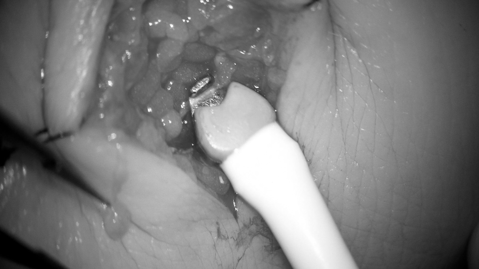
Lymphatic vessel being measured using the 0.7 mm probe.
An electrical excitation causes the Flowprobe transducers to emit plane waves of ultrasound, which intersect the vessel/duct under study in both upstream and downstream directions and then bounce off the fixed acoustic reflector on the opposite side of the vessel/duct. The waves again intersect the vessel and are received by the opposing transducers where they are converted into electrical signals. From these signals, the Flowmeter derives an accurate measure of the “transit time” it takes for the waves of ultrasound to travel from one transducer to the other. Once a repeatable, reproducible flow waveform was seen on the monitor (AureFlo; Transonic Europe), the flow was observed for 15 seconds. The mean flow value after 15 seconds of this reproducible flow was noted.
The exact same procedure was followed to measure the blood flow in the subdermal vein. Subsequently, the LVA was performed (Fig. 2), and anastomotic flow was measured at the site of the recipient vein following the same method described above (Fig. 3).
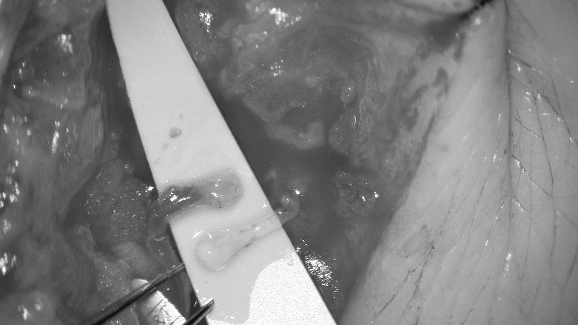
Lymphatic vessel and vein before formation of the anastomosis.
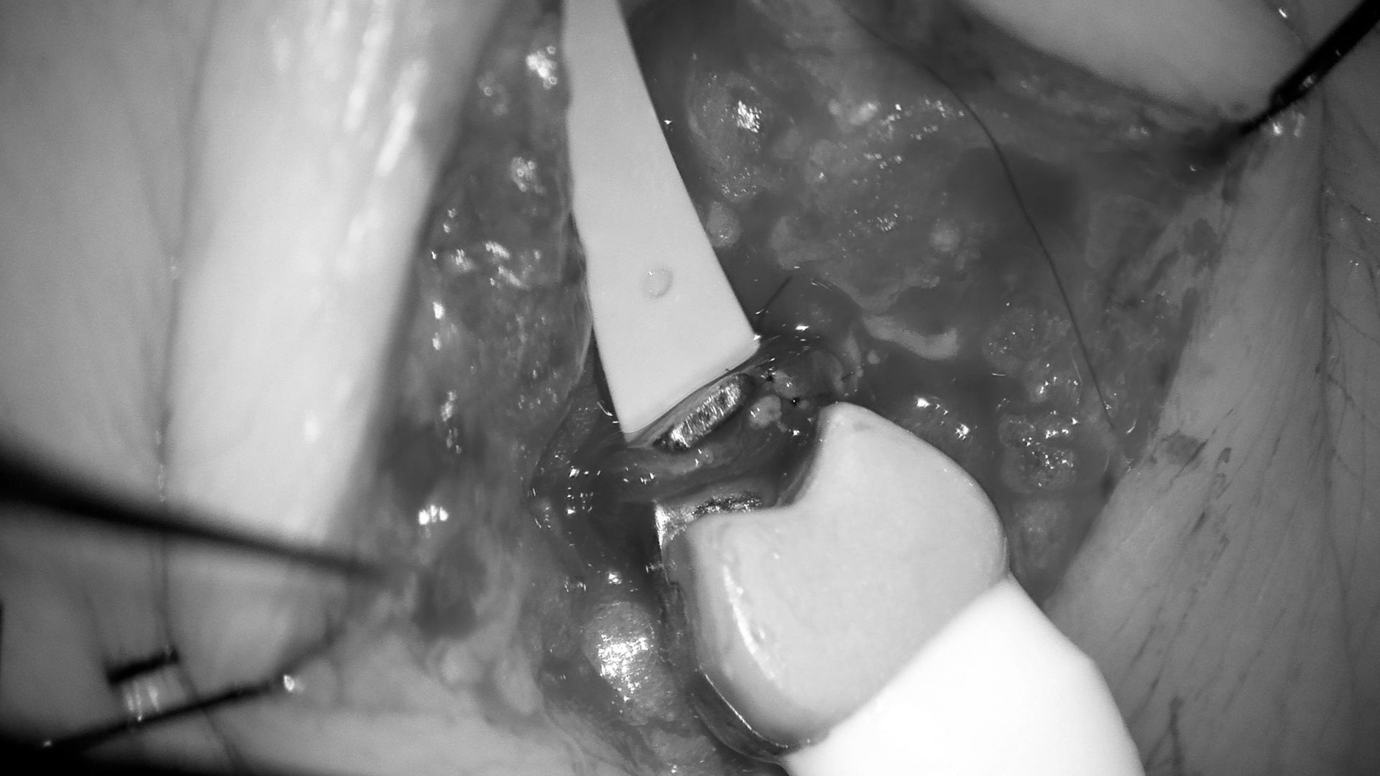
Lymphaticovenous anastomosis being measured using the 0.7 mm probe.
Flow values were given in mL/min and were reported for subdermal veins, lymphatic collectors, and LVA. An estimated diameter of the lymphatic vessel and the subdermal vein used for the LVA was given by the operating plastic surgeon and was reported as well.
To avoid unnecessary extension of the duration of the surgery, the measurement was completed in one LVA only, regardless the total number of LVA performed per incision.
Outcomes
Changes of the lymphatic flow values before (measured at the lymphatic collecting vessel) and after (measured at the recipient vein) the LVA were measured and described.
Statistical analysis
Paired sample t-test was used to determine the significance of the differences between the means before and after anastomosis. Independent sample t-test was performed to compare the mean between upper and lower extremities. One-way analysis of variance was used to compare the mean flow of patients with different ICG stages of lymphedema. Results were analyzed with SPSS Statistics 25 using an alpha level of 0.05 to determine significance.
Results
Patient characteristics
In total, 25 patients were identified (21 females and 4 males) with a mean age of 57.7 years (range 35–81 years) and a mean body mass index of 27.4 kg/m2 (range 20.8–35.5 kg/m2). All included patients presented secondary lymphedema except one patient, who suffered from primary lymphedema. ISL stage IIA was present in 17 patients and stage IIB in 8 patients. ICG lymphangiography showed ICG stage II in 1 case, stage III in 14 cases, and stage IV in 9 cases. ICG stage was not reported in one patient. Upper extremity lymphedema was present in 20 patients, lower extremity lymphedema in 4 patients, and both the abdomen and lower extremity lymphedema in 1 patient. The mean time since the onset of lymphedema was 7.7 years (range 1–23 years).
Flow rates
Blood flow in the subdermal veins ranged from 0.02 to 0.88 mL/min (mean 0.26 ± 0.24 mL/min). Lymph flow before anastomosis ranged from 0.02 to 0.80 mL/min (mean 0.25 ± 0.19 mL/min) and from 0.02 to 0.86 mL/min (mean 0.27 ± 0.22 mL/min) after LVA. The difference between the means was not statistically significant (p = 0.340). Flow increased after anastomosis in 11 patients (mean difference 0.19 mL/min), whereas decrease in flow after anastomosis was noticed in 11 other patients (mean difference 0.11 mL/min). Lymphatic flow values were equal before and after anastomosis in one patient (0.18 mL/min). In two patients, lymph flow after anastomosis was not measured due to technical failure. Mean diameters of lymphatic collectors and subdermal veins used for LVA were 0.50 and 0.55 mm, respectively. Patient characteristics, flow values in mL/min, and diameters are presented in Table 1.
Patient Characteristics, Flow Values in Milliliter/Minute, and Diameters in Millimeter
Estimated value.
A, abdomen; F, female; ICG, indocyanine green; ISL, International Society of Lymphology; LE, lower extremity; LV, lymphatic vessel; LVA, lymphaticovenous anastomosis; M, male; NR, not reported; UE, upper extremity.
Despite equal diameters of both lymphatic collectors and subdermal veins used for LVA in the upper and lower extremities, the mean flow values in the lymphatics and LVA were significantly higher in the upper extremities compared with the lower extremities (Table 2). Furthermore, there was a decrease in flow in patients with ICG stage IV in comparison with ICG stage III, without reaching statistical significance (Table 3).
Comparison of Flow (Milliliter/Minute) and Vessel Diameter (Millimeter) of Upper and Lower Extremities
Bold values denote statistical significance at the p < 0.05 level.
Mean Flow Velocity in Patients with Different Indocyanine Green Stages in Milliliter/Minute
Mean values of ICG stage II were left out of the comparison since this was a single case.
Discussion
The current study described lymph flow values before and after LVA, measured in 25 lymphedema patients using the Transonic transit-time ultrasound Microvascular Flowprobe. The mean lymph flow slightly increased after anastomosis; however, this difference did not show statistical significance. The lymph flow after LVA was significantly higher in the upper extremity than in the lower extremity. No significant differences were found in the lymph flow depending on the ICG stage.
Assessment of lymph transport has proven challenging in the past decades. Literature on lymphatic flow is scarce, and adequate measurement methods have been lacking. Direct and indirect measurement methods described unalike results, reference values were not available, and therefore, no cutoff values could be assessed to determine the presence of lymphedema. Furthermore, most of the knowledge on this subject is known from animal studies and few clinical studies were available. Due to this reason, values obtained in those studies could not be extrapolated to humans because of obvious differences in the body volume and upright body position. For these reasons, comparison of our findings with most previous work would not be valid.
A combination of intrinsic lymphatic pumping and extrinsic forces causes lymph fluid propulsion. 28 Olszewski and Engeset 29 recorded spontaneous contractility with intralymphatic pressure tracing through cannulation of leg lymphatics. The mean flow values of 0.25 ± 0.04 mL/h during rest and 1.62 ± 0.64 mL/h during pulsation were measured in superficial lymphatic vessels in nonedematous human legs. Movements of the leg and foot significantly increased flow, due to skeletal muscle contractions functioning as an extrinsic pump.28,30 Using lymphoscintigraphy, Modi et al. 31 showed a decrease in intrinsic lymphatic pump force in patients with secondary lymphedema by comparing velocities of labeled lymph fluid in uncuffed healthy (8.9 ± 5.8 cm/min), cuffed healthy (7.6 ± 10.5 cm/min), and cuffed lymphedematous (3.2 ± 8.9 cm/min; p = 0.004) limbs. Using the same technique, Stanton et al. 32 found reduced lymph flow in lymphedematous arms. Studies conducted with MRL confirmed this reduced flow (0.30–1.48 cm/min).13–15
It is known that accumulation of interstitial fluid in combination with partial outflow obstruction in lymphedema causes elevation of the afterload and consequently increases lymphatic capillary pressure.11,33,34 Fischer et al. 10 used fluorescence microlymphography to measure flow velocity of lymphatic capillaries in the dorsum of the foot. In healthy subjects, the median velocity during the first 45 seconds after dye injection was 0.51 mm/s [0.27;0.61] and 9.7 μm/s [6.9;14.2] in resting conditions, 1 minute after injection. In accordance with the aforementioned, they found higher initial filling velocities (0.89 ± 0.43 mm/s) in patients with primary lymphedema in comparison with controls. 35
All indirect methods are subjected to increase of the interstitial pressure as a result of dye injection, which will consequently increase flow velocities and thus affect outcomes.9–11 Furthermore, velocities were calculated from the lymph advancement during a given time, and therefore, these methods might be vulnerable to measurement errors. By contrast, direct methods allowed to measure the volume of lymph fluid that passes a certain point per time unit.
TTUT is a technique that provides real-time quantitative lymphatic flow values. Although this is an invasive method that can only be used during surgery, integrity of the vessel is preserved. Chen and Zhao 24 were the first to describe TTUT in the context of lymphatic surgery. Flow values between 0 and 1.2 mL/min in the lymphatics and between 0.22 and 1.4 mL/min in the LVA were observed, which were comparable to the results of the current study. Diameters and flow rates of subdermal veins were comparable to those of lymphatic vessels, confirming patency of both veins and lymphatics and allowing an appropriate end-to-end anastomosis.
Observations in the present study might suggest that lymph flow of the lower extremities is lower compared with the upper extremity; however, only five patients with lower extremity lymphedema were included, of which only four LVA were measured. Furthermore, outliers in the upper extremity group were observed both before (0.76 and 0.80 mL/min) and after (0.86, 0.70, and 0.62 mL/min) LVA.
Previous literature described differences between the lymphatic system in the upper and lower extremities due to gravity, which increases capillary filtration pressure during upright position. Stanton et al. 36 found a denser network of lymphatics in the lower extremity compared with the upper extremity. They suggested that the lymphatic vessels might adapt to higher lymph fluid production because of increased filtration pressures of the leg. 30 Likewise, Mellor et al. 37 showed an increased density of lymphatic collectors in edematous arms in comparison with normal arms. Lymphangiogenesis is thought to occur in case of accumulation of lymph fluid to enhance lymphatic drainage. However, a significantly lower lymph flow in the lower extremity was not described previously.
There are certain limitations to this study. Physiological tremor of the surgeon during the measurement might influence the outcomes. To address this limitation, the flow value was reported once a repeatable, reproducible flow waveform was seen on the monitor for at least 15 seconds. Furthermore, clinical outcomes could not be correlated with flow values in the current study, since multiple anastomoses were performed in most patients and only one anastomosis was measured. However, the aim of the current study was to report possible changes in the lymph flow caused by the LVA procedure, hence descriptive. Nevertheless, this was the largest population in which lymph flow was directly measured using TTUT, and the values described in this study could be a basis for future investigations of lymph flow and lymphedema.
Conclusion
Transonic transit-time ultrasound (TTUT) Microvascular Flowprobe is a suitable instrument to measure real-time quantitative lymphatic flow in both lymphatic collecting vessels and LVA. Patency of lymphatic collectors and LVA can be confirmed peroperatively using this technique. Significantly higher lymph flow velocities were found in upper extremities in comparison with lower extremities, both before and after LVA. Further studies should be performed to evaluate lymph flow values and clinical correlation.
Ethics Statement
This study was approved by the local ethical committee of University Hospital Maastricht/Maastricht University (reference number METC 2019-1156).
Footnotes
Author Disclosure Statement
No competing financial interests exist.
Funding Information
This research did not receive any specific grant from funding agencies in the public, commercial, or not-for-profit sectors.