
Abstract
Foot-and-mouth disease (FMD) is caused by FMD virus (FMDV) is a highly contagious disease of ruminants, which is primarily controlled by vaccination. The monitoring of antisera after vaccination is currently depending on liquid-phase blocking ELISA (LPBE). Recently, bacterium-original FMD virus-like particle (VLP) showed the potential as vaccine candidates. In this study, to minimize the risk of live virus involvement, the Escherichia coli original VLP of FMDV serotype O were used as the immunogen for monoclonal antibodies (Mabs) production and the capture antigen in the development of a solid-phase competition ELISA (SPCE). The samples with a percentage inhibition of >50% were considered positive in the SPCE assay. The concordance rate of the Mab-based SPCE compared with the LPBE for clinical serum samples test was 93.4%, and with a high agreement (kappa = 0.892) with LPBE in antibody duration monitoring. Results indicated that the VLP-based SPCE had high specificity and sensitivity, which provides an alternative method for postimmunization antibody evaluation of FMDV serotype O.
Introduction
Foot-and-mouth disease (FMD) caused by the FMD virus (FMDV) is economically the most significant viral disease of cattle worldwide.(1) FMDV is a single-stranded positive-sense RNA virus belonging to the genus Aphthovirus within the family Picornaviridae. The viral capsid consists of four structural proteins, VP1, VP2, VP3, and VP4. The VP1, VP2, and VP3 are present on the surface of the virus, whereas VP4 is located internally.(2) Seven serotypes of FMDV have been identified, including type A, O, C, Asia 1, and Southern African Territories 1, 2, and 3 (SAT1, SAT2, and SAT3).
FMDV serotype O topotype Middle East–South Asia PanAsia lineage strain was responsible for pandemic in Asia, South Africa, and Europe from 1998 to 2001.(3) In China, FMD outbreak was predominant due to FMDV serotype O Mya-98 lineage since 2010.(4,5) The application of inactivated vaccine and serological surveillance after immunization are prevailing key strategies for FMD prevention and control.(6) Therefore, serological monitoring of immune status is important for evaluating the efficacy of inactivated vaccines.
Virus neutralization test (VNT) and enzyme-linked immunosorbent assay (ELISA) were recommended by the Office International des Epizooties (OIE) for FMDV antibody detection,(1) and ELISA has the advantages of being faster, cell cultures independent and without interaction with live virus compared with VNT. The polyclonal antibody-based indirect sandwich ELISA,(7) the liquid-phase blocking ELISA (LPBE)(8,9) and solid-phase competition ELISA (SPCE)(10,11) have been utilized for the diagnosis of FMD and virus serotyping. However, antisera have some proportion of false-positive reactions or unexpected cross-reactivity owing to complex components,(12) and the monoclonal antibody (Mab)-based ELISAs showing the advantages of homogeneity and specificity for FMD diagnosis and virus typing.(13,14)
In the past two decades, virus-like particle (VLP) of FMDV were evaluated as the vaccine candidate with the high protective immune responses and improved biosafety, including the serotype O(15–17) and Asia I.(18,19) Therefore, the sensitivity and specificity of an SPCE method, which was based on Escherichia coli original VLP of FMDV serotype O and two Mabs produced from the VLP immunization, were evaluated.
Materials and Methods
VLP of recombinant FMDV serotype O for mice immunization
The recombinant VP1, VP2, VP3, and VP4 of FMDV serotype O (based on the strain O/CHN/Mya98/33-P; GenBank: JQ973889.1) were prepared according to the method as previously described.(18) The purified recombinant VP1, VP2, VP3, and VP4 were reassembled into VLP. The morphological characteristics of the VLP were visualized by a transmission electron microscope (TEM).(18)
Mabs production
Mabs were prepared according to the previous report.(20) In brief, 10 female BALB/c mice were immunized with VLP of FMDV serotype O (100 μg/mouse) with complete Freund's adjuvant (50% v/v) through hypodermic injection. This procedure took place three times at a 14-day interval. The antibody levels of serum samples in immunized mice were evaluated using LPBE Kit for Detecting Antibodies of Foot and Mouth Disease Virus Type O (Lanzhou, Lanzhou Veterinary Research Institute, China). The cultured myeloma cells and lymphocytes recovered from the immunized mice with high antibody titers were used for the fusion procedure. After the screening of Mabs production, the positive hybridomas were submitted to successive passages. The supernatant containing single clones were tested by LPBE Kit for the verification of Mab production.
Conjugation of Mab with horseradish peroxidase
Mouse IgG in ascites was purified using protein G resin (GenScript, Nanjing, China). In brief, the resin was equilibrated with binding/wash buffer (20 mM Na2HPO4 and 0.15 M NaCl, pH 8.0) and then be loaded with ascites that preliminary purified by saturated ammonium sulfate. After washing with binding/wash buffer, the Mab was eluted by elute buffer (0.1 M glycine, pH 2.5) and neutralized to pH 7.4 by neutralization buffer (1.0 M Tris-HCl, pH 8.5). The purified mouse Mab was enzymatically conjugated with horseradish peroxidase (HRP; Sigma-Aldrich, St. Louis, MO) by a sodium metaperiodate (NaIO4)-based assay.(21)
Mabs characterization
The specificity of selected Mabs was analyzed by indirect immunofluorescent assay (IFA) in the 293T cells with transient expression of VP1, VP2, or VP3. In brief, the pCAGGS plasmid harboring the genes of VP1, VP2, or VP3 were constructed and confirmed by sequencing (pCAGGS-VP1, pCAGGS-VP2, and pCAGGS-VP3). The 293T cells were seeded in 6-well plates (3.0 × 10⁵/well) and incubated for 24 h, and then the cells were transfected with three recombinant pCAGGS plasmids using Lipofectamine 2000 (Qiagen, Valencia, CA) according to the manufacturer's instructions. At 48 h post-transfection, the cells were fixed with 80% acetone (4°C, 30 min) and washed twice with phosphate-buffered saline (PBS; 0.01 M, pH 7.2). After that, the fixed cells were incubated with the Mabs produced in this study (dilution of 1:500) at 37°C for 1 h and washed twice with PBS, then the cells were incubated at 37°C for 1 h with fluorescein isothiocyanate-conjugated goat anti-mouse IgG (Sigma-Aldrich, St. Louis, MO) at the dilution of 1:400. After washing with PBS, images were captured by a fluorescence microscope (ECLIPSE Ts2; Nikon, Japan). Mock-infected cells were used as controls.
Serum samples
A total of 1000 serum samples that collected from FMD serotype O immunized pigs, which were tested negative (n = 500) and positive (n = 500) by LPBE Kit, were used to determine the cutoff value of the SPCE. A total of 181 pig serum samples collected clinically were used for concordance rate comparing between LPBE and SPCE. Serum samples (n = 48) collected from FMD serotype O immunized pigs from preimmune to 6 months postimmunization were tested by SPCE and LPBE for antibody duration monitoring. Positive serum samples of FMDV serotype O or A, porcine circovirus type 2 (PCV2), porcine reproductive and respiratory syndrome virus (PRRSV), porcine pseudorabies virus (PRV), classical swine fever virus (CSFV), porcine parvovirus (PPV), porcine epidemic diarrhea virus (PEDV), porcine transmissible gastroenteritis virus (TGEV), and porcine rotavirus (PRoV; three samples for each virus) were obtained from National Research Center for Veterinary Medicine. The experimental protocols of serum samples collection were approved by the Institutional Animal Care and Use Committee of the National Research Center for Veterinary Medicine.
SPCE development
The procedure of SPCE was as follows (100 μL/well were used throughout; plates washed with PBS [pH 7.4] containing 0.05% Tween 20 three times after each step). A checkerboard titration was conducted to determine the best pairing of coating concentration. Stripwell Polystyrene Flat Bottom 96-Well Plate (Corning, Kennebunk, ME) were coated with capture Mab (1.875–7.5 μg/mL) in bicarbonate buffer (pH 9.6) and incubated overnight at 4°C. After blocking with 5% bovine serum albumin overnight at 4°C, VLP of FMDV serotype O (100 μg/mL) were added. Next, the positive and negative serum samples (dilution of 1:5) of FMDV serotype O were added to the wells and incubated overnight at 4°C, then the HRP-labeled Mab (twofold dilution from 1:4000 to 1:64,000) in blocking buffer was added. The plates were washed before substrate (3,3′,5,5′-tetramethylbenzidine) in citrate phosphate buffer (pH 5.0) was added. After 15 min of incubation, the reaction was stopped by adding 2 M sulfuric acid. The optical density of each well was read using a spectrophotometer (Biotek, Winooski, VT) with a 450 nm filter.
The percentage of inhibition (PI) of each sample was calculated using the following formula:
Antibody detection of clinical samples using SPCE
For cutoff level setting of PI value, concordance rate comparing between LPBE and SPCE, and the duration of antibody monitoring, the serum samples collected clinically were tested by SPCE method followed the steps as described in the SPCE development section, except that the serum samples with known background were replaced with the clinical serum samples.
Statistical analysis
Frequency distribution of the tested serum samples for PI value setting in SPCE assay and the kappa value assessment between the SPCE and the LPBE were analyzed by the R software (version 3.5.1).
Results
Mabs production by FMDV serotype O VLP immunization
The VLP assembled in vitro by VP1, VP2, VP3, and VP4 were confirmed with sodium dodecyl sulphate-polyacrylamide gel electrophoresis (Fig. 1A) and TEM (Fig. 1B) with a diameter of 27–30 nm. The purified recombinant VLP were used for mice immunization. Three Mabs, 6G6, 7D2, and 5D3, were obtained after subclone screening of Mabs production in positive hybridomas, and antibody titer of 7D2 (1:11,520) and 6G6 (1:23,040) significantly higher than 5D3 (1:5760) in LPBE assay.
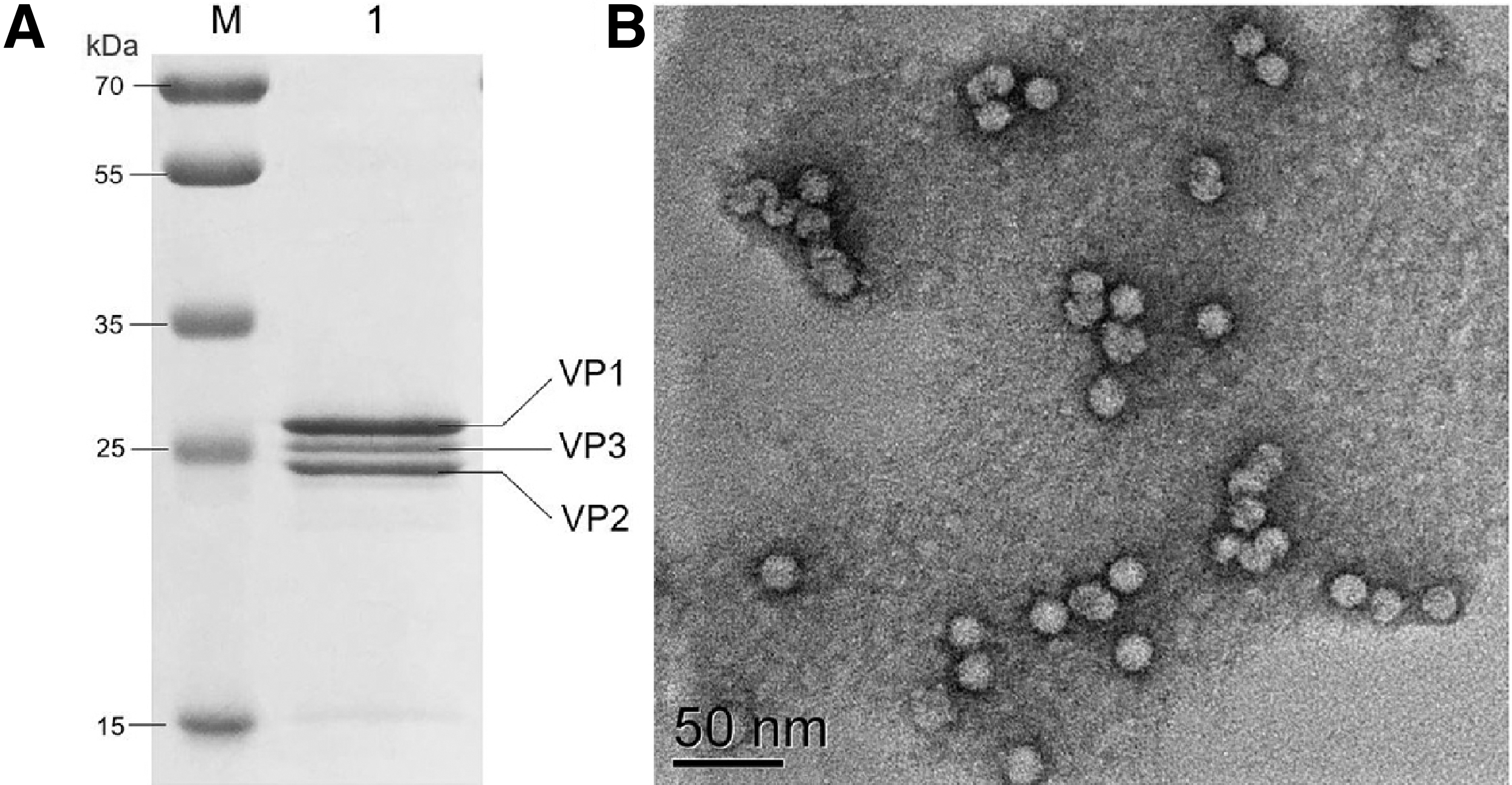
Expression and assembly of VLP of FMDV serotype O.
Mabs characterization
In antigen identification, the results of IFA using the 293T cells with VP1, VP2, or VP3 of FMDV serotype O transient expression showed that 5D3 and 6G6 reacted with VP2 of FMDV serotype O and 7D2 reacted with VP1, none of the three mAbs reacted with VP3 protein (Fig. 2).
Characterization of FMDV serotype O-specific Mabs. Mabs 5D3, 6G6, and 7D2 were analyzed by immunofluorescent assay using the 293T cell with recombinant VP1, VP2, and VP3 proteins of FMDV serotype O expression by transient transfection. Scale bar 50 μm. Mab, monoclonal antibody.
Different Mab pairs were evaluated by SPCE using the positive and negative serum samples identified by LPBE. As shown in Table 1, the results of SPCE using Mab pair of 6G6 (as the capture Mab) and 7D2 (as the detection Mab) were correlated well with LPBE for positive (10/10) and negative (10/10) identification. Thus, Mabs 6G6 and 7D2 were selected as capture antibody and detection antibody in SPCE development, respectively.
Mabs Pairs Screening of Capture Mab and Detection Mab for SPCE Development
ELISA, enzyme-linked immunosorbent assay; Mab, monoclonal antibody;
For specificity assay, serum samples of specific-pathogen-free (SPF) pigs and the positive serum samples of common viral diseases from pigs, including FMDV serotype O, FMDV serotype A, PCV2, PRRSV, PRV, CSFV, PEDV, TGEV, PRoV, and PPV, were analyzed using SPCE. Only the positive serum of FMDV serotype O showed the blocking rate >50% (88.97% ∼ 96.55%), and rest of the positive serum samples showed the blocking rates between 1.03% and 26.33%, which indicated the high specificity of the SPCE in antibody detection of FMDV serotype O (Fig. 3A).
Development of SPCE based on FMDV-VLP.
Development of the SPCE
For the standardization of SPCE protocol, the optimum concentrations of 6G6 and HRP-labeled 7D2 were fixed after conducting a checkerboard titration. The optimal working condition of the 6G6 and HRP-labeled 7D2 in SPCE were 0.45 μg/mL and the dilution of 1:16,000, respectively.
To determine the cutoff level of PI value for the SPCE, a total of 1000 pig serum samples (negative: 500 samples; positive: 500 samples) were examined. After constructing a one-sided 95% confidence interval for the blocking rates, the cutoff values of positive and negative serum samples were 49.6% and 43.5% (Fig. 3B), respectively. Thus, the cutoff value was fixed at the PI value of 50% to avoid false positives in the clinical application.
Performance of the SPCE
To check the suitability of SPCE for the measurement of antibody response against FMDV serotype O, serum samples (n = 181) collected clinically were tested by SPCE and LPBE. As shown in Table 2, the concordance rate of clinical serum samples between SPCE and LPBE was 93.4%. For antibody duration monitoring, serum samples (n = 48) collected from six pigs immunized with FMD serotype O from preimmune to 6 months postimmunization were tested by SPCE and LPBE. As illustrated in Figure 4, the virus antibody titers obtained from the tested serum samples using LPBE correlated well with the blocking rate determined by the SPCE in antibody duration monitoring (kappa = 0.892), indicating high efficiency of the SPCE in immunoassay.
The application of SPCE in antibody duration monitoring. The results of LPBE are shown as antibody level (1/x), and the results of SPCE are shown as blocking rate (%).
Comparison of the Performance of the SPCE with That of the LPBE for the Antibody Detection of Clinical Samples
LPBE, liquid-phase blocking ELISA.
Discussion
To control the FMD, different vaccination strategies are effective,(6) as far the epidemiological and serological survey mainly rely on ELISA; therefore, it is necessary to increase the reliability to measure the titer of FMDV antibody in the herd. Traditionally, inactivated vaccines produced by FMDV-infected cells are widely used to control FMD. In the past few years, FMDV-VLP showed high immune efficacy compared with traditional inactivated vaccines and exhibited its potential as a promising vaccine candidates,(18,19) which implicit the need to evaluate serum antibody level of the VLP prepared vaccine. In China, the incidences of FMD predominantly due to FMDV serotype O Mya-98 lineage.(4,5) Therefore, an SPCE method for specific detection of FMDV serotype O antibodies was developed based on the E. coli original VLP and its Mabs.
To replace the use of inactivated FMDV vaccine would be suggested to reduce the biosecurity risks for immunity evaluation by FMDV challenge (PD50 test)(1) and antibody detection.(22) In this study, the essential diagnostic antigen in SPCE method was the type O-specific FMDV-VLP, the VLP assembled in vitro based on the viral proteins VP1, VP2, and VP3 expressed in the bacterial system of E. coli, which indicated improved biosafety and cost-efficiency without involvement of live virus and to be more convenient in mass antigen production,(18,23) demonstrating that the assay may contribute to the evaluation of herd immunization with inactivated FMD vaccines or serotype-specific VLP.
Quality control of FMD vaccines must ensure the consistency of different batches by using easier and more practical methods. The standardized homologous antigens expressed in E. coli in this study is highly serotype specific for FMDV and easy for purification, which indicates the possibility of replacing the high-performance liquid chromatography-based(24,25) methods for 146S quantification in vaccine production by VLP-based SPCE for the no requirement of specialized equipment. Moreover, the Mabs of FMDV serotype O produced by FMDV-VLP immunization applied in this SPCE method showed a high specificity in antigen detection, suggesting an advantage of distinguishing serotype-specific antibodies than the polyclonal antibody-based(26) method for 146S quantification, especially for the VLP type antigens.
In the future, the VLP-based SPCE methods for antibody detection of other FMDV serotypes can be developed according to this strategy, which can enrich the methods for detecting antibody levels triggered by other serotype FMD vaccines.
Footnotes
Author Disclosure Statement
No competing financial interests exist.
Funding Information
This study was supported by the Special Project of Industrial Cluster in Self-Created Zone in Zhengzhou, Luoyang, and Xinxiang Cities (181200211700).