
Abstract
Immunotoxins, as a class of antitumor agents, consist of tumor-selective ligands linked to highly toxic protein molecules. This type of modified antibody has been designed for the therapy of cancers and a few viral infections. In this study, we designed immunotoxin consisting of mouse programmed cell death protein-1 (PD1), which genetically fused to diphtheria toxin (DT) subunit A (DT386). DNA construct was cloned, expressed in a bacterial system, purified, and confirmed by western blotting. The immunotoxin potency in the treatment of tumorous C57BL/6 mice was evaluated. Immunotoxin was injected intratumoral to mice, and through eight injections, 67% of the tumor volume of the test group started shrinking dramatically. On the contrary, the tumor size of the control group, treated with phosphate-buffered saline, continued its growth. The successful targeting of solid tumor cells by PD1-DT immunotoxin demonstrates the potential therapeutic utility of these conjugates.
Introduction
Cancer is a type of malignancy characterized by uncontrolled development of abnormal cells caused by external and internal factors such as unhealthy diet, genetic mutations, and hereditary hormones.(1) Despite the relative effectiveness of common and standard methods of cancer treatment, including surgery, radiotherapy, and chemotherapy, the results in some patients show no improvement and the mortality rate continues.(2) Removal of solid tumors by surgery is not ordinarily appropriate, and the remaining cells result in recurrence of the tumor.(3) Also, treatment by radiotherapy is nonspecific and normal cells are affected as well.(4) Although chemotherapy shows a good response at first, after a while, cancer cells appear to be stable to chemical agents.(5) On the contrary, the toxicity of chemotherapy drugs is nonspecific and causes side effects.(6) Over the past 30 years, targeted cancer therapeutics have been developed and used clinically to discover more about the possibility of targeted therapy.(7) Identification and removal of malignant cells by antibodies were brought up more than a century ago by Paul Ehrlich.(7) Now, many tumor-targeting antibodies such as trastuzumab, cetuximab, and rituximab are approved as a group of therapeutic monoclonal antibodies (Mab). These are capable of detecting cancer cells surface receptors and block signaling pathways, then destructing cancer cells by inducing an innate immune response and activating cytotoxic T cells.(8)
One promising method for the treatment of cancer is the use of fusion proteins that result from binding fragments of antibody or ligand to a toxin, which are referred to as immunotoxin.(9) Diphtheria toxin (DT), a ribosome inhibitor protein, is one of the bacterial toxins which is used as a toxin moiety in several potent immunotoxins. (10) This toxin is synthesized as a single-chain peptide and is secreted externally. (11) When the toxin is broken down by trypsin, it comes in the form of two active peptides (subtypes A and B) that are bound by a disulfide bond. Subunit B detects and receives receptors on the surface of the host cell, thereby stimulates toxin entry into the host cell and releases subunit A into the host cell, then disables cell transcription by elongation factor-2 (EF-2) inhibition. (12)
Programmed cell death protein-1 (PD1), a 50–55 kDa protein belonging to type I transmembrane glycoprotein, was identified as a protein that stimulates programmed antigen-specific T-cell death.(13) The two known PD-1 ligands are PD-L1 and PD-L2, which are present on the surface of activated cancer cells and inhibit immune response upon binding of PD-1.(14) Due to the increased expression of PD-L1 in various cancers, the construction of a fusion protein containing PD1 and DT can create a high potent immunotoxin for targeting cancer cells.
The present study was designed to express recombinant PD1-DT immunotoxin for tumor treatment. This immunotoxin was constructed by fusion of mouse PD-1 with DT. The antitumor ability of this fusion protein in the treatment of cancerous C57BL/6 mice was evaluated.
Materials and Methods
Immunotoxin gene construction
At first, to make the immunotoxin, two DNA fragments encoding PD-1 and DT subunit A (DT386) were fused according to Figure 1. The resulting plasmid was named pET26-PD1-DT and was transformed into Escherichia coli BL21. The cloning process was confirmed by colony-polymerase chain reaction (PCR), restriction digestion, and DNA sequencing analysis.
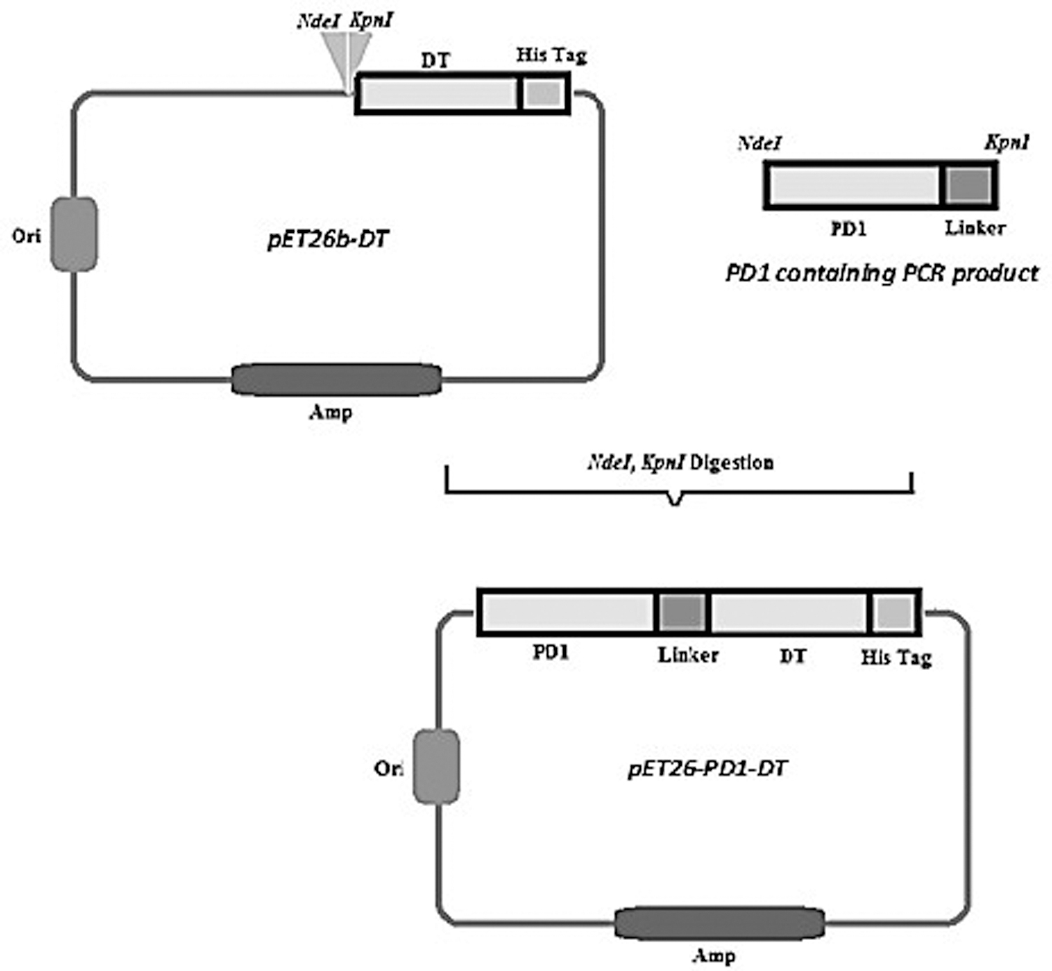
Schematic representation of the immunotoxin DNA construct. PCR was performed with M13 (forward) and specific (reverse) primers, for PD1 gene amplification. The PD1 PCR product and pET26b-DT, which contained the DT gene, were digested with Kpn I and Nde I restriction enzymes and ligated and transformed into Escherichia coli. The resulting plasmid was named pET26-PD1-DT. DT, diphtheria toxin; PCR, polymerase chain reaction; PD1, programmed cell death protein-1.
Recombinant expression and purification
Transformed cells were cultured in LB broth supplemented with 100 mg/mL kanamycin in a shaker incubator. Recombinant protein expression was induced by 1 mM IPTG (isopropyl-b-D-thiogalactopyranoside) at an optical density of 0.6–0.8 (600 nm) for 12 hours at 37°C. Also, optimization of protein expression using different IPTG concentrations (0.1, 0.2, 0.5, 1, 1.5, and 2 mM) and incubation time after induction (3, 5, 8, 12, 18, and 24 hours) were evaluated.
Induced bacteria were harvested and cytoplasmic proteins were extracted by an ultrasonic homogenizer. Cell lysates were centrifuged at 12,000 rpm for 10 minutes at 4°C. The sediment was resuspended in 10 mM Tris-HCl (pH 8.0) containing 8 M urea, and samples were loaded into the Ni-NTA column. Unbound proteins were removed with wash buffer (10 mM Tris-HCl, 8 M urea, pH 6.3). Finally, bounded proteins were eluted by decreasing the buffer pH. The recombinant fusion protein was dialyzed (12 kDa cutoff dialysis bag) against phosphate-buffered saline (PBS), and analyzed by 12% sodium dodecyl sulfate–polyacrylamide gel electrophoresis (SDS-PAGE). Also, western blotting was performed with rabbit anti-His, followed by goat anti-rabbit IgG HRP (Horseradish peroxidase) conjugated and the color developed by DAB (3,3′ diaminobenzidine tetrahydrochloride) a substrate for detecting peroxidase.
Tumor treatment
Mouse model
C57BL/6 mice were purchased from the Pasteur Institute of Iran and used as a cancer model. Twelve mice per cage were housed in standard ventilated cages containing food and water and were maintained according to the laboratory animal care protocol. All applicable international, national, and institutional guidelines for the care and use of animals were followed.
Treatment
About 106 TC-1 cells were resuspended in 200 μL of PBS and subcutaneously injected into the shaved right flank of mice. About 7 days after cell inoculation, a tumor mass began to form. The tumor treatment started when the tumor volume reached 50 mm3. The TC-1 tumor-bearing mice were randomly divided into two groups. One (test group) received 150 μL (100 μg/μL) of immunotoxin and the other (control group) received 150 μL of PBS subcutaneously at 1-week intervals. Injections were given subcutaneously around the tumor. Treatment was continued for 8 weeks and the rate of mortality and tumor size was monitored.
Result assessment
Tumor size was monitored once a week using a caliper according to the equation: V = L × W2 × 0.52.(15) Where V: volume, L: length, and W: width.
Tumor volume change was monitored according to relative tumor volume (RTV) formula: tumor volume on day x/tumor volume on day 0.
Results
Immunotoxin gene construction
PD-1 gene was amplified by PCR using M13 (forward) primer and specific primer (reverse), which contained Kpn I and linker sequence (Fig. 2A), and subsequently was digested and cloned to a pET26b-DT plasmid containing DT gene. To confirm the success of the ligation process, colony PCR (Fig. 2B) and confirmation digestion (Fig. 2C) were done.
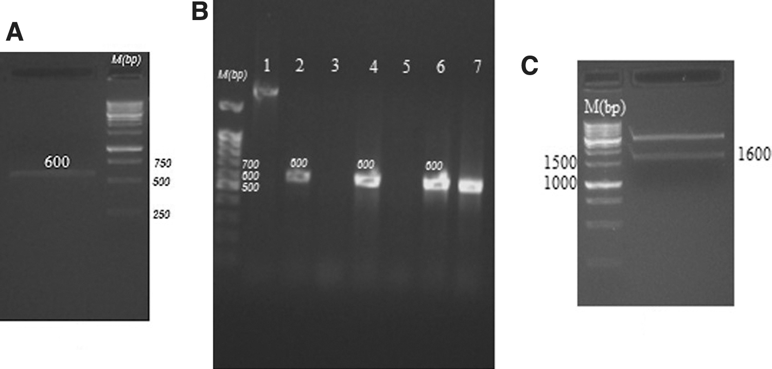
Immunotoxin gene construction and confirmation. PD-1 and linker gene amplification
Recombinant expression
The pET26-PD1-DT plasmid was transformed into BL-21 E. coli. Protein expression was optimized by two independent variables, incubation time after induction and IPTG concentration. According to Figure 3, the best protein expression occurs at 5–18 hours incubation after induction, 1, and 1.5 mM IPTG concentrations. Western blotting was performed using an anti-His tag antibody to confirm protein expression. According to Figure 3C, a 60 kDa protein band appears, which corresponds to the size of the immunotoxin.
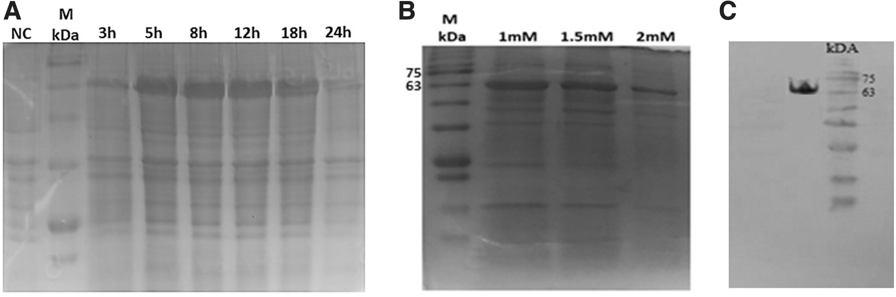
Recombinant immunotoxin expression. Expression optimization of the time after induction
Tumor treatment
Using TC1 cell, a tumor model was developed in C57BL/6 mice. Twelve mice were divided into two groups. For 8 weeks with 1-week intervals, PD1-DT immunotoxin was injected into the test group and PBS to the control group. The tumor size was evaluated weekly and the average tumor volume was calculated (Fig. 4A). Accordingly, the mean tumor volume on the eighth week in the test and control groups were 88 and 2847 mm3, respectively, which indicates the positive effect of PD1-DT immunotoxin on inhibiting tumor growth. The difference in tumor size in the fifth to eighth weeks was statistically significant (p < 0.05). Figure 4B and C show tumor size in two mice of the test and control groups. During the study, all mice were alive.
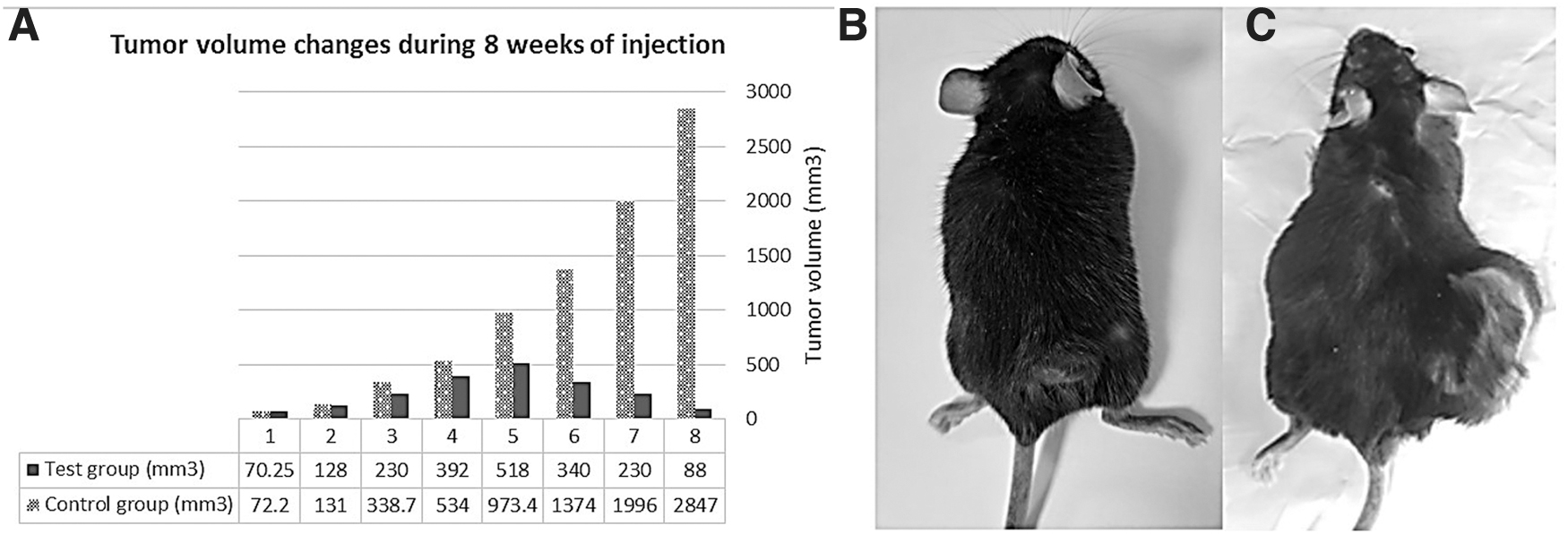
Inhibitory effect of PD1-DT immunotoxin on tumor size. Tumor volume changes during 8 weeks of injection [The difference in the fifth to eighth weeks was statistically significant (p < 0.05)]
Discussion
One of the cancer treatment aspects that has received a lot of attention in recent years is the use of immunological switches or immune checkpoints. In this regard, efforts are made to inhibit immunological switches, such as CD28–cytotoxic T lymphocyte-associated antigen 4 (CTLA4) interaction and PD1–PDL1, to prevent the immune system from switching off against tumor tissue.(16) Anti-PD1/PDL1 immunotherapies are one of the most critical anticancer immunotherapies as the growth in the number of anti-PD1/PDL1 clinical trials has increased from 1 in 2006 to 2250 in 2018 worldwide.(17)
Recently, immunotoxin in the Mab family as a new target therapy is a different treatment that was developed for cancer therapy with fewer side effects. Because various conventional treatments such as chemotherapy, radiation therapy, and surgery cannot decrease the rate of mortality, and these methods dramatically reduce the life quality of the treated patients and cause nausea, hair loss, and severe energy loss.(18,19) In immunotoxin therapy, the cytotoxic toxin is delivered selectively to tumor cells, and toxin moieties are highly effective in comparison to radiation- and chemical agents.
The first attempt in immunotoxins development was reported by Yamaizumi et al. that confirmed the effects of DT on mammalian.(20) Thorpe introduced the concept of using antibodies to direct toxin to one specific cell target. The strategy involved the use of chemical binding agents to bind toxins to these antibodies, and thus “at the same time” the immunotoxin was born.(21–23) The first FDA-approved immunotoxin-based drug, Ontak (denileukin diftitox), is a recombinant DNA-derived cytotoxic protein composed of the DT and the human interleukin-2, which is approved for treating T cell lymphoma.(24,25)
In this project, in addition to using the PD1 as a binding part, we used DT, a potent toxin with the ability to inhibit protein synthesis. This toxin is preferable in terms of how it enters the cancer cell through nonentry into the Golgi apparatus and the endoplasmic reticulum and direct entry from the endosome into the cytoplasm and inhibits protein synthesis. We evaluated the ability of PD1-DT immunotoxin in C57BL/6 mice and the process of tumor inhibition in these mice. It has been proved that PD1-DT structure can inhibit 67% of tumor growth compared to controls.
In conclusion, here, we proposed a new immunotoxin, including PD1-DT for cancer therapy, and the ability of this immunotoxin in tumor growth inhibition was evaluated in an animal model. This recombinant immunotoxin was produced in the bacterial expression system, which is a cheap and available expression system and can be used as an inexpensive drug to treat cancer in the future.
Footnotes
Author Disclosure Statement
No competing financial interests exist.
Funding Information
This work was financially supported by the Pasteur Institute of Iran and the National Institute for Medical Research Development (NIMAD), grant no. 943314.