
Abstract
In a study of 39 isolates of Edwardsiella piscicida made from Korean aquaculture sites, sul genes were detected in 16 isolates and dfr genes in 19. Ten isolates were shown to contain both sul and dfr genes. MIC and disc diffusion zones assays were performed to measure the phenotypic susceptibilities of the 39 isolates. Normalized resistance interpretation was applied to these data to categorize isolates as either fully susceptible or as manifesting reduced susceptibility. The standard CLSI protocols specify the use of a mixture of sulfamethoxazole/trimethoprim (20:1) in both MIC and disc diffusion tests. Using the CLSI MIC protocol, 100% of the isolates containing dfr genes, but only 75% of the isolates containing sul genes, were categorized as manifesting reduced susceptibility. Using the CLSI disc diffusion protocol, only 58% of the isolates containing dfr genes and 69% of those containing sul genes were categorized as manifesting reduced susceptibility. When the single agent trimethoprim was substituted for the combined mixture in both the MIC and disc diffusion protocols, 100% of the dfr-positive isolates were categorized as NWT. When the single-agent sulfamethoxazole was substituted, the analysis of the MIC characterized 100% and the disc zone data 94% of the sul-positive isolates as manifesting reduced susceptibility. It is argued that the use of trimethoprim and sulfamethoxazole as single agents in phenotypic susceptibility tests would provide more meaningful data than the currently recommended use of these two agents combined.
Introduction
T
Epidemiological cutoff values are protocol- and species-specific values that allow the categorization of isolates either as a fully susceptible member of their species (WT) or as manifesting reduced susceptibility (NWT). In contrast to clinical breakpoints, epidemiological cutoff values are not affected by the conditions or properties of any therapeutic administration of the agent against infections by the relevant bacterium. 1 Equally they are not affected by the variations in the pharmacokinetics or pharmacodynamics of the agent or the bacterium in any infected host.
Genotypic data are not considered in the calculation of epidemiological cutoff values, however, EUCAST defines a microorganism as NWT with respect to an agent, if it possesses an acquired or mutational resistance mechanism to that agent, 1 and CLSI have stated that WT isolates can be assumed to lack these mechanisms. 2 Thus, the extent to which their application categorizes as NWT isolates, which possess genes that encode resistance mechanisms, is a significant property of any epidemiological cutoff value.
The relatively low cost of the sulfonamide agents is a major factor contributing to the widespread use of these antimicrobial agents in Asian agriculture and aquaculture and has led to significant concentrations of these agents, particularly sulfamethoxazole, being detected in tropical Asian waters.3,4 A number of studies have reported significant frequencies of sulfonamide-resistant bacteria and/or resistance genes in these aquatic environments.3,5–9 In aquaculture, sulfonamides are frequently administered together with trimethoprim and resistance to trimethoprim, often in association with class 1 integrons, has also been regularly reported in bacteria isolated from the aquatic environment.3,10–15 These observations suggest that investigations of resistance to sulfonamides and trimethoprim should be included in any programs designed to monitor antibiotic susceptibilities of bacteria isolated from aquatic animals.
Currently, the only standard protocols for assessing the antibiotic susceptibility of bacteria isolated from aquatic animals are those that have been produced by CLSI. The standard protocol for disc diffusion is provided in the guideline VET03-A 16 and that for MIC assays is provided in VET04-A2. 2 Both standard protocols recommend that susceptibility to potentiated sulfonamides (sulfamethoxazole/trimethoprim and ormetoprim/sulfadimethoxine) should be established using the two agents in combination.
Minogue et al. 17 compared the performance of the combined sulfamethoxazole/trimethoprim discs recommended in the standard CLSI disc diffusion protocol with that of discs containing sulfamethoxazole alone. They used both discs in a study of 7 isolates of Aeromonas salmonicida that contained sul1 genes and 9 that did not. They demonstrated that when discs containing sulfamethoxazole alone were used, the sul1 containing isolates manifested no inhibition zones and were clearly separable from the isolates, in which this gene was not detected, for which the mean zone size was 28 mm.
In contrast, when discs containing a mixture of sulfamethoxazole/trimethoprim were used, the distribution of zone sizes for isolates that contained the sul1 gene overlapped with, and could not be separated from, the zones obtained with these discs for strains that did not contain this gene. Thus, using the combined disc recommended by the CLSI protocol, the sul1 containing isolates were categorized as WT but, using the single sulfamethoxazole discs, they manifested a phenotype of reduced susceptibility and were categorized as NWT.
This work was undertaken to further investigate the performance characteristics of susceptibility tests for sulfamethoxazole and trimethoprim in studies of 39 Edwardsiella piscicida isolates. The E. piscicida isolates used in this work had previously been classified as E. tarda. 18 However, recent developments in the taxonomy of the Edwardsiella have led to the separation of isolates previously classified as E. tarda into three species E. tarda, E. piscicida, 19 and E. anguillarum 20 and the development of appropriate molecular methods for their identification to the species level.
The species E. piscicida was originally proposed following the study of 12 isolates from fish made in Asia and Europe and that had previously been classified as E. tarda. 19 In a retrospective reexamination of isolates made from US catfish farms during 2007–2012, previously classified as E. tarda, all 44 were reclassified as E. pisicida. 21 Although extensive retrospective reanalysis has yet to be performed, the presently available data18,19 suggest that many of the isolates previously classified as E. tarda, that had been isolated from fish, may well be reclassified as E. piscicida. The isolates grouped as E. anguillarum would, so far, appear to be associated with diseased eels. 20
The phenotypic susceptibility of the 39 Edwardsiella piscicida isolates to sulfamethoxazole and trimethoprim separately and to both agents in combination was investigated using CLSI disc diffusion 16 and MIC protocols. 2 These strains were also examined for the presence of three sul genes, seven dfr genes, and for the gene intI1, and the correlations between the genotypes of the isolates and their categorization by the various phenotypic methods were investigated.
Materials and Methods
Bacterial strains
The 39 E. piscicida isolates studied were a subset of those studied by Lim et al. 18 Twenty-one strains were isolated from olive flounder between 1999 and 2004, 17 were isolated from eels or eel pond water between 2011 and 2012, and one was isolated from turbot. All isolates were stored at −80°C in tryptic soy broth (TSB; Difco) containing 1% NaCl and 10% glycerol and cultured on tryptic soy agar (TSA; Difco) supplemented with 1% NaCl and incubated for 24 hours at 28°C. The taxonomic status of all isolates was confirmed by amplifying the fimbrial subunit for E. piscicida using species-specific primers (EPF:5′-CTTTGATCATGGTTGCGGAA-3′, EPR:5′-CGGCGTTTTCTTTTCTCG-3′) and thermal conditions previously described by Griffin et al. 21
Antibiotic resistance genes detection
Genomic DNA was extracted from all isolates using Accuprep® Genomic DNA Extraction Kit (Bioneer, Korea) according to the manufacturer's instructions. For detection of antibiotic resistance genes (ARGs) related to sulfonamide and trimethoprim, PCR was performed using primer pairs listed and thermal conditions described in references in Table 1. Each 20 μl of reaction mixture consisted of 1 μl of genomic DNA used as template, 1 μl of each primer (10 μM), and 17 μl of sterile DW with AccuPower® PCR PreMix (Bioneer, Korea). PCR products were identified by electrophoresis in 1% (w/v) agarose gel stained with ethidium bromide and visualized by ChemiDoc™ MP System (Bio-Rad).
Whole-genome sequence analysis of the ETW41 isolate
The whole-genome sequence (WGS) was determined for the isolate ETW41 that had been obtained from eel farm water. Using disc diffusion data, this isolate had been categorized as NWT with respect to chloramphenicol, florfenicol, erythromycin, sulfamethoxazole, tetracycline, and trimethoprim. 18 The isolate was grown in tryptic soy broth (TSB; Difco) for 24 h at 28°C. Genomic DNA was extracted using DNeasy Blood and Tissue kit (Qiagen, Streetville, ON, Canada) according to the manufacturer's instructions. Following library preparation, whole-genome sequencing was conducted with PacBio RSII P6-C4 chemistry platform (Pacific Biosciences, Menlo Park, CA) using a single-molecule real-time cell. De novo assembly of generated raw sequences was performed in Hierarchical Genome Assembly Process (HGAP3), resulting in two contigs with 3,982,211 bp and G + C content of 59.49%.
The classification of isolate ETW41 as a member of the species E. piscicida was confirmed by calculating its average nucleotide identity (ANI) with the reference strains E. piscicida C07-087, E. anguillarum ET080813, and E. tarda FL95-01using EDGAR version 2.2 (http://edgar.computational.bio). 22
Gene prediction and annotation were predicted using the Prokka pipeline of Prokaryotic Genome Annotation System (http://vicbioinformatics.com). Resistance Gene Identifier (RGI) in the Comprehensive Antibiotic Resistance Database (CARD) (https://card.mcmaster.ca/analyze/rgi) 23 and the repository of antibiotic resistance cassettes (RAC) (http://rac.aihi.mq.edu.au/rac) 24 were used to find ARGs and cassettes in the WGS of the strain ETW41. ARGs in detected integron cassettes in this study were confirmed through BLASTp in UniProt (http://www.uniprot.org/blast) and visualized using CLC main workbench 7.7.3 (Qiagen, Arahus, Denmark). The two large contigs contained in the genome have been deposited at GenBank under the accession numbers CP019440 and CP019441.
Phenotypic susceptibility assays
All phenotypic susceptibility tests were performed at 28°C with incubation for 24 hours. The MIC assays were performed according to the broth microdilution protocols provided in VET04-A2. 2 The MICs for the combined sulfamethoxazole/trimethoprim (SXT) were performed using Sensititre CMP1MSP plates (Trek Diagnostic Systems; ThermoScientific.com/microbiology) that contained a range of concentrations from 0.015/0.3 mgL−1 to 1/19 mgL−1. For the single agents the microdilution assay plates were prepared in the laboratory. For each agent, overlapping twofold dilutions were prepared. The range of concentrations in these plates was 0.1875–256 μg/ml for trimethoprim (TRI) and 1–128 μg/ml for sulfamethoxazole (SUL).
The disc diffusion assays were performed according to the protocols provided in VET03-A. 16 The discs used were obtained from Oxoid (Cambridge, UK) and contained 25 μg sulfamethoxazole (SUL), 5 μg trimethoprim (TRI), and a combination of 23.75 μg sulfamethoxazole and 1.25 μg trimethoprim (SXT).
Normalized resistance interpretation analysis
Epidemiological cutoff values (COWT) were determined for the log2 transformed MIC data by the normalized resistance interpretation (NRI) method of Kronvall. 25 For the disc diffusion data, NRI analysis was performed according to the method described by Smith and Kronvall. 26 For both the log2 MIC and disc diffusion data sets, NRI analysis was performed using the MS Excel spreadsheet available online at www.bioscand.se/nri.
The categories established by NRI analysis using sulfamethoxazole/trimethoprim in combination were termed SXTWT and SXTNWT. Those established by using trimethoprim as a single agent were termed TRIWT and TRINWT and those established by using sulfamethoxazole as a single agent were termed SULWT and SULNWT.
Results
Detection of sul, dfr, and int genes
Genes relevant to reduced susceptibility to both sulfamethoxazole and trimethoprim were detected in 10 (26%) of the 39 isolates (sul+ and dfr+) (Table 2). In addition, 6 (15%) isolates contained genes relevant only to reduced susceptibility to sulfamethoxazole (sul+ and dfr−), and 9 (23%) contained genes relevant only to reduced susceptibility to trimethoprim (sul− and dfr+). Overall, the sul1 gene was detected in 10 (26%) of the isolates and sul2 in 7 (18%). One isolate was shown to contain both sul1 and sul2, and the gene sul3 was not detected in any of the isolates. With respect to trimethoprim, dfrA5 was detected in 23% of the isolates, dfrA12 in 21%, and dfrA7 in 5%. The genes dfrA1, dfrA14, dfrA17, and dfr18 were not detected in any isolates. A total of five different genotypic patterns were observed (Table 2).
The genes sul3 and dfrA1, dfrA14, dfrA17, and dfr18 were not detected in any of the 39 isolates.
The gene intI1 was detected in all the 19 dfr+ isolates and in none of the 20 dfr- isolates. It was also detected in all the 10 isolates, in which sul1 genes were detected, but in only one of the seven isolates, in which sul2 was detected. The data obtained in this work from 18 of the 19 intI1-positive isolates studied demonstrate that intI1 genes, the dfr genes, and the sul genes occurred in the same cell. They cannot demonstrate that the dfr and sul genes are located within a class 1 integron. However, analysis of the WGS obtained from one of the intI1-positive E. piscicida isolates, ETW41, revealed that the genes dfrA12 (497 bp), aadA2 (779 bp), and sul1 (839 bp) were located within a class 1 integron (Fig. 1). In total, 41 genes potentially associated with antibiotic resistance were identified in the WGS of this isolate. These included genes for resistance to chloramphenicol (catII), erythromycin (mphA), florfenicol (floR), sulfonamides (sul1, sul2), tetracycline (tetA), and trimethoprim (dfrA12). The ANI of isolate ETW41 was 99.46% with reference strain E. piscicida C07-087, 95.14% with E. anguillarum ET080813, and only 85.4% with E. tarda FL95-01. These data confirm the classification of this isolate as a member of the species E. piscicida.

Class 1 integron with cassette genes (dfrA12, aadA2, and sul1) carried by Edwardsiella piscicida ETW41.
Studies with trimethoprim as a single agent
The zones produced using 5 μg trimethoprim discs showed a clearly separated bimodal distribution (Fig. 2). Twenty-one isolates gave no zones and 18 gave zones between 33 and 42 mm. The COWT, calculated by NRI analysis of these data, was 29 mm with a standard deviation of the normalized WT distribution of 3.4 mm. Application of this cutoff value categorized the 18 isolates as TRIWT. The 21 isolates categorized as TRINWT included all 19 of the isolates, in which dfr genes were detected.
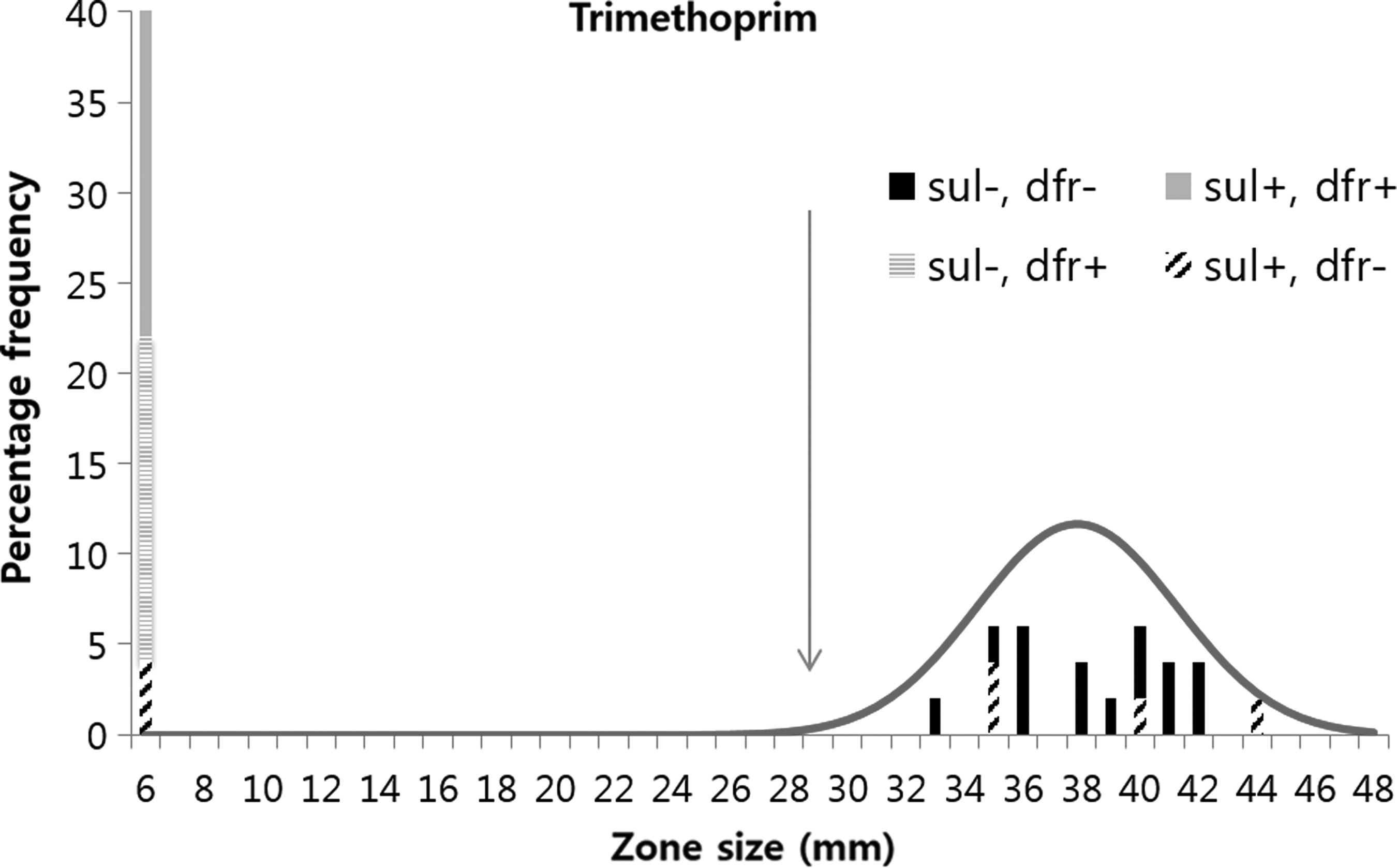
Percentage frequency of zone sizes for 39 E. piscicida isolates obtained using discs containing 5 μg trimethoprim.
The trimethoprim MIC values fell into two clearly separated groups. Twenty-one isolates gave values >256 μg/ml, and the other 18 isolates gave values ≤0.185 μg/ml. As the MIC values for the majority of these 18 isolates lay below the range of concentrations tested, NRI analysis could not be performed. Visual examination of these zone data would suggest that a TRIWT cutoff of ≤0.185 μg/ml would be reasonable, and when this value was applied, there was a 100% agreement in the categorizing of isolates on the basis of the disc and MIC data.
In tests performed with trimethoprim alone, there was a 100% agreement in the categorizing of isolates on the basis of the disc and MIC data. On the basis of the MIC and disc diffusion data, all 19 isolates, in which dfr genes had been detected, were categorized as TRINWT (Table 3). However, two of the 20 isolates, in which dfr genes had not been detected, were also categorized as TRINWT by both phenotypic tests.
SXT indicates test performed with combined sulfamethoxazole/trimethoprim.
SUL indicates test performed with sulfamethoxazole alone.
TRI indicates test performed with trimethoprim alone.
Studies with sulfamethoxazole as a single agent
In the diffusion assays with 25 μg sulfamethoxazole discs, 17 isolates gave no zones, the zones for 22 isolates ranged from 16 to 37 mm (Fig. 3). NRI analysis calculated a COWT of ≥11 mm with a standard deviation for the normalized distribution of WT observations of 5.8 mm. Applying this cutoff, 15 of the 16 isolates (94%), in which sul genes were detected, were categorized as SULNWT.
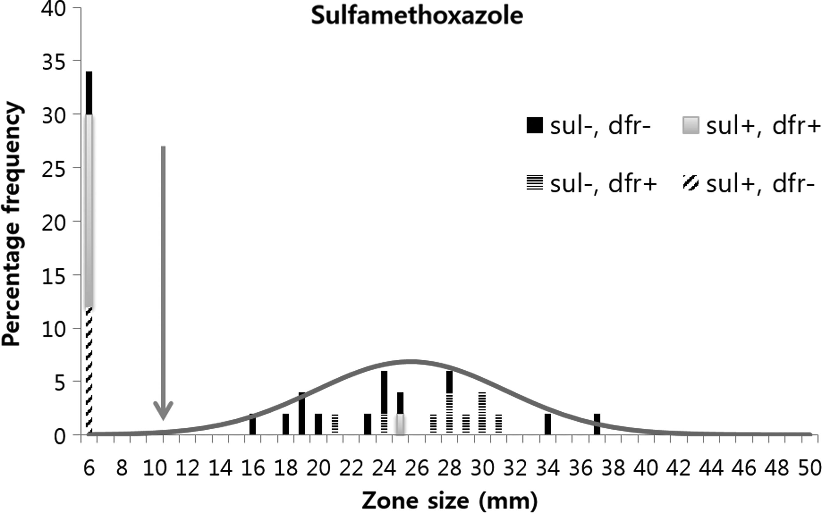
Percentage frequency of zone sizes for 39 E. piscicida isolates obtained using discs containing 25 μg sulfamethoxazole.
In the sulfamethoxazole MIC assays, 18 isolates gave values ≥128 μg/ml, and 21 gave values between 1.5 μg/ml and 16 μg/ml (Fig. 4). NRI analysis calculated a COWT of ≤16 μg/ml with a standard deviation of the normalized WT distribution of 0.86 log2 μg/ml. Application of this COWT categorized 21 isolates as SULWT. These SULWT isolates were composed of 12 of the 14 sul− and dfr− isolates and all 9 of the sul− and dfr+ isolates. The distribution of the MIC values for these two genotypes differed slightly (Fig. 4), suggesting that possession of a dfr gene may result in a small reduction in sulfamethoxazole susceptibility. However, possibly because of the small numbers of observations, NRI analysis was not able to generate a COWT that separated these two genetic groups on the basis of the distribution of their MIC values.
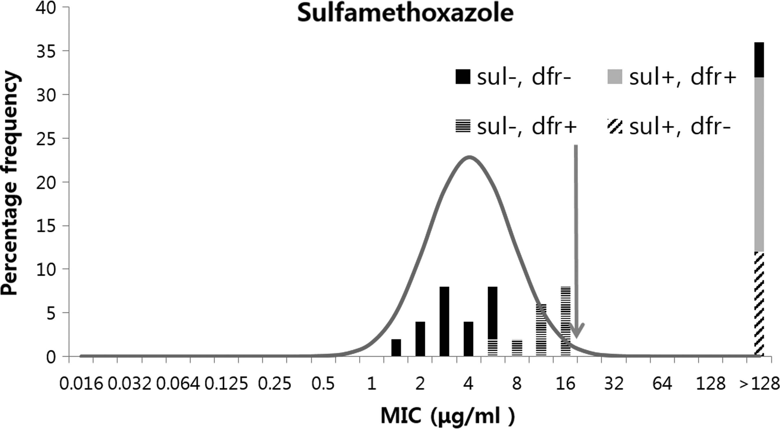
Percentage frequency of MIC values for sulfamethoxazole against 39 E. piscicida isolates.
In tests performed with sulfamethoxazole alone, there was a 97% agreement in the categorizing of isolates on the basis of the disc and MIC data. On the basis of the MIC data, all the 16 isolates, in which sul genes had been detected, were categorized as SULNWT, but on the basis of the disc data, only 15 of the 16 of these isolates were categorized as SULNWT. However, in two isolates that were categorized as SULNWT by the analysis of the data from both of these phenotypic tests, sul1, sul2, and sul3 genes were not detected.
Studies with sulfamethoxazole/trimethoprim combined
In the disc diffusion studies with the combined sulfamethoxazole/trimethoprim discs, 12 isolates gave no zones. The zones for the remaining 27 were spread out over a wide range from 15 to 46 mm (Fig. 5). Visually there was no evidence indicating the presence of more than one modal group in the distribution of the zones recorded from these 27 isolates. NRI analysis calculated a COWT of ≥15 mm, and application of this COWT categorized 27 isolates as SXTWT. The standard deviation of the normalized distribution of the zones recorded for these 27 isolates was 7.5 mm. The isolates categorized as members of the SXTWT group were, however, genetically heterogeneous (Fig. 5).
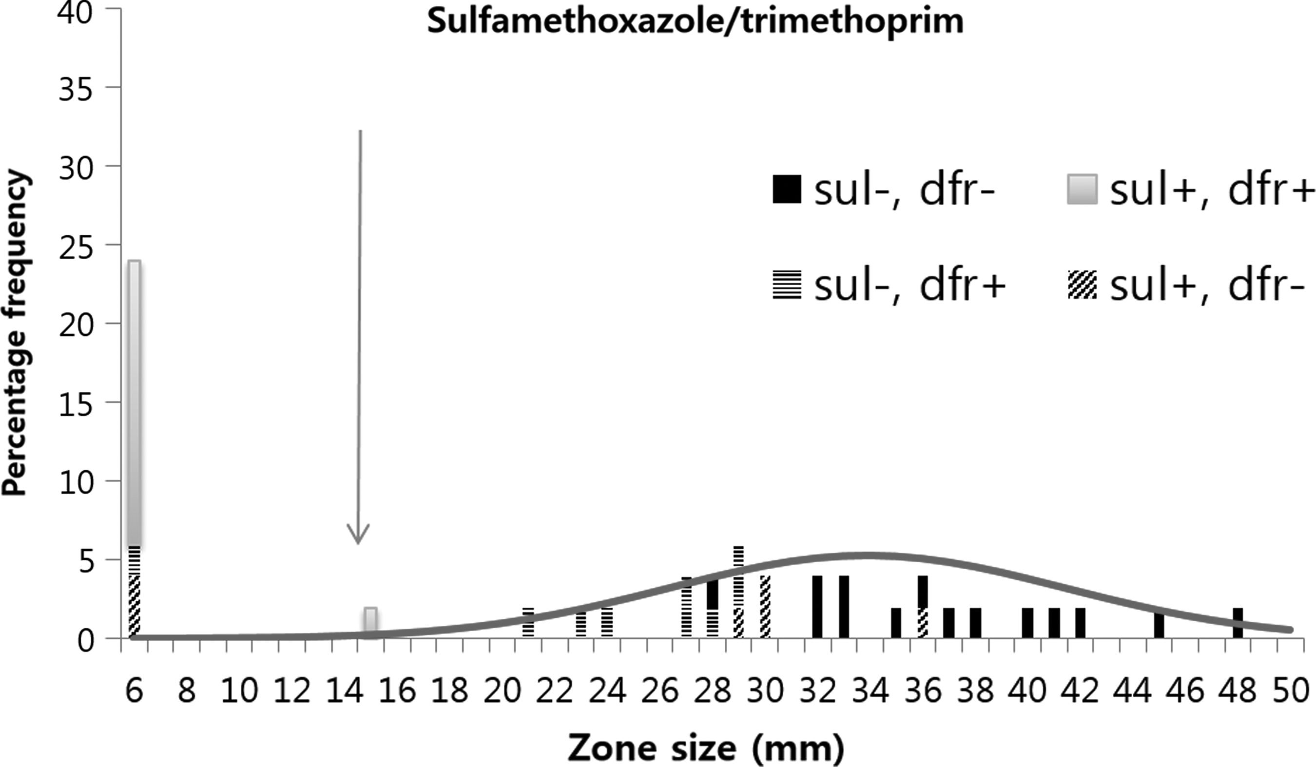
Percentage frequency of zone sizes for 39 E. piscicida isolates obtained using discs containing 23.75 μg sulfamethoxazole and 1.25 μg trimethoprim.
This group contained all 14 of the sul− and dfr− isolates, but it also included 4 of the 6 sul+ and dfr− isolates, 8 of the 9 sul− and dfr+ isolates, and 1 of the 10 sul+ and dfr+ isolates. The median values of the zone sizes for the three genotypic groups sul− and dfr−, sul+ and dfr−, and sul− and dfr+ were 36, 30, and 27 mm, respectively, indicating that the presence of either sul genes or dfr genes might be associated with a slight reduction in zone sizes. The extent of overlap in the distributions of the zones for these three genotypic groupings had the result that they could not be separated using the test protocol used in this work.
Nine of the 10 isolates in which both sul and dfr genes were detected were categorized as SXTNWT on the basis of the analysis of the data obtained with the combined discs. Overall, however, these studies failed to categorize 31% (5 of 16) of the sul-containing isolates and 48% (7 of 19) of the dfr-containing isolates as SXTNWT (Table 3).
In the combined sulfamethoxazole/trimethoprim MIC data, 21 isolates gave MIC values for SXT >1/19 μg/ml and the remaining 18 gave MIC values between 0.015/0.3 and 0.06/1.19 μg/ml (Fig. 6). NRI analysis calculated a COWT of ≤0.12/2.38 μg/ml with a standard deviation of the normalized WT distribution of 0.58 log2 μg/ml. Application of this cutoff value categorized 18 isolates as SXTWT. Examining the genotypes of the isolates categorized as SXTWT indicated heterogeneity within this group. The SXTWT group contained all 14 of the sul− and dfr− isolates, but it also included 4 of the 6 sul+ and dfr− isolates.
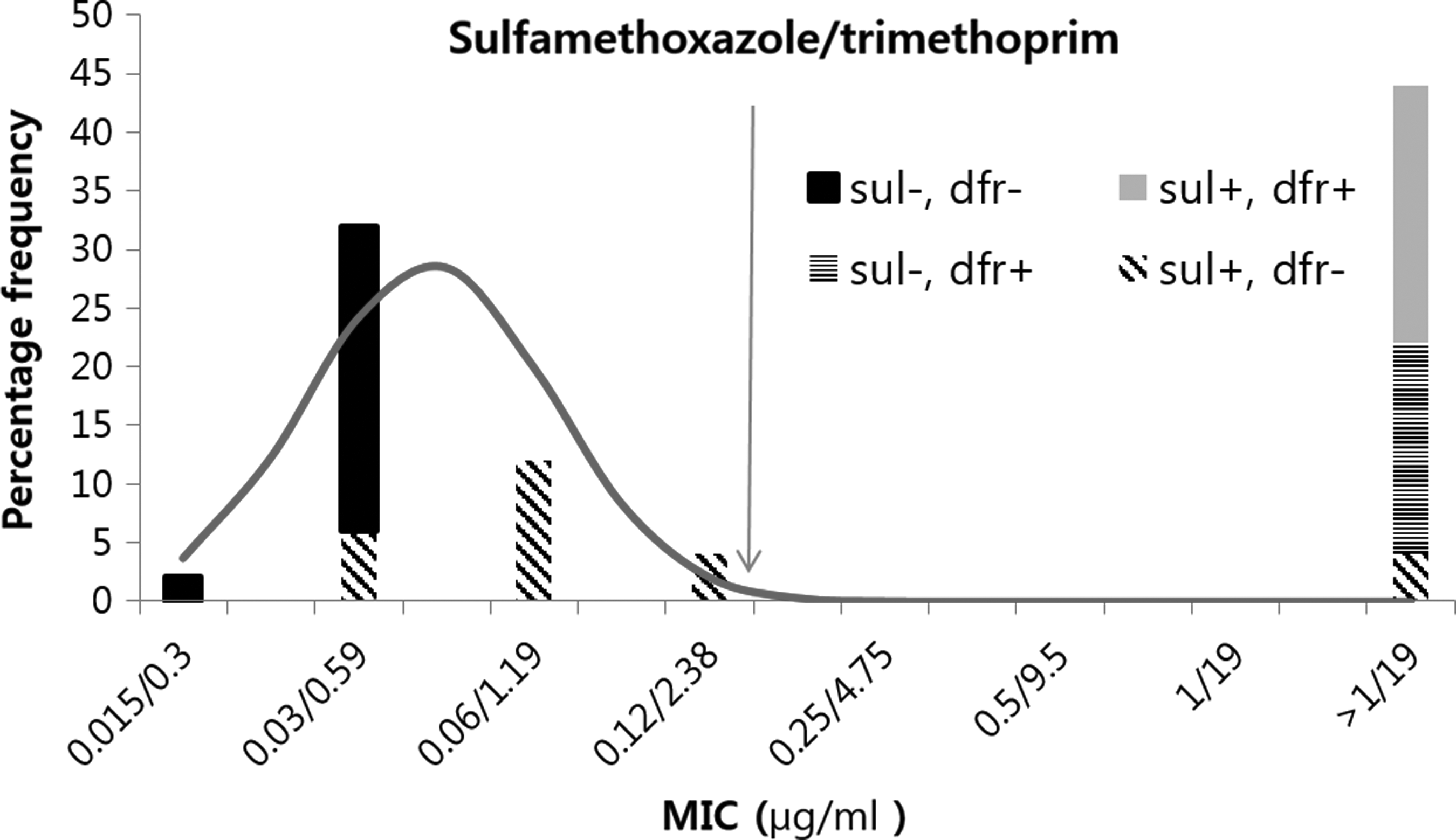
Percentage frequency of MIC values for combined sulfamethoxazole and trimethoprim (20/1) against 39 E. piscicida isolates.
All of the 10 isolates in which both sul and dfr genes were detected and all of the 9 isolates in which as dfr genes were detected in the absence of any of the sul genes were categorised as SXTNWT on the basis of the analysis of the MIC data obtained with the combined agents. Overall, however, these studies failed to categorize 4 of 16 (25%) of the sul-containing isolates as SXTNWT (Table 3).
Discussion
Phenotypic studies
In this work, the application of the epidemiological cutoff values calculated from the data generated in single-agent studies categorized the vast majority (100% for MIC and 96% for disc) of isolates, in which the relevant genes were detected as manifesting a phenotype of reduced susceptibility and categorized them as NWT. The reverse was not, however, true. With respect to both agents, relevant dfr or sul resistance genes were not detected in all the isolates that manifested reduced phenotypic susceptibility.
When trimethoprim was used as a single agent in phenotypic tests, two isolates, in which no dfr genes were detected, were categorized as manifesting reduced susceptibility (TRINWT). Both these isolates were collected from olive flounder in 2003, and disc diffusion assays 18 demonstrated that they shared the same susceptibility to 17 other antibiotics. It is, therefore, possible that these two isolates might be clonally related. In this work, the presence of only seven dfr genes (dfrA1, dfrA5, dfrA7, dfrA12, dfrA14, dfrA17, and dfr18) was investigated. However, in addition to these genes, another 15 genes have been reported to encode reduced susceptibility to trimethoprim (https://ardb.cbcb.umd.edu/resis_goterm.shtml). Thus, it is entirely possible that the reduced susceptibility of these two isolates might have been encoded by one of the other dfr genes.
When sulfamethoxazole was used as a single agent, two isolates, in which no sul genes were detected, were categorized as manifesting reduced susceptibility in phenotypic tests (SULNWT). These isolates were made from eel farms in the same area in 2011 and shared the same disc diffusion profiles for 17 other antibiotics. 17 It is possible, therefore, that they might be clonally related. 17 A failure to detect sul genes in isolates that were phenotypically resistant to sulfamethoxazole was also reported by Hoa et al. 7 They reported that sul1, sul2, and sul3 were not detected in 32% of 127 isolates from the aquatic environment that they selected on media containing 60 μg/ml sulfamethoxazole. In commentating on this result, they noted that in addition to these three sul genes, the phenotype of reduced susceptibility to sulfonamides has been reported to be mediated by alterations of the chromosomal DHPS gene folP. 27
When sulfamethoxazole and trimethoprim in combination were used in MIC tests, all isolates, which had been categorized as TRINWT and all isolates in which dfr genes had been detected, were shown to manifest reduced susceptibility and were categorized as SXTNWT. However, 4 of the 16 isolates (25%) that had been categorized as SULNWT, on the basis of their reduced susceptibility in single-agent MIC studies with sulfamethoxazole, in which sul genes had been detected, did not manifest reduced susceptibility in these combined-agent MIC tests. These four isolates, categorized as SXTWT, possessed a sul2 gene, but not any of the dfr genes examined, and all belonged to genotypic group C (Table 2).
The two other members of genotypic group C were, however, categorized as SXTNWT. These two isolates both manifested reduced susceptibility to trimethoprim and, as discussed above, may have possessed a dfr gene that is not studied in this work. Thus, the data generated in MIC tests using combined agents lacked the sensitivity to allow the separation of fully susceptible isolates from those that possessed a sul+ or dfr− genotype.
The categorization of isolates on the basis of the data generated, when discs containing sulfamethoxazole and trimethoprim in combination were used, they showed major discrepancies both with categorizations based on single-agent tests and with the genotypic data (Table 3).
The nine isolates classified in genotypic group A (Table 2), in which dfr genes (dfrA5) were detected but in which sul genes were not detected, were categorized as TRINWT on the basis of single-agent tests with trimethoprim. However, eight of these nine isolates did not manifest reduced susceptibility in studies using combined discs and, on this basis, were categorized as SXTWT.
The six isolates classified in genotypic group C (Table 2), in which sul genes (sul2) were detected but in which dfr genes were not detected, were categorized as SULNWT on the basis of single-agent tests with sulfamethoxazole. However, four of these six isolates did not manifest reduced susceptibility in studies using combined discs and, on this basis, were categorized as SXTWT. These four isolates were those that were also categorized as SXTWT on the basis of the data generated by MIC tests using sulfamethoxazole and trimethoprim in combination.
Thus, the data generated in tests using combined-agent discs lacked the sensitivity to allow the separation of fully susceptible isolates from those that possessed a sul+; dfr−; sul−; or a dfr+ genotype. The data generated using single-agent discs did allow this separation and this raises serious questions as to the value of the use of combined discs. Consideration of the precision of the data generated in tests using combined discs suggests further reasons for questioning their value.
The standard deviations of the normalized WT distributions calculated by NRI analysis provide a measure of the precision of disc diffusion data, 28 and it has been argued that the cutoff values generated from data showing abnormally high standard deviations should be rejected. 29 The standard deviation of the data generated in the disc test using combined discs in this work was 7.5 mm. This value can be compared with the mean value of 2.6 ± 0.71 mm recorded for the standard deviations of 16 disc data sets obtained using other antibiotics but tested under the same conditions.21,30 Thus, the standard deviation of the data generated by the combined discs indicates a low precision and, therefore, that the COWT calculated from them and the isolate categorization based on its application must be considered suspect.
Genotypic studies
Recent studies demonstrated the potential for WGS of bacterial strains to become a valuable tool in the investigation of antibiotic resistance.31,32 However, in 2017, a EUCAST subcommittee reported this application of WGS data. 33 They concluded that the published data for using WGS to infer susceptibility was either “poor or nonexistent.” They recommended that priority should be given to studies that allowed the comparison of data deriving from WSG with those derived from the application of epidemiological cutoff values to phenotypic data.
With respect to the genus Edwardsiella, there have been few studies that allow the comparison of genotypic and phenotypic methods of investigations of antimicrobial susceptibility. One E. tarda isolate categorized as phenotypically resistant to chloramphenicol and tetracycline was shown to harbor the genes catA3 and tetA. 34 A study of the MIC values for 64 E. ictaluri isolates 35 was followed by a genotypic study of eight of these isolates that had been classified as resistant to oxytetracycline. 36 They detected tetA genes in all eight strains and dfhr1 and sul2 genes in the six of them that had been categorized as phenotypically resistant to sulfamethoxazole and trimethoprim.
In this work, there was, for six agents, an agreement between the phenotypic resistances and the WGS data for isolate ETW41. In the PCR studies on the 39 E. piscicida isolates, there was, however, a less than 100% agreement between the phenotypic and genotypic analyses. With respect to trimethoprim, dfr genes were detected in only 90% (19/21) of the isolates that were categorized as NWT, on the basis of the phenotypic tests using single agents, and with respect to sulfamethoxazole, the similar figure was 88% (16/18). As argued above, these less than 100% agreements might well be a result of the limited number of genes studied in this work. It is possible that the use of WGS data would resolve these anomalies. It can be concluded that, at present, the available data for Edwardsiella fall short of the EUCAST requirements, 33 but they do indicate the potential of genotypic methods.
Conclusions
The analysis of the E. piscicida data obtained in this work demonstrates that susceptibility tests that use sulfamethoxazole/trimethoprim in combination provide unreliable indications as to the categorization of isolates made on the basis of tests using either sulfamethoxazole or trimethoprim as separate agents. Also, importantly, they did not generate data that allowed a reliable prediction of the presence of genes known to reduce susceptibility to either agent.
The current CLSI standard susceptibility assay protocols2,16 recommend the use of sulfamethoxazole and trimethoprim in combination. The data presented in this work demonstrate, however, that phenotypic susceptibility tests using trimethoprim and sulfamethoxazole as single agents provide a more effective method of identifying isolate that possesses mechanisms that encode resistance to these agents than tests that use both agents combined. It is, therefore, argued that the standard CLSI testing protocols for susceptibility testing of bacteria isolated from aquatic animals2,16 should be modified to recommend the use of the two agents separately. This conclusion is not a new one. Waterworth 37 reported in 1978 that the World Health Organization was recommending at that time that, in using disc diffusion methods to test susceptibility to potentiated sulfonamides, the susceptibilities to the two components should be determined independently.
Footnotes
Acknowledgments
The NRI method was used with permission from the patent holder, Bioscand AB, TÄBY, Sweden (European Patent No. 1383913, US Patent No. 7,465,559). This work was supported by the National Institute of Fisheries Science (NIFS R2018062).
Disclosure Statement
No competing financial interests exist.