
Abstract
This study examined the performance of serum ubiquitin C-terminal hydrolase (UCH-L1) in detecting traumatic intracranial lesions on computed tomography (CT) scan (+CT) in children and youth with mild and moderate TBI (mmTBI) and assessed its performance in trauma control patients without head trauma. This prospective cohort study enrolled children and youth presenting to three level 1 trauma centers after blunt head trauma and a Glasgow Coma Scale (GCS) score of 9–15 as well as trauma control patients with GCS 15 that did not have blunt head trauma. The primary outcome measure was the presence of intracranial lesions on initial CT scan. Blood samples were obtained in all patients within 6 h of injury and measured by enzyme-linked immunosorbent assay ELISA for UCH-L1 (ng/mL). A total of 256 children and youth were enrolled in the study and had serum samples drawn within 6 h of injury for analysis; 196 had blunt head trauma and 60 were trauma controls. CT scan of the head was performed in 151 patients and traumatic intracranial lesions on CT scan were evident in 17 (11%), all of whom had a GCS of 13–15. The area under the receiver operating characteristic curve (AUC) for UCH-L1 in detecting children and youth with traumatic intracranial lesions on CT was 0.83 (95% confidence interval [CI], 0.73–0.93). In those presenting with a GCS of 15, the AUC for detecting lesions was 0.83 (95% CI, 0.72–0.94). Similarly, in children under 5 years of age, the AUC was 0.79 (95% CI, 0.59–1.00). Performance for detecting intracranial lesions at a UCH-L1 cut-off level of 0.18 ng/mL yielded a sensitivity of 100%, a specificity of 47%, and a negative predictive value of 100%. UCH-L1 showed good performance in infants and toddlers younger than 5 years and performed well in children and youth with a GCS score of 15. Before clinical application, further study in larger cohort of children and youth with mild TBI is warranted.
Introduction
A
Ubiquitin C-terminal hydrolase (UCH-L1) is found copiously in neurons and, attibuted to its specific expression in neurons, has been used as a histological marker. It is an enzyme that is involved in the removal and recycling of ubiquitin molecules from degraded proteins and links together ubiquitin molecules in tagging proteins for disposal. UCH-L1 has been recently assessed as a serum biomarker in adults for mild and moderate TBI (mmTBI) 15 –18 and more recently in pediatrics. 19 –21 In a 2012 study in mTBI patients, serum UCH-L1 distinguished mTBI patients from trauma control patients (without TBI) and detected intracranial lesions on CT with a sensitivity of 100% and also predicted neurosurgical intervention in 100% of cases. 22 Most recently, the time course of UCH-L1 was examined in 584 trauma patients. UCH-L1 was detectible within 1 h of injury, reached a peak at 8 h, and decreased steadily over 48 h. 18
Based on the important function of UCH-L1 in neurons, its abundance in the central nervous system (CNS) and its association with relevant acute measures of injury in adults, this study assessed whether UCH-L1 was significantly elevated in serum of children and youth with mmTBI compared to other trauma patients without mmTBI. Additionally, this study examined the relationship between UCH-L1 levels and traumatic intracranial lesions on CT scan.
Methods
Study design
This prospective, controlled cohort study enrolled a convenience sample of children and youth (birth–21 years of age) with blunt head trauma presenting to the ED within 6 h of injury with a Glasgow Coma Scale (GCS) score of 9–15. Trauma control patients without head trauma were enrolled simultaneously from the ED within 6 h of injury.
Study setting and population
Study sites included the EDs of three level 1 trauma centers: a pediatric level 1 trauma center in Philadelphia, Pennsylvania; a pediatric level 1 trauma center in Orlando, Florida; and an affiliated adult level 1 trauma center in Orlando, Florida. This study was approved by the respective institutional review boards of each institution. Informed consent was obtained from patients and/or their legal authorized representatives before enrollment, and assent was obtained for children between the ages of 7 and 18 years.
Eligibility for the study was determined by the treating physician based on the history of blunt head trauma presenting to the ED within 6 h of injury with an initial GCS of 9–15. Eligibility was also prospectively verified by the research team before enrollment. Head trauma patients were categorized into children with and without TBI symptoms (loss of consciousness [LOC], amnesia, disorientation, or change in behavior) based on the American Congress of Rehabilitation Medicine definition. 23 Head CT scans were performed at the discretion of the treating physician. Patients were excluded if they: 1) had syncope or seizure preceding their head trauma; 2) had known chronic psychosis, neurological disorder, or active CNS pathology; 3) were pregnant; 4) were incarcerated; 5) had spinal cord injury; or 6) had hemodynamic instability.
Trauma control patients included patients with GCS 15 presenting to the ED with a traumatic mechanism of injury that did not have blunt head trauma and had a normal mental status post-injury (as verified by the research team) and had no evidence of acute brain injury or hemodynamic instability. These patients were carefully screened to ensure they had no LOC, no amnesia, and no alteration in sensorium or behavior at any time post-injury. Mechanisms of injury included falls, motor vehicle collisions (MVCs), and sports injuries. Trauma controls were enrolled during the same period as head trauma patients. The purpose of including trauma controls was to examine biomarker levels in patients who were exposed to traumatic forces without direct blunt head trauma.
Study protocol
All initial patient assessments were made by emergency physicians board certified in either pediatric or adult emergency medicine and trained by a formal 1-h session on evaluating patient eligibility. At the time of enrollment, the study team carefully reassessed every patient to ensure each patient met inclusion and exclusion criteria. A single blood sample was obtained from each head trauma and non-head-injured trauma control shortly after arrival to the ED and within 6 h of the reported time of injury. A blood sample of 2.5–5.0 mL (based on weight) was placed in a serum separator tube and allowed to clot at room temperature before being centrifuged. Volume was based on recommendations of the National Institutes of Health pediatric TBI biospecimens and biomarkers workgroup. 24 Serum was placed in bar-coded aliquot containers and stored at −70°C until transport to a central laboratory, where samples were analyzed in batches using sandwich enzyme-linked immunosorbent assays (ELISAs) for UCH-L1. Lab personnel conducting the ELISA assays were blinded to the clinical data.
After assessment and treatment in the ED, patients were either discharged home or admitted to the hospital based on severity of their injuries and patient management was not altered by the study. Patients underwent a standard CT scan of the head according to the judgment of the treating physician. CT examinations were interpreted by board-certified radiologists who recorded location, extent, and type of brain injury. Radiologists were blinded to the study protocol, but had the usual clinical information. All physicians, investigators, and research personnel were blinded to the serum biomarker results.
Outcome measurements
The primary outcome measure was the presence of intracranial lesions on initial CT scan. Only children who had actual CTs performed were included in this analysis; no surrogate measures were used. Intracranial lesions on CT included any acute traumatic intracranial lesions visualized on CT scan, as defined by any traumatic intracranial lesion including intracranial hemorrhage (epidural, subdural, or subarachnoid hemorrhage) or contusion, cerebral edema, diffuse axonal injury, midline shift of intracranial contents or signs of brain herniation, or pneumocephalus. 6 Isolated skull fractures were assessed separately given that the force required to injure the skull may be enough to release biomarkers into the circulation. The secondary outcome measure included the performance of the biomarkers in trauma controls (without head trauma) versus head trauma patients.
Earlier biomarker studies of myelin basic protein, neuron-specific enolase, and S100β in children have shown differential expression of these markers by age, 25 so we evaluated the performance UCH-L1 in children of different ages by subdividing them into blocks of 5 years: birth–five years (early childhood); 5–10 years (late childhood); 10–15 years (early adolescence); and 15–21 years (late adolescence/early adulthood).
Statistical analysis
Descriptive statistics with means and proportions were used to describe the data. For statistical analysis, biomarker levels were treated as continuous data, measured in ng/mL and expressed as means (±95% confidence interval [CI]). Data were assessed for equality of variance and distribution. Logarithmic transformations were conducted on non-normally distributed data. Group comparisons for different trauma groups were performed using analysis of variance with multiple comparisons using Games-Howell's post-hoc test. Receiver operating characteristics (ROC) curves were created to detect intracranial lesions on CT scan. Estimates of the area under these curves (AUCs) were obtained (AUC = 0.5 indicates no discrimination and an AUC = 1.0 indicates a perfect diagnostic test). UCH-L1 cutpoints were selected based on the ROC curve to maximize the sensitivity and correctly identify as many patients with CT lesions as possible. Performance was also assessed by sensitivity, specificity, and positive (PPV) and negative predictive values (NPV) with 95% CIs. All analyses were performed using the statistical software package, PASW (version 17.0; IBM Corporation®, Somers, NY).
Biomarker analysis
Serum UCH-L1 levels were measured in duplicate for each sample using a validated ELISA platform (Banyan Biomakers Inc., San Diego, CA). The lower limit of quantification for this assay is 0.100 ng/mL and upper limit of quantification is 9 ng/mL. The limit of detection is 0.045 ng/mL. Any samples yielding a signal over the quantification or calibrator range were diluted and reassayed.
Results
A total of 256 children and youth were enrolled in the study and had serum samples drawn within 6 h of injury for analysis; 196 had blunt head trauma and 60 were trauma controls. Of the 196 patients with blunt head trauma, 148 had TBI symptoms and 48 did not. CT scan of the head was performed in 151 patients and traumatic intracranial lesions on CT scan were evident in 17 (11%), all of whom had a GCS 13–15. A CT scan of the head was performed in 86% of head trauma patients with TBI symptoms and in 48% head trauma patients without TBI symptoms. A CT scan was also performed in 1 trauma control patient, despite the lack of head trauma, and it was negative. The flow diagram in Figure 1 describes the distribution of enrolled patients. The mean age of enrolled patients was 12 years with a range from 2 weeks to 21 years and 66% were male. The distribution of clinical characteristics in each group is presented in Table 1. There were no statistically significant differences in age, sex, race, or admission rate between head trauma patients and trauma controls (Table 1).
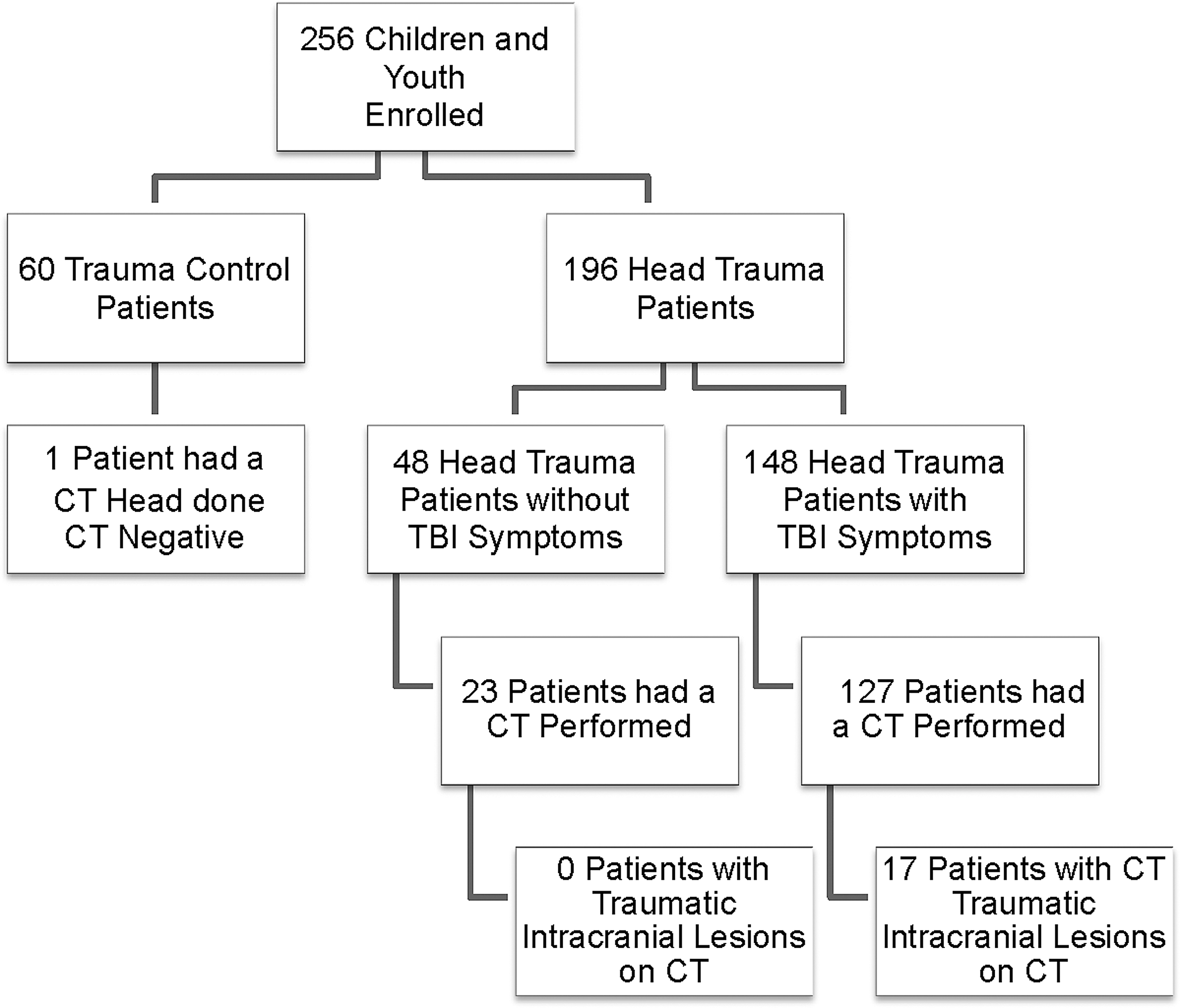
Flow diagram of enrolled patients. Flow diagram showing the number of all TBI and control patients enrolled. CT, computed tomography; TBI, traumatic brain injury.
The table describes data from children and youth enrolled in each of the three groups. Because of rounding, percentages may not add up to 100.
SD, standard deviation; GCS, Glasgow Coma Scale; ED, emergency department; TBI, traumatic brain injury.
Both the head trauma and trauma controls had serum samples drawn within 6 h of injury with the average time from injury to serum sample collection at 3.5 h (95% CI, 3.3–3.7). The average time to serum collection for head trauma patients was 3.3 hours (95% CI, 3.1–3.5) and for non-head-injured trauma controls it was 4.1 hours (95% CI, 3.7–4.5). UCH-L1 was detectible within an hour of injury. The distribution of UCH-L1 levels in children and youth with head trauma within 6 h post-injury is shown in Figure 2.
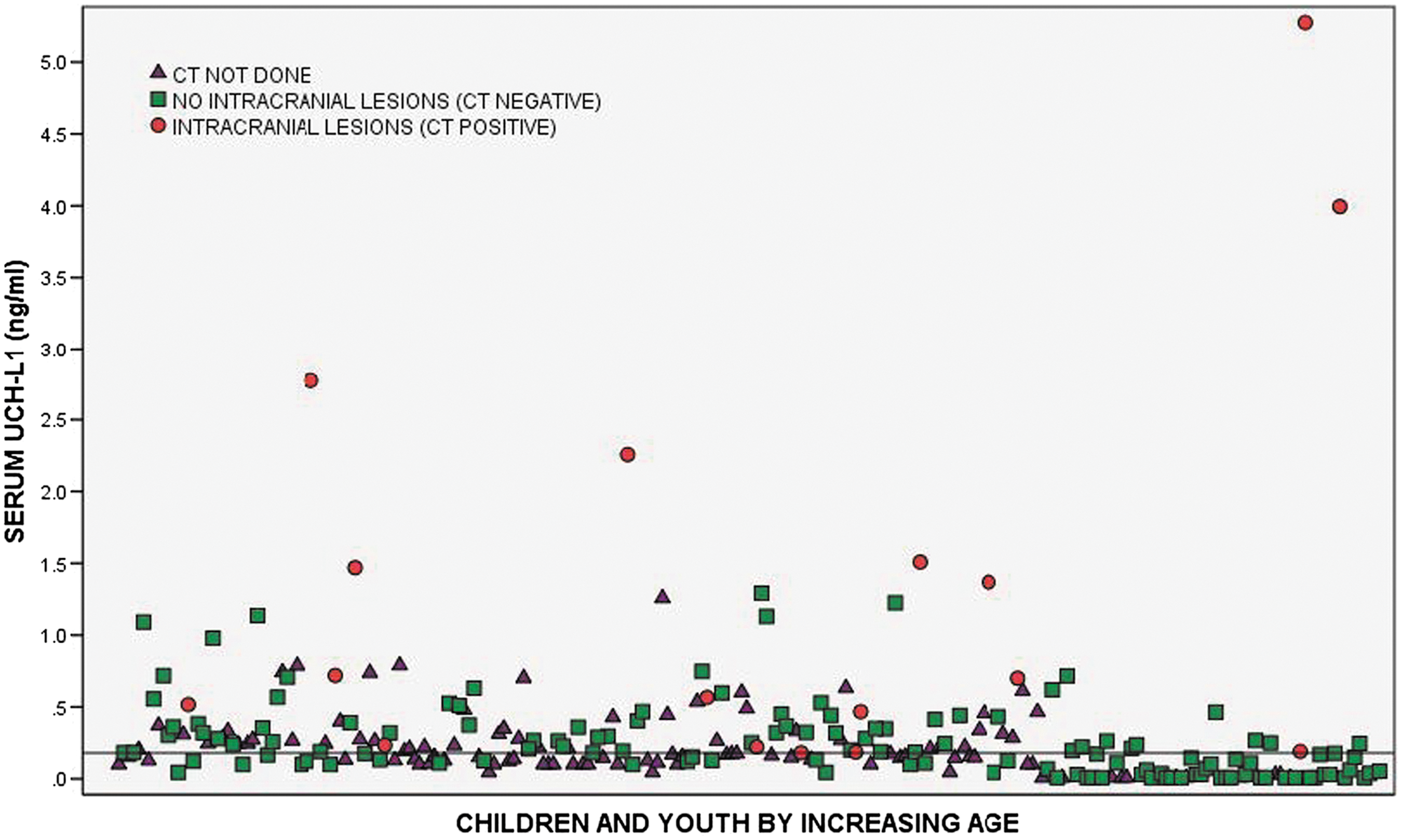
Scatterplot of UCH-L1 levels measured within 6 h of injury in children and youth with head trauma. Dots represent levels of UCH-L1 (ng/mL) in individual patients by increasing age. Scatter plot symbols represent: 1) children without CT imaging performed; 2) children with imaging but negative scans (no intracranial lesions); and 3) children with positive CT imaging (intracranial lesions). The reference line represents the UCH-L1 threshold of 0.18 ng/mL selected to determine sensitivity and specificity for predicting the need for CT scan. There is one value greater than 0.55 ng/mL (not shown), from a patient without an intracranial injury. CT, computed tomography; UCH-L1, ubiquitin C-terminal hydrolase.
In Figure 3, levels of UCH-L1 are compared between four groups of participants: 1) trauma controls; 2) head trauma patients without TBI symptoms; 3) head trauma patients with TBI symptoms (CT negative); and 4) patients with intracranial lesions (CT positive). All patients with positive CTs had symptoms of TBI. After adjusting for multiple comparisons, there were statistically significant differences in UCH-L1 concentrations between each of the groups relative to presence of CT lesions (p < 0.001). Median UCH-L1 levels increased incrementally from trauma controls (0.15; interquartile range [IQR], 0.10–0.28), to head trauma without TBI symptoms (0.18; IQR, 0.04–0.32), to head trauma patients with TBI symptoms (CT negative; 0.20; IQR, 0.1–0.35), and were highest in those with intracranial lesions (0.70; IQR, 0.23–1.89). There was also a statistically significant difference between trauma controls and head trauma patients (p = 0.041). There was no correlation between age and levels of UCH-L1 in trauma controls (p = 0.79).
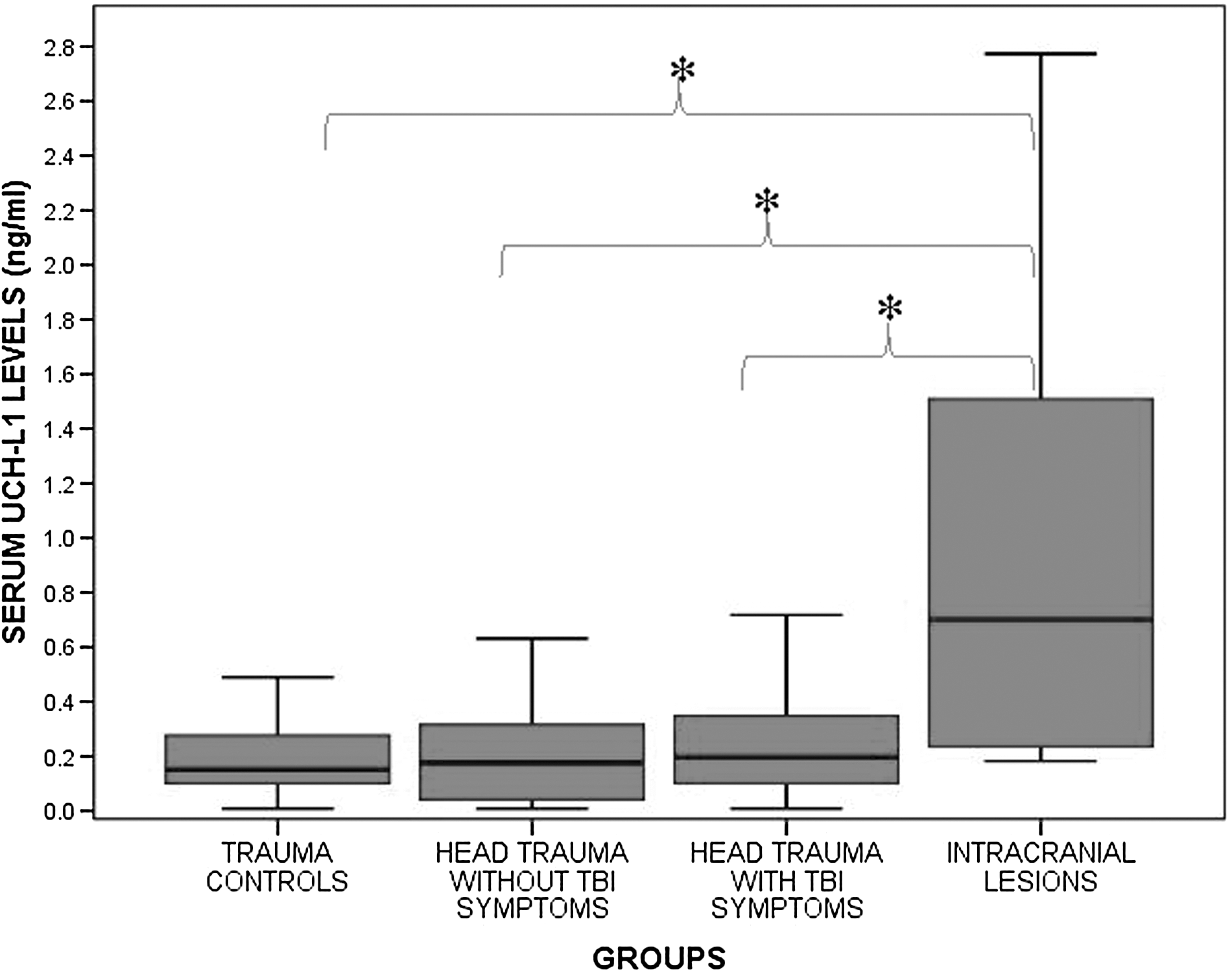
Comparison of serum levels of UCH-L1 drawn within 6 h of injury in four groups of participants. Groups included: 1) trauma controls (orthopedic and MVC; n = 60); 2) head trauma patients without TBI symptoms (n = 48); 3) head trauma patients with TBI symptoms (CT negative; n = 131); and 4) patients with intracranial lesions (CT positive; n = 17). After adjusting for multiple comparisons, there were statistically significant differences between each of the groups relative to the intracranial lesions group (indicated by the asterisk “
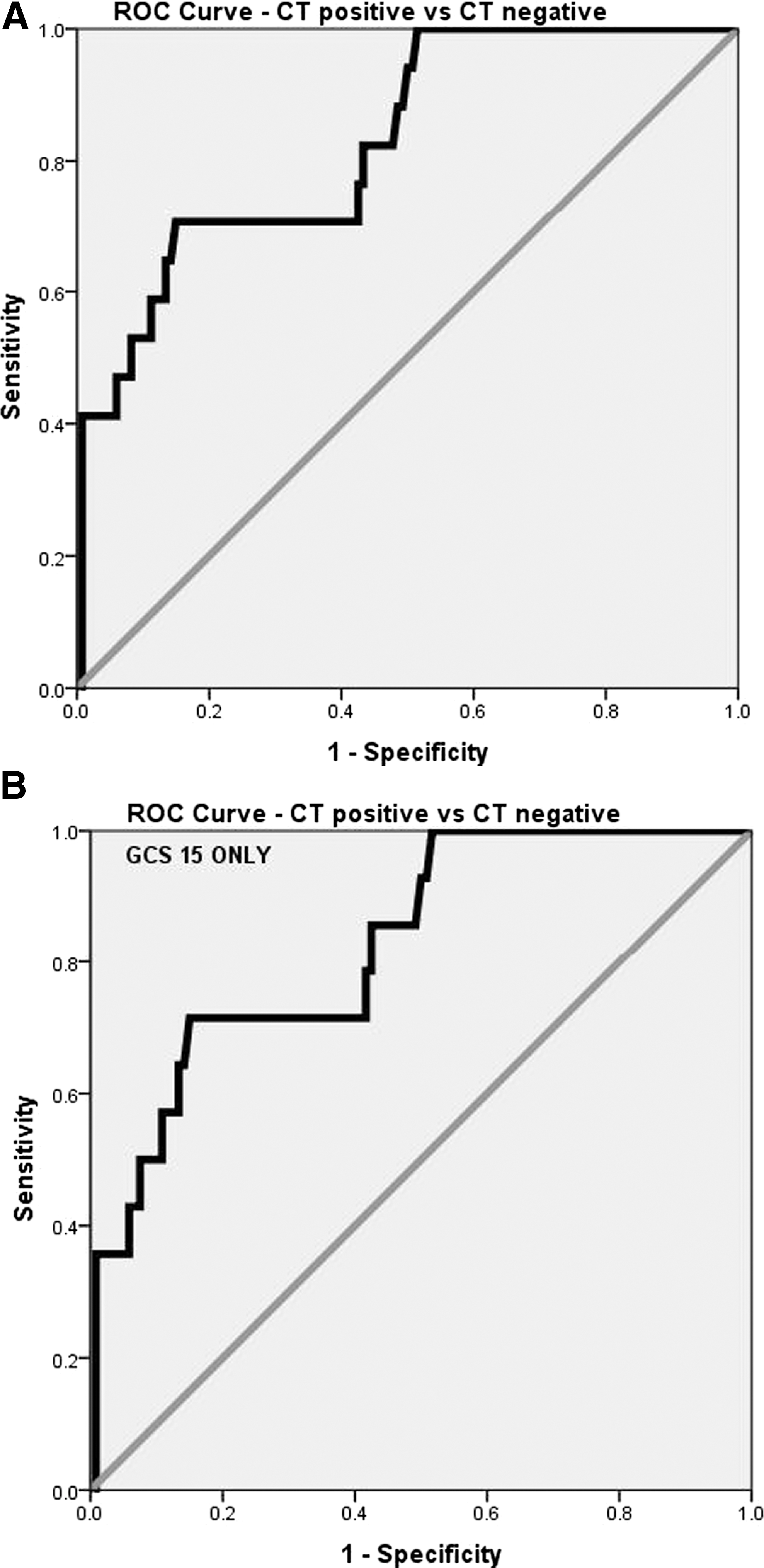
When serum levels of UCH-L1 were compared in children and youth with traumatic intracranial lesions on CT scan (CT positive; n = 17) to those without CT lesions (CT negative; n = 134), median UCH-L1 levels were significantly higher in those with intracranial lesions (0.70; IQR, 0.23–1.89) than those without lesions (0.18; IQR, 0.06–0.36) among all children who had CTs performed (p < 0.001). The AUC was calculated from the ROC curves constructed to assess the performance of early UCH-L1 levels (within 6 h of injury) in predicting traumatic intracranial lesions on CT. The AUC for discriminating between CT scan positive and CT scan negative was 0.83 (95% CI, 0.73–0.93; Fig. 4A). When patients with GCS 15 were assessed independently, median UCH-L1 levels were significantly higher in those with CT scan lesions (0.63; IQR, 0.23–1.48; n = 14) than those without (0.18; IQR, 0.06–0.35; n = 120; p < 0.001). The AUC for detecting intracranial lesions in those with GCS 15 was 0.83 (95% CI, 0.72–0.94; Fig. 4B). Moreover, when isolated skull fractures were combined with intracranial lesions for the analysis, the AUC was 0.79 (95% CI, 0.68–0.90).
(
Children and youths were subdivided to assess the performance of UCH-L1 for detecting intracranial lesions on CT by age. Among patients with head trauma, the highest proportion of CT scans (85%) was performed in the 15- to 21-year age group. However, 73% of children from birth to 5 years also had CT scans performed after head trauma (Table 2). AUCs for predicting intracranial lesions in each age category spanned from 0.59 to 1.00 and are shown in Table 2. For children younger than 5 years, the AUC was 0.79 (95% CI, 0.59–1.00).
Children and youths were separated into four categories based on age in years: birth–5 years; 5–10 years; 10–15 years; and 15–21 years to assess the performance of UCH-L1 for detecting intracranial lesions on CT.
UCH-L1, ubiquitin C-terminal hydrolase; CT, computed tomography; ROC, receiver operating characteristic; CI, confidence interval.
Cut-off points for UCH-L1 were derived from the ROC curves for detecting intracranial lesions on CT scan to maximize the sensitivity and to correctly classify all traumatic intracranial lesions. Classification performance for detecting intracranial lesions on CT at a UCH-L1 cut-off level of 0.18 ng/mL yielded a sensitivity of 100%, a specificity of 47%, and an NPV of 100% (Table 3). When isolated skull fractures were considered together with intracranial lesions, performance was almost identical with a sensitivity of 100%, a specificity of 49%, and an NPV of 100% (Table 3). UCH-L1's performance in children and youth with an unaltered mental status (GCS 15) remained notably consistent in predicting intracranial lesions, with a sensitivity of 100% and a specificity of 48%.
UCH-L1, ubiquitin C-terminal hydrolase; NPV, negative predictive value; PPV, positive predictive value; CT, computed tomography; GCS, Glasgow Coma Scale.
In Figure 5, patients were categorized by severity of CT head lesions and compared to those who did not have a head CT performed. Levels of UCH-L1 increased with severity of CT lesions: no lesions (n = 98); scalp hematomas (n = 29); skull/facial fractures (n = 7); and intracranial lesions (n = 17; p < 0.001). Those who did not have a CT performed had the lowest levels of UCH-L1 compared to any other group (n = 105; p < 0.001). The reference line represents the UCH-L1 threshold of 0.18 mg/mL selected to determine sensitivity and specificity for predicting the need for CT scan. Most of the patients who did not have a CT performed were below the UCH-L1 threshold of 0.18 ng/mL, and all patients with no injuries were below the threshold (Fig. 5).
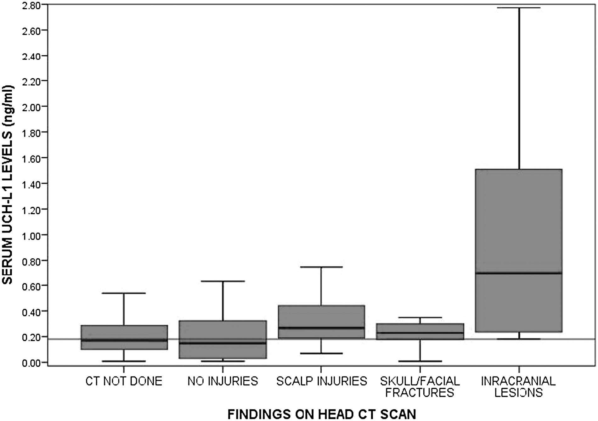
Box plot of levels of UCH-L1 in patients without a head CT performed compared to different lesions on head CT. Patients have been distributed by severity of CT head lesions and compared to those who did not have a head CT performed. Levels of UCH-L1 increased with severity of CT lesions: no lesions (n = 98), scalp hematomas (n = 29), skull/facial fractures (n = 7), and intracranial lesions (n = 17; p < 0.001). Those who did not have a CT performed had the lowest levels of UCH-L1 compared to any other group (n = 105; p < 0.001). The reference line represents the UCH-L1 threshold of 0.18 mg/mL selected to determine sensitivity and specificity for predicting the need for CT scan. CT, computed tomography; UCH-L1, ubiquitin C-terminal hydrolase.
Discussion
This prospective study assessed the performance of UCH-L1 within 6 h of trauma in a cohort of children and youth, with and without head trauma, presenting to two pediatric level 1 trauma centers and one adult level 1 trauma center. UCH-L1 was considerably higher in children and youth with traumatic intracranial lesions on head CT, compared to those with no lesions, with a sensitivity of 100% and a specificity of 49%. In children and youth presenting with a GCS score of 15, UCH-L1 performed consistently and predicted intracranial lesions with a sensitivity of 100% and a specificity of 48% at a cut-off level of 0.18 ng/mL. This cutoff was selected in order to optimize sensitivity for detecting traumatic intracranial lesions on CT so that intracranial lesions would not be missed. Based on this UCH-L1 cut-off level, 64 (42%) CTs could have potentially been avoided. This is among the largest published studies to date assessing UCH-L1 in children and youth with mmTBI in an ED setting.
UCH-L1 performed well in infants and toddlers 5 years of age and younger with an AUC of 0.79 and a sensitivity of 100% and specificity of 32%. This finding has significant implications for the management of infants and toddlers with potential brain injury. Not only is head trauma very prevalent in this age group, 26 but injury severity can be particularly difficult to assess clinically because many infants and toddlers are either nonverbal or unable to provide an accurate history. The CT ordering rate was 73% in this age group. Therefore, alternate diagnostic strategies to reduce ionizing radiation from head CTs, such as blood-based biomarkers, are crucial to improve care.
Using trauma patients without head trauma as a comparison group, instead of uninjured controls, allowed us to assess UCH-L1's brain specificity in a population that had been exposed to significant trauma. The forces that induced injuries in trauma control patients paralleled that of TBI patients, with the exception that trauma controls lacked both blunt head trauma and TBI symptoms. UCH-L1 was higher in children and youth with head trauma than trauma controls who did not strike their head. When patients were categorized by severity of CT head lesions concentrations of UCH-L1 increased with severity of CT lesions from no lesions to scalp hematomas to skull/facial fractures and finally intracranial lesions. All patients with no injuries were below the threshold for predicting the need for CT scan. These findings are comparable to the results of UCH-L1 in adult studies that have used similar control groups. 22
In an ED setting, most head trauma patients are evaluated within an hour or two of injury. In this study, UCH-L1 was detectable within an hour post-injury and was also detectable at 6 h. This is consistent with time course of UCH-L1 in adults with mTBI where UCH-L1 rises rapidly post-injury, peaks at 8 h, and decreases steadily over 48 h. 18 This suggests that UCH-L1 is an early marker of injury.
There was a CT ordering rate of close to 50% in children with head trauma patients who did not exhibit TBI symptoms. Despite this, there was not a single positive CT in this group. Clinical judgement (based on a number of clinical and nonclinical factors) leads to the decision to use diagnostic imaging. Therefore, there is an opportunity, through the use of a blood biomarker (blood test), to provide clinicians with an important piece of information that could potentially be used to reduce unnecessary CT scans to reduce the risk of ionizing radiation in children with head trauma. Current clinical decision rules for determining the need for CT children have a wide range of sensitivities (84–100%) and specificities (44–85%). A blood test to detect intracranial lesions could add a layer of objectivity to clinical decision making and perhaps become a useful adjunctive tool to clinical decision making.
Given that initial GCS scores in the ED in this population can be unreliable, we chose to include children with a GCS of 9–15. The classification of a TBI as mild or moderate can change based on neuroimaging results and the presence of factors altering mental status, such as intoxication, medications, and other injuries. Although we studied TBI patients from GCS 9 to 15, we included focused analyses of those with a GCS score of 15 and carrying a diagnosis of “concussion.” This has implications for concussion management, where a point-of-care test could be used to detect TBI immediately at the scene of injury in settings such as in the ambulance, on the playing field, or on the battlefield, 10 especially given that UCH-L1 is known to have such a rapid rise post-injury. 18
Two recent studies in children and youth examined the role of glial marker, glial fibrillary acidic protein (GFAP), in mild traumatic injury and found GFAP to have AUCs of 0.82–0.85 along with sensitivities of 94–100% and specificities of 47–55%. 11,12 This is similar to what we found in this current study, with UCH-L1 yielding an AUC of 0.83 and a sensitivity and specificity of 100% and 47%, respectively. Other studies conducted in children have compared these two biomarkers and have found conflicting evidence, with one showing GFAP as the better biomarker 20 and the other showing UCH-L1 as superior. 21 Both these studies were limited by very small sample sizes of 25 (mTBI) and 45 (mild to severe) TBI patients respectively, with the latter article including mostly severe TBI.
If these findings can be further validated, UCH-L1's association with the presence of intracranial lesions on CT scans could help emergency physicians reduce the number of CTs performed and it could be incorporated into guidelines for neuroimaging decisions and decisions to transfer patients to higher levels of care.
Limitations
Although these data are encouraging, the authors recognize that there are limitations to this study. Patients were enrolled as a convenience sample because the research team could not be on duty 24/7. Despite this, patients were recruited consecutively when research assistants were on duty, including on weekends and nights, so a representative sample could be enrolled.
The current study was performed in a substantial cohort of children and youth post-trauma; however, the sample was small when we separated the children into age categories. UCH-L1 demonstrated some variability in performance across age categories for detecting intracranial lesions on CT, particularly in the 10- to 15-years group. Small sample size in each of the categories likely contributed to this variability. Despite this, the sensitivity was still 100% in this group, but specificity was 28%. Future studies with larger cohorts will be required to reassess this finding.
Additionally, the CIs around the sensitivity and specificity for detecting intracranial lesions were wide and reflected the relatively small number of children and youth with lesions on CT in the cohort (17). Again, a much larger number of children will be required to test the precision of the biomarker.
This study addressed severity of injury in the acute care setting and did not describe long-term outcome in these patients. Further, future studies should define UCH-L1 in an uninjured population of children and youth at different ages. We did not find any differences in UCH-L1 levels in children and youth of different ages in our trauma control group.
We cannot confirm that UCH-L1 is brain specific and may be released from other organs during injury. However, in this study, levels of UCH-L1 in trauma controls were significantly lower than TBI patients. Studies conducted in adults comparing mTBI to trauma controls have shown good discrimination. 22 Alternatively, the mechanisms of injury in the trauma control patients may be such that acceleration/deceleration forces are exerted on the brain and lead to the release of UCH-L1. Further study is needed to examine this.
Conclusion
This study introduces UCH-L1 as a valuable candidate biomarker for detecting traumatic intracranial lesions on CT in children and youth with suspected mTBI, and the findings appear consistent with the work conducted in adults. UCH-L1 showed good performance in infants and toddlers younger than 5 years and performed well in children and youth with a GCS score of 15. Before clinical application, further study in a larger cohort of children and youth with mTBI is warranted to validate these findings.
Footnotes
Acknowledgments
The project described was supported, in part, by Award Number R01NS057676 from the National Institute of Neurological Disorders and Stroke (NINDS). The content is solely the responsibility of the authors and does not necessarily represent the official views of the NINDS or the National Institutes of Health.
Author Disclosure Statement
Dr. Papa is a scientific consultant of Banyan Biomarkers, Inc., but receives no stocks or royalties from the company and will not benefit financially from this publication.