
Abstract
Abstract
Gastric cancer is the third leading cause of cancer-related mortality worldwide. Recent evidence points to importance of cross talk between cancer cells and the surrounding stroma on gastric cancer progression. Tumor microenvironment biomarkers thus represent a new opportunity for diagnostics innovation. Reactive stromal fibroblasts selectively express the fibroblast activation protein alpha (FAP-α), a homodimeric integral membrane gelatinase that belongs to the serine protease family. We report here that FAP-α expression is significantly elevated in gastric cancer samples by more than fivefold (p < 0.05), using transcriptome data from The Cancer Genome Atlas. Notably, the greatest FAP-α upregulation was observed in the poorly differentiated group (p < 0.001). Moreover, elevated FAP-α expression levels correlated with adverse clinical–pathological characteristics, such as diffuse histological subtype (p < 0.001), advanced pathological stage (p < 0.01) and poor survival. Functional annotation analysis demonstrated that FAP-α upregulation was associated with activation of biological processes implicated in tumor progression, including cell migration and angiogenesis pathways. These observations underscore the possible prognostic significance of FAP-α in gastric cancer and its potential as a novel biomarker for personalized medicine. We caution, however, that further multiomics, biochemical, and animal studies are necessary to ascertain the role of FAP-α as a causative and mechanistic biomarker. Based on pathway analyses, we hypothesize that gastric cancer patients exhibiting FAP-α upregulation might presumably benefit from antiangiogenic drugs in addition to standard therapeutic regimens. We call for future research focusing on the tumor microenvironment biomarkers in clinical oncology.
Introduction
G
The tumor stroma is composed of several nonmalignant cell types, including fibroblasts, inflammatory cells, endothelial cells, adipocytes, and noncellular component such as extracellular matrix (ECM), together building up the tumor microenvironment (Bhowmick et al., 2004; Mueller and Fusenig, 2004). Recent advances of next-generation sequencing studies have shown that global transcriptomic profiling of tumor tissues can identify different stromal reaction signatures and thus represents a valuable tool to subgroup tumors for prognostic and predictive purposes in various human cancers, including gastric cancer (Isella et al., 2015; Lenz et al., 2008; Uhlik et al., 2016). Therefore, it is of great interest to identify the key players contributing to gastric cancer progression in the stromal compartment.
Fibroblast activation protein alpha (FAP-α), an important tumor microenvironment regulator, is secreted by activated stromal fibroblasts (Mathew et al., 1995). Overexpression of FAP-α has been implicated in several human malignancies, including breast cancer, ovarian cancer, pancreatic adenocarcinoma, colon cancer, lung cancer, melanoma, and lymphoma (Henry et al., 2007; Kraman et al., 2010; Lee et al., 2005; Lv et al., 2016). For instance, it was reported that FAP-α promotes ovarian cancer cell proliferation, drug resistance, and invasiveness through integrin receptors and small guanosine triphosphatase Rac1 (Xing et al., 2010; Yang et al., 2013). Similarly, elevated FAP-α expression was shown to be associated with suppressed lymphocyte-dependent immune response and poor survival of nonsmall cell lung cancer and pancreatic adenocarcinoma (Liao et al., 2013; Patsouras et al., 2015). However, the role of FAP-α in gastric cancer is still unknown.
We report here an integrated analysis of transcriptome using data from The Cancer Genome Atlas (TCGA) (Cancer Genome Atlas Research, 2014) and that FAP-α expression is significantly elevated in gastric cancer samples. The prognostic values of FAP-α were studied in multiple independent clinical cohorts. Finally, we went a step further to characterize the biological processes associated with FAP-α upregulation in gastric cancer.
Materials and Methods
FAP-α expression analysis in the TCGA data set
Whole genome messenger RNA expression data and clinical information of 366 gastric cancer samples were obtained from TCGA database (http://cancergenome.nih.gov) (Cancer Genome Atlas Research Network, 2014). Two additional clinical data sets, Ooi et al. (2009) and the Cristescu et al. (2015), were retrieved from the Gene Expression Omnibus database under the accession numbers GSE15459 and GSE62254. The studies are summarized in Table 1, and details about the data sets are given below.
GEO, Gene Expression Omnibus; TCGA, The Cancer Genome Atlas.
Level 3 data comprising expression values in gastric cancer were downloaded through the TCGA portal on November 19, 2015. As part of TCGA project, the purpose of the study was to comprehensively evaluate the molecular and clinical characteristics of gastric cancer. Primary gastric cancer tissues not treated with prior chemotherapy or radiotherapy were collected.
Data set GSE15459 was produced by Ooi et al. (2009). The purpose of the study was to identify major oncogenic pathways in gastric cancer with significant relationships to patient survival. The gastric cancer samples were collected from three cohorts: Peter MacCallum Cancer Centre (Australia), National Cancer Centre, (Singapore), and Leeds Institute of Molecular Medicine (United Kingdom). All samples were processed uniformly.
Data set GSE62254 was described by Cristescu et al. (2015) for the purpose of establishing clinically relevant molecular subtypes for gastric cancer. Three hundred primary independent gastric cancer specimens were procured at the time of total or subtotal gastrectomy at Samsung Medical Center, Korea.
Ethics review and approval
This research utilized a public database for analyses and does not contain studies with human participants or animals.
Patient stratification
For comparative analysis of clinical–pathological parameters and survival analysis, gastric cancer patients from the TCGA data set were stratified as depicted in Figure 2A. Patients with lower than median expression level of FAP-α were defined as FAP-αLow group, while patients with higher than or equal to the median one were considered as FAP-αHigh group.
Gene Set Enrichment Analysis
Gene set enrichment analysis (GSEA) was performed using R package “fgsea” from Bioconductor. Parameters used for the analysis were as follows. The msigdb.v5.1.symbols.gmt gene set was used for running GSEA and 1000 permutations were used to calculate the p value. The Enrichment Map plugin was used to generate networks derived from the GSEA results in cytoscape (http://cytoscape.org). Network visualization was done by the same plugin.
Statistical analysis
Student's t-test and one-way analysis of variance (ANOVA) test were used to test the significance of differences. Tukey's procedure tested pair-wise differences for significant ANOVAs. Comparative analysis of clinical–pathological parameters was evaluated using the Chi-squared test or Fisher's exact test. Survival analysis was performed using Kaplan–Meier analysis. The log-rank test was used to assess the statistical significance between stratified survivals groups. The results were considered statistically significant when p values were <0.05. All p values are indicated in the figures.
Results
FAP-α expression was upregulated in human gastric cancer and associated with neoplastic stages
To gain insight into the pathological role of FAP-α in gastric cancer progression, we explored FAP-α expression pattern in gastric cancer samples by analyzing the transcriptomic data from the TCGA database. We found that FAP-α expression levels were strongly elevated in gastric cancer tissue compared with tumor surrounding tissue (5.1-fold median difference, p < 0.0001) (Fig. 1A). Comparison of paired samples of gastric cancer tissue and adjacent noncancer tissue revealed the same trend (Fig. 1B).
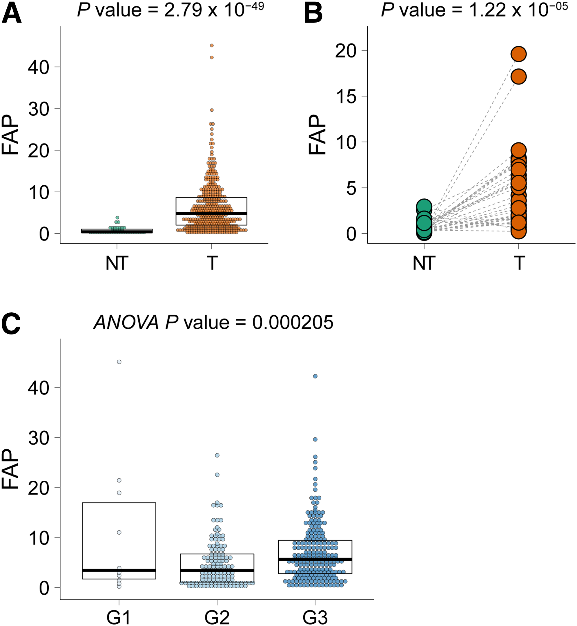
FAP-α expression levels are upregulated in human gastric cancer.
Seeking additional cues for how FAP-α expression accumulates during disease progression, we further compared its levels in gastric cancer samples at different neoplastic stages. Notably, the expression levels of FAP-α varied significantly in gastric cancer samples of different stages. Patient samples at G3 stage exhibited roughly twofold higher FAP-α expression in relative to patient samples at G1 or G2 stage as determined by ANOVA and subsequent Tukey post hoc analysis (Fig. 1C). Consistently, according to the immunohistochemistry-based data available at Human Protein Atlas database (www.proteinatlas.org), 9 out of 11 gastric cancer specimens display moderate–strong FAP-α expression. These results indicate that FAP-α upregulation might play a critical role in the malignant progression of human gastric cancer.
FAP-α expression levels correlate with clinical–pathological features of gastric cancer
Key gene expression levels derived from primary tumors have been shown to predict response to therapy, distant metastasis, and poor survival (Blanco and Kang, 2011; Kato et al., 2008). To investigate whether FAP-α expression was associated with clinical–pathological features in gastric cancer, patient samples were divided into two groups, FAP-αHigh and FAP-αLow patients, according to median expression value as illustrated in Figure 2A. FAP-αHigh gastric cancer was more frequent in adenocarcinoma type than intestinal type in term of the histologic subtypes (Fig. 2B). In addition, FAP-α expression levels robustly stratified patients at more advanced stages from at preliminary stages for pathological stage (p < 0.01) and pathological T stage (p < 0.001) (Fig. 2C, D).
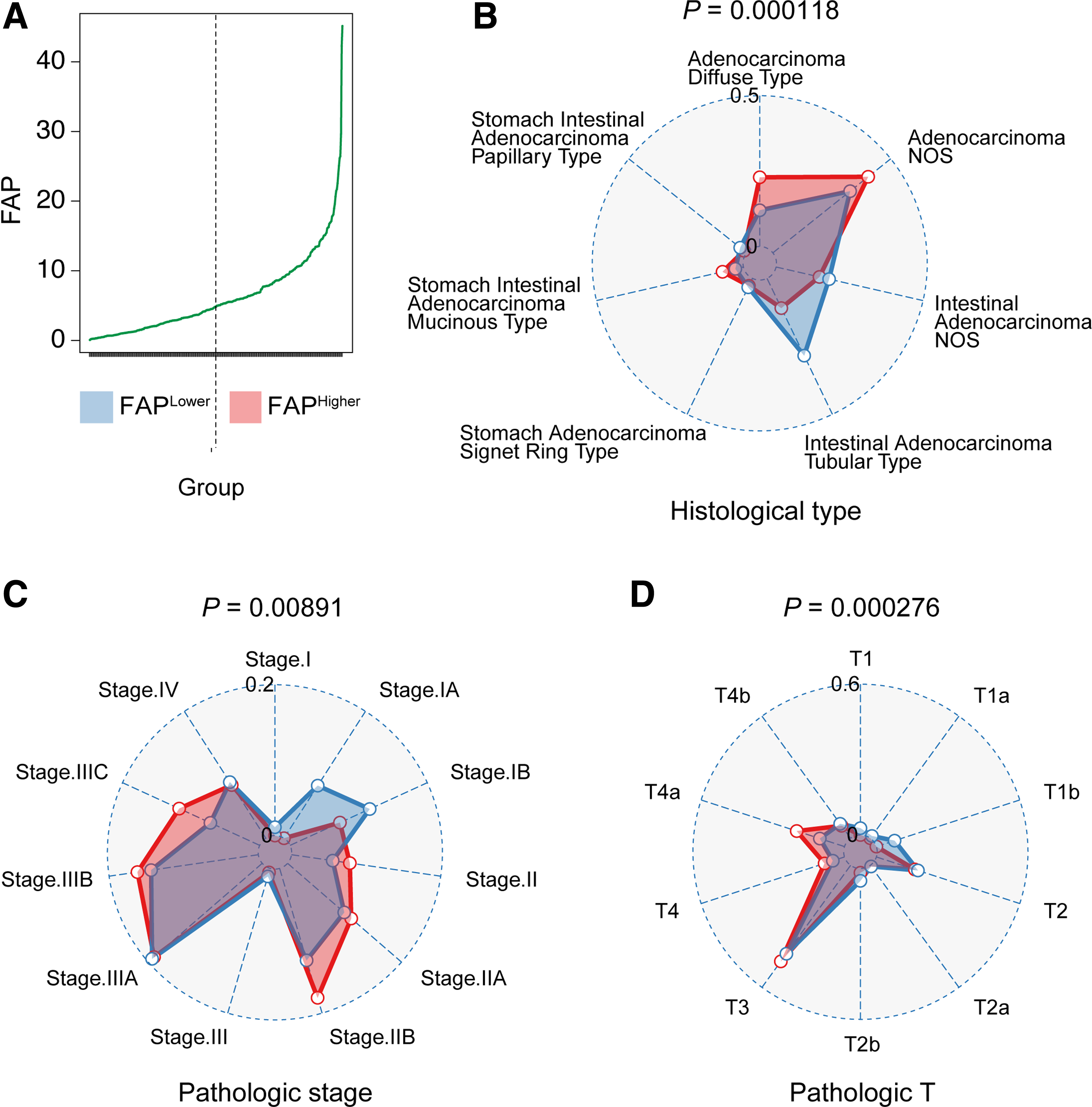
FAP-α upregulation correlates with the adverse clinical–pathological characteristics in gastric cancer samples.
Elevated expression of FAP-α is associated with adverse clinical outcome
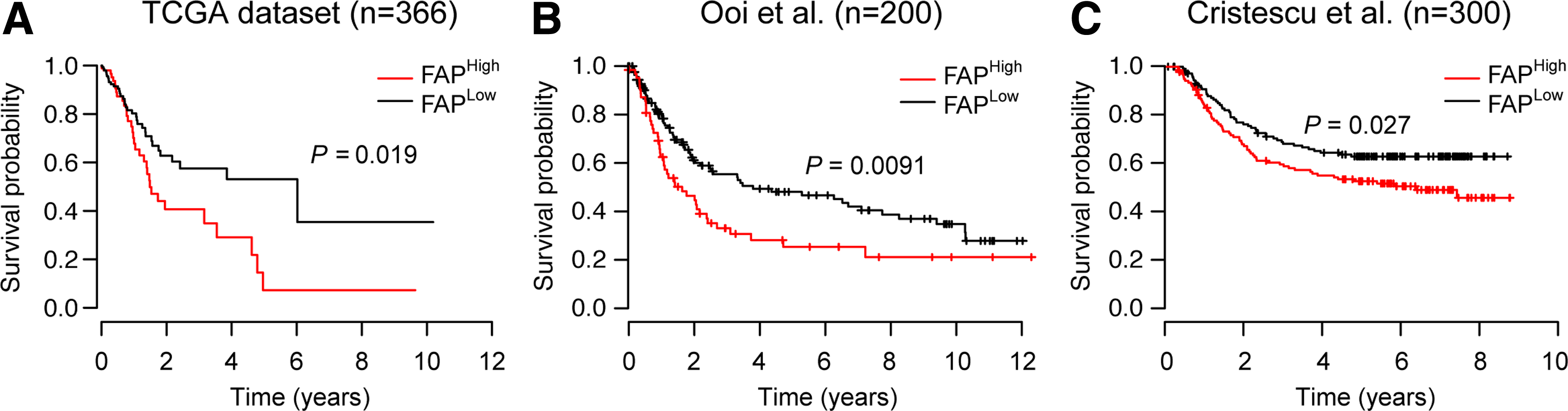
Next, we assessed the association of FAP-α expression with prognosis of gastric cancer patients through Kaplan–Meier survival curve analysis with a log-rank test. FAP-αHigh gastric cancer patients demonstrated significantly shorter overall survival compared with FAP-αLow gastric cancer patients (Fig. 3A). The median survival time for FAP-αHigh patients was 1.6 years, significantly shorter than 6.0 years of FAP-αLow patients (log-rank test p = 0.019). Investigation of another two independent gastric cancer cohorts demonstrated similarly significant association between FAP-α expression levels and patient overall survival (log-rank test p = 0.0091 for Ooi et al. cohort and 0.027 for Cristescu et al. 2015 cohort) (Fig. 3B, C). Taken together, these results imply that FAP-α might be a useful independent prognostic biomarker for gastric cancer patients.
FAP-αHigh gastric cancer samples possess characteristics of stromal cell-associated signaling pathways
To define the molecular characteristics of FAP-αHigh versus FAP-αLow patients, we compared the transcriptomic profiles by GSEA. FAP-αHigh gastric cancer samples showed enrichment of signaling pathways implicated in development and carcinogenesis, including immune responses, angiogenesis and morphogenesis, cell migration and neuron development, cell differentiation and cell growth (Fig. 4A). It is notable that signaling pathways of cell migration and angiogenesis have been shown to be closely associated with activation of stromal fibroblasts in various cancer types (Fig. 4B) (De Wever and Mareel, 2003; Kitadai, 2010). These results highlight the relevance of FAP-α expression and activation of stromal cell-related signaling pathways in gastric cancer pathology.
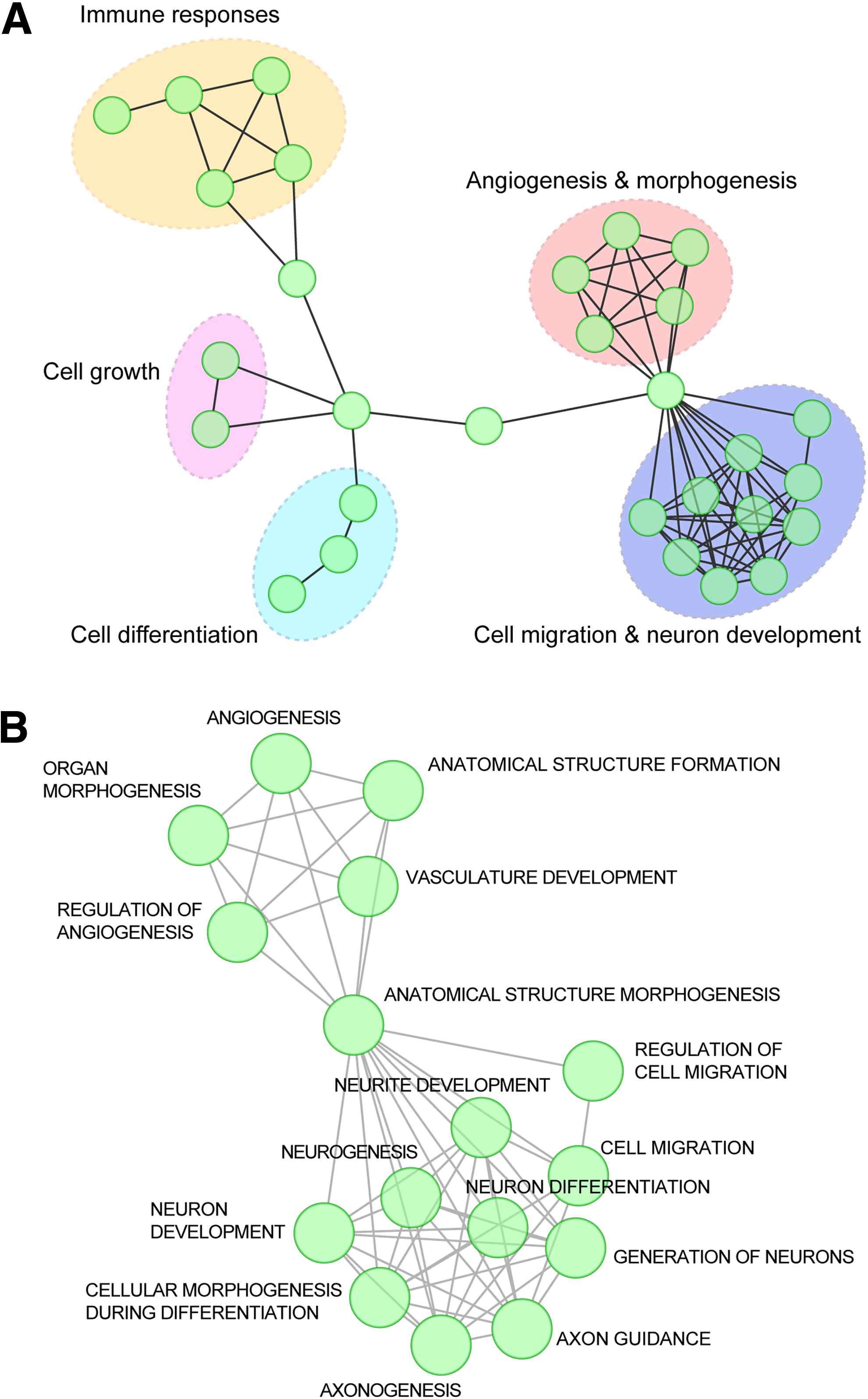
Biological processes activated in FAP-αHigh gastric cancer samples by GSEA.
The role of FAP-α in mediating signaling pathways of cell migration and angiogenesis was further investigated by comparing the FAP-αHigh versus FAP-αLow samples with additional officially annotated gene sets. Agreed with the above results, enrichment of cell migration pathway-related genes was more dominant in FAP-αHigh gastric cancer samples in relative to FAP-αLow gastric cancer samples (Fig. 5A). The same holds true in terms of angiogenesis pathways. Two angiogenesis-related gene sets, Lu-tumor angiogenesis-up and PID-lymphangionesis pathway, were significantly enriched in FAP-αHigh gastric cancer samples (Fig. 5B). Together, these results support the idea that FAP-αHigh cancer samples tend to be more aggressive, probably due to the activation of stromal compartment-mediated cell migration and angiogenesis signaling pathways.
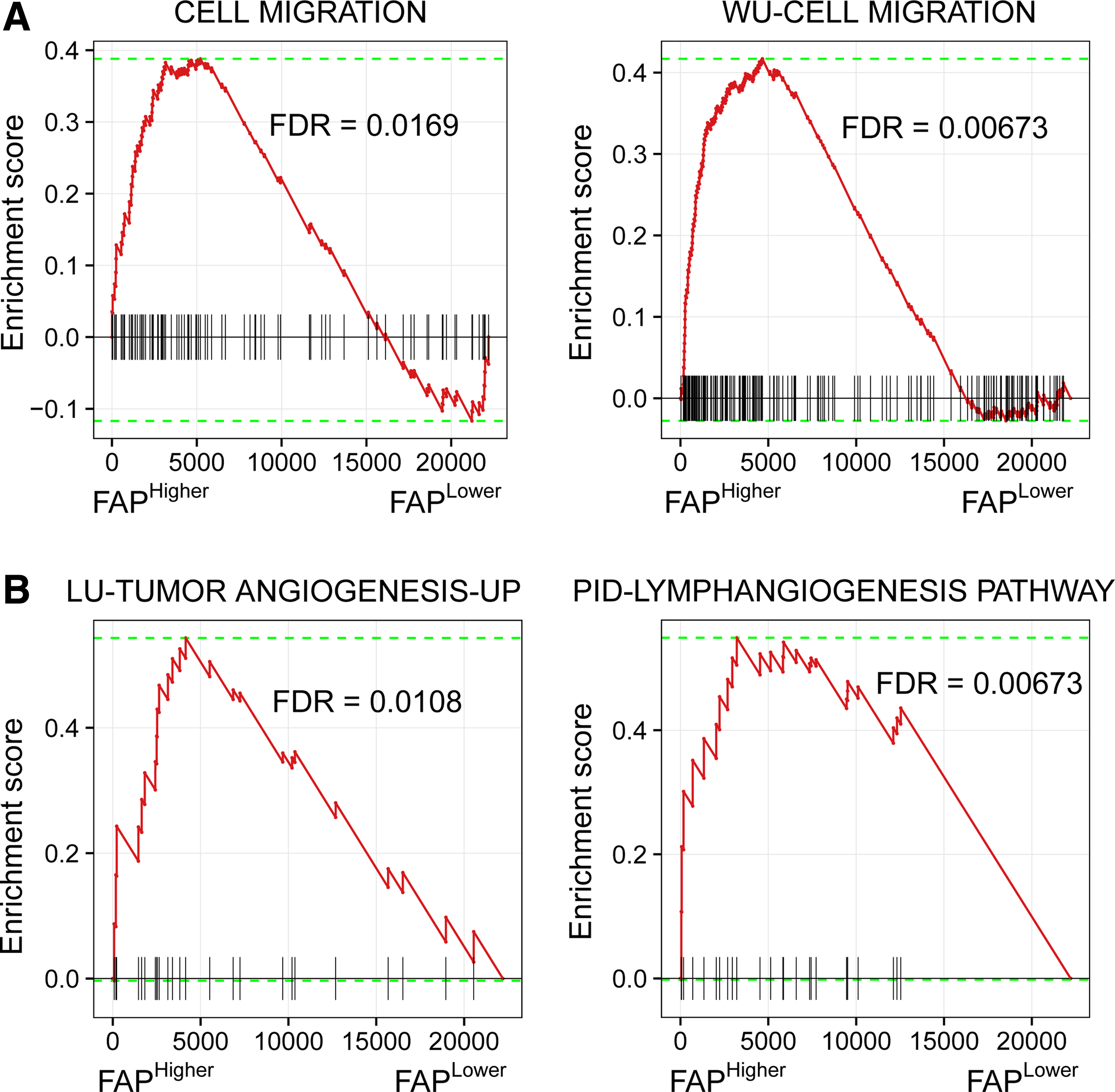
Gastric tumors with higher FAP-α expression exhibit enrichment of gene signature of cell migration and angiogenesis pathway.
Discussion
In our study, we found that FAP-α was significantly unregulated in gastric cancer tissue. Elevated levels of FAP-α correlated to higher pathological T stage, diffuse histological subtype, and poor prognosis of patients. These results suggested that FAP-α might be closely related to the cancer development and be the potential biomarker for decision-making and patient surveillance of gastric cancer.
Tumor surrounding microenvironment is activated for continuous paracrine communication and supports carcinogenesis (Quail and Joyce, 2013), but the regulatory mechanisms are not fully understood. FAP-α has been reported to be elevated in stromal compartments of many epithelial tumors and plays an important role in regulating tumor growth and metastasis (Ariga et al., 2001; Tchou and Conejo-Garcia, 2014). In a mouse model of fibrosarcoma, FAP-α has been shown to induce tumor cell motility and spontaneous metastases (Tansi et al., 2016). Recently, Yang et al. (2016) showed that FAP-α can promote STAT3 activation and CCL2 expression to trigger the induction of cancer-associated fibroblasts with an inflammatory phenotype. Our work showed that FAP-α expression was closely related to the cell migration and tumor growth signaling pathways, which suggested that the FAP-α plays an important role in regulating tumorigenesis and development.
Antitumor strategies targeting tumor microenvironment have been proven to be effective in several preclinical mouse models of human cancer. Study showed that depletion of FAP-α expressing stromal cells repressed the tumor progression and increased tumor antigen-specific T-cell responses concomitantly (Zhang and Ertl, 2016). Similarly, alterations of tumor microenvironment through disruption of FAP-α activity could affect pancreatic and other cancer cell invasiveness by disrupting the organization of stromal ECM (Lee et al., 2011). Inhibition of FAP-α can also inhibit mammary carcinoma cell growth by regulating the tumor microenvironment in a mouse model (Cai et al., 2013). Our analysis revealed that higher levels of FAP-α in the gastric cancer tissues were linked to poor prognosis. Thus, FAP-α silencing or pharmacological inhibition might also influence the growth of tumor and could be the therapeutic target for further anticancer research in gastric cancer.
Taken together, we uncovered the close correlation between expression levels of FAP-α, the tumor stromal microenvironment-related gene, and gastric cancer progression. We also showed the regulatory signaling pathways related to FAP-α upregulation in gastric cancer. These results indicated that FAP-α might not only be an effective prognostic biomarker of gastric cancer but also might exert its function as the regulator of many signaling pathways for gastric cancer formation and development. Our study comprehensively showed the significance of FAP-α expression in gastric cancer patients and gave a new insight into the function of tumor microenvironment regulator in gastric cancer for further prognostic and clinical study.
Conclusions and Future Directions
The present analysis revealed that FAP-α upregulation in gastric cancer samples correlates with clinical–pathological characteristics and predicts a worse survival outcome. Pathway analysis implied activation of stromal cell-related signaling pathways involved in tumor progression, including cell migration and angiogenesis pathways. Our work provides evidence that FAP-α expression levels can be used to stratify gastric cancer patients into low- and high-risk groups and to predict their clinical outcomes. Based on the pathway analysis, we suggest that gastric cancer patients exhibiting FAP-α upregulation might benefit from antiangiogenic therapy in addition to standard regimens.
Footnotes
Acknowledgments
H.B.Z. and B.J.F. conceived the project. M.M.H. and C.J.Q. collected the data, performed the computational coding, and conducted data analysis. M.M.H. drafted the article. All authors were involved in writing the article and had final approval of the submitted and published versions. This work was supported by the Natural Science Foundation of China (Grant No. 21505053), Science and Technology Planning Project of Guangdong Province, China (Grant Nos. 2015A030401045, 2016A030310089).
Author Disclosure Statement
The authors declare that no conflicting financial interests exist.