
Abstract
Introduction
Ozone is a proven antimicrobial agent that has been used extensively in the medical field for many years. 7,8 An ozone-delivery system, developed recently (HealOzone; Kavo, Biberach, Germany), has allowed the application of gaseous ozone to a precise area on the tooth surface under controlled conditions because of the antimicrobial potential of ozone against common oral pathogens. 4 Baysan and Linch 8 reported that ozone application either for 10 or 20 sec was effective in killing the great majority of microorganisms in root carious lesions in vitro. The 10-sec application was also capable of reducing the numbers of Streptococcus mutans and S. sobrinus in vitro. 4 Further studies demonstrated the effects of ozone, delivered in the same manner, on the microbial flora and clinical severity of primary root caries. 9 In addition, the efficacy of the use of ozone for the management of root caries was proven in a longitudinal study for a period of 12 months. 9 The mechanism of action of HealOzone was related to ozone's strong antimicrobial properties and its ability to oxidize proteins associated with the dental caries process. Ozone could be effective on these microorganisms by rupturing their membranes and blocking enzymatic pathways. 10
The disinfecting effect of lasers has been well known for a long time. It was shown that the Nd:YAG laser, due to its wavelength (1,064 nm) and pulsed action, had the highest bactericidal effect among all lasers presently available on the market. 11 The wavelength of the Nd:YAG laser is highly absorbed by the pigmented cell membranes of the cariogenic bacteria, resulting in permanent bacterial membrane destruction by sudden heating on the cell membranes and selective bacteria reduction, without any thermal side effects to the tissues and adjacent structures by using controlled parameters. 5 Besides, the Nd:YAG laser is able to eliminate pathogenic bacteria in even deeper layers of dentin, up to 1,000 μm. 12 This provides a distinct advantage, because bacteria can travel up to 1,000 μm into the tubules. 12
In the adhesive dentistry field, many efforts have been directed to improving adhesion to tooth tissues, enhancing bonding durability, and simplifying adhesive procedures. To this end, adhesive systems using self-etch technology were introduced. 13 Self-etch adhesives are extremely fast and simple to apply in clinical situations, which saves clinical time and dramatically reduces the technique sensitivity of the bonding procedure. 14 Current self-etch adhesive systems are generally divided into two types. 15 The first type is termed “self-etching primer” (two-step self-etch) systems and combines the demineralizing agent and primer with a separate adhesive resin. The most recent type combines all components into one liquid, and these have been referred to as “all-in-one” or “one-step self-etch” systems. Although low technique sensitivity and consistent performance are expected to be achieved with one-step self-etch adhesives because of their simplified application procedures, some previous studies indicated controversies on the performance of these newly introduced adhesives. 16
Some concerns have been expressed regarding potential adverse effects of application of cavity disinfectants before bonding procedures in performing adhesive restorations. The aim of this in vitro study was to evaluate the effects of ozone and Nd:YAG laser pretreatment on the shear-bond strength of either one-step or two-step self-etch adhesives to coronal and root dentin.
Materials and Methods
The materials used in the study are shown in Table 1.
In total, 60 intact human cuspids extracted for periodontal reasons were scaled with a periodontal scaler to remove organic debris before cleaning with water/pumice slurry. The teeth were stored in 0.5% chloramine solution for ≤1 month and left in distilled water for 24 h at 4°C before use. Each tooth was examined under a stereomicroscope (Leica DM-IL Stereo-microscope; Heerbrugg, Switzerland) at × 25 magnification to eliminate those with cracks or hypoplastic defects. The superficial enamel and cementum were abraded from the buccal tooth surfaces with a 180-grit SiC paper under running water to expose both coronal and root dentin within the same tooth. The prepared surfaces were polished with 220-, 320-, and 400-grit SiC papers under copious amounts of water for 10 sec each and finally with a 600-grit SiC paper for 60 sec, to create a standard and clinically relevant smear layer. All specimens were examined under stereomicroscope at × 25 magnification to ensure no remnant of enamel or cementum and no pulp exposure. Each tooth was mounted in a Plexiglas mold with autopolymerizing acrylic resin (Meliodent; Heraeus/Kulzer, Hanau, Germany) to position the flattened surface of the tooth parallel to the base. The specimens were placed in distilled water to reduce the temperature increase from the exothermic polymerization reaction of the embedding resin. After ultrasonic cleaning with distilled water for 3 min to remove the debris, the surfaces were washed and dried with oil-free compressed air. The teeth were then randomly allocated into three groups (n = 20). Control group: The specimens in this group received no pretreatment. Specimens with polished surfaces served as the control. Ozone group: A circle with a diameter of 5 mm was marked on the coronal and root dentin surfaces of each specimen to demarcate the operation site. The ozone-delivery system (HealOzone, KaVo, Biberach, Germany) was a portable apparatus with an ozone generator (Fig. 1). The concentration of ozone produced was 2,100 ppm, and the flow rate was 615 ml/min. Ozone was applied onto the demarcated area on coronal and root dentin surfaces with a disposable silicone cup (5-mm diameter) adapted to the handpiece for 30 sec each, according to manufacturer's recommendations. Nd:YAG group: The coronal and root dentin surfaces to be treated with Nd:YAG laser were also demarcated, as in the Ozone group. The Nd:YAG laser device (Fidelis III; Fotona Medical Lasers, Ljubljana, Slovenia) had a wavelength of 1,064 nm with a 300-μm quartz fiberoptic delivery system. The coronal and root dentin surfaces were manually irradiated to simulate the clinical conditions, in scanning movements, perpendicular to the surface, ∼1 mm away from dentin surface for 30 sec. The Nd:YAG laser was operated with the parameters of 1.5 W, 15 Hz, 100 mJ per pulse, 141.5 J/cm2, and 100 μsec pulse duration.
5
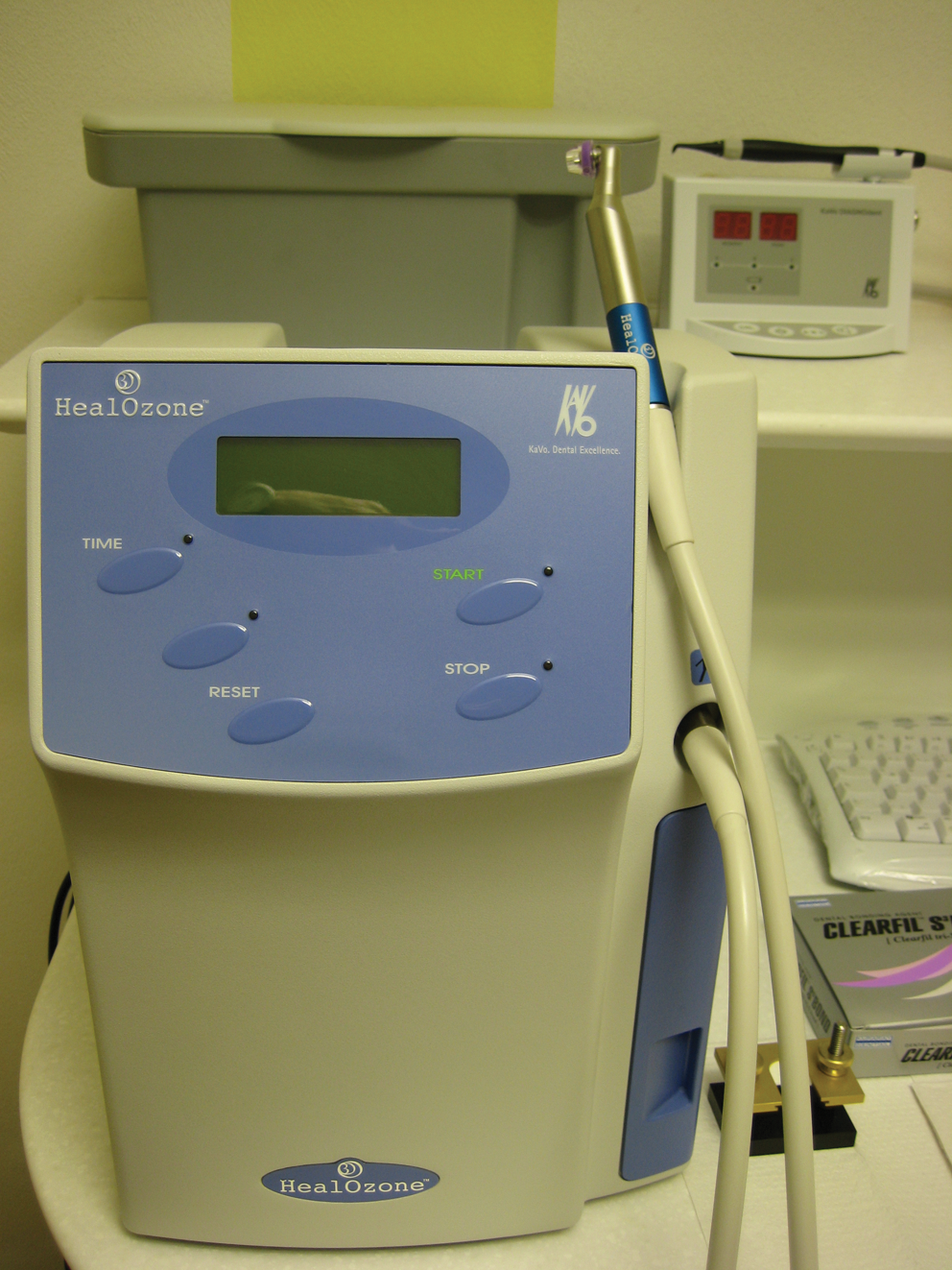
The ozone delivery system (HealOzone) used in this study.
Immediately after pretreatments, coronal and root dentin of 10 teeth from each group were treated with a two-step self-etch adhesive (Clearfil SE Bond; Kuraray Medical, Tokyo, Japan; SE) according to the manufacturer's instructions. The primer was applied to dentin surfaces by using a sponge supplied by the manufacturer for 20 sec. The primed dentin surface was then dried with oil-free compressed air. The bonding agent was applied, air spread until a homogeneous layer was observed on the surface, and light cured for 10 sec by using an LED curing light with a light intensity of 800 mW/cm2 (Bluephase C8; Ivoclar Vivadent, Schaan, Liechtenstein).
The remaining 10 teeth of each group were treated with a one-step self-etch adhesive (Clearfil Tri-S Bond; Kuraray Medical; S3) according to the manufacturer's instructions. The adhesive was applied to the dentin surface for 20 sec, air blown for 5 sec to remove water and solvent, and light cured for 10 sec.
After the respective pretreatment and adhesive sequences, a Teflon jig (Ultradent, Salt Lake City, UT) with an inner diameter of 2.3 mm and a height of 3 mm was attached to the prepared dentin surfaces. Resin composite (Clearfil Majesty Esthetic, Kuraray Medical; A3 shade) was placed as two increments (1.5-mm thick each), and each increment was light cured for 20 sec. After curing, the Teflon jig surrounding the composite was carefully removed. The specimens were stored in distilled water at 37°C for 24 h, and then loaded by a metal rod parallel with and close to the bonding interface at 1 mm/min in the shear mode until rupture occurred, by using a universal testing machine (Lloyd, Hampshire, Southampton, UK). Shear-bond strength (SBS) values were calculated as the ratio of fracture load to bonding area and expressed in MPa.
The fractured surface of each specimen was examined with a stereomicroscope (DM-IL Stereo-microscope; Leica, Heerbrugg, Switzerland) at 80 × magnification to determine the mode of failure. The failure mode was classified as either adhesive (between dentin and adhesive), cohesive (within the adhesive or dentin), or mixed (a combination of adhesive and cohesive failures).
The data obtained from SBS testing and failure mode evaluation were subjected to statistical analyses for differences between and within the groups. The normality of data distribution and the homogeneity of group variances were evaluated before the selection of statistical tests. Because the variances of coronal dentin groups were not homogeneous, the data of this group were subjected to Welch ANOVA followed by the Dunnet T3 test. The variances of the root dentin groups were homogenous, so the data obtained from this group was evaluated with ANOVA followed by Tukey HSD test. The differences between dependent variables within each group (coronal and root dentin) were determined with the Student t test. The differences among the groups for failure mode distribution were evaluated with the χ 2 test. In all the tests, the level of significance was set at P < 0.05, and calculations were handled by the SPSS 12.0 software for Windows (SPSS, Chicago, IL).
Results
For coronal dentin, the Control/SE group showed significantly higher SBS values than the Control/S3, Ozone/S3, and Nd:YAG/S3 groups (p < 0.05). Although the SBS values of the Control/SE group were higher than those of the Ozone/SE and Nd:YAG/SE groups, the differences among these groups were not significant (p > 0.05). No significant differences were observed among S3 groups (p > 0.05) (Table 2).
Data with the same superscript are not statistically different.
For root dentin, Ozone/SE demonstrated higher values than Nd:YAG/S3 (p < 0.05).
Comparisons of SBS to coronal and root dentins in each group did not exhibit significant differences (p > 0.05), except the Control/SE group, which showed higher values for coronal dentin (p < 0.05) (Table 3).
Student-t Test (p = 0.05).
The most frequently observed failures were adhesive in nature for S3-applied groups both for coronal and root dentin. In SE-applied coronal dentins, dominant failure types were adhesive (80%) for the Nd:YAG/SE group and mixed (60%) for the Ozone/SE groups, whereas the Control/SE group exhibited equivalent adhesive (50%) and mixed (50%) failure rates. For root dentin, most of the failures were adhesive in the Nd:YAG/SE (60%), Ozone/SE (70%), and Control/SE (90%) groups. However, the failure-mode distributions among all the groups were not statistically different (p > 0.05).
No statistically significant differences were found between coronal and root dentins of the Control/S3, Ozone/SE, Ozone/S3, Nd:YAG/SE, and Nd:YAG/S3 groups (p > 0.05). Nevertheless, in Control/SE group, coronal dentin exhibited 50% adhesive and 50% mixed failures, whereas the root dentin showed 90% adhesive failures (p < 0.05) (Table 4).
Chi-square test (p = 0.05).
Discussion
The present study compared the in vitro SBS of two different self-etch adhesive systems to human coronal and root dentin treated with either Ozone or Nd:YAG laser. The results showed that the type of adhesive presented a greater influence on SBS for coronal dentin, whereas the effects of pretreatments and different dentin regions were very low for the criteria assessed.
Brännström 17 reported that the cause of pulpal damage is infection rather than operative procedures or the materials used in restorative dentistry. Infection beneath the restorations is the greatest threat to the pulp. Microbial activity may result in increased pulp sensitivity, pulpal inflammation, and secondary caries. 18 Caries excavation alone leads to a significant reduction in microorganisms, with 60% of hard dentin samples from the floor of the cavity still infected with an average of 102 CFU of bacteria. The histologic and microbiologic experiments, performed to determine whether viable organisms remain on the dentinal surface at the termination of routine cavity preparation, showed that only a proportion of the teeth were sterile after preparation. 19 Because bacteria left in the dentin can live for a long time, removal of the remaining bacteria in the cavity by using a disinfection procedure should reduce or eliminate postoperative complications. 1
The antimicrobial effect of ozone on the oral microbiota has been investigated in both in vitro 4,20 and in vivo 9 studies. Polydorou et al. 21 reported that, the application of ozone with a concentration of 2,100 ppm for 80 sec on an in vitro–infected dentinal cavity model was successful in reducing the numbers of microorganism. This is important for minimally invasive dentistry applications, 22 as the limited dentin removal could lead to a considerable number of residual bacteria within the cavity walls. However, the effect of ozone application on dental hard tissues before restoration has been poorly investigated. Ozone had been reported not to negatively influence the microleakage and penetration of a sealant, 23 but the implications of ozone applications on human dentin before bonding procedures have not been clarified.
For the past decades, the inhibiting effect of oxygen on resin composites has been widely studied. 24 Oxygen inhibition causes numerous deleterious effects including slow polymerization rates, long induction periods, low conversion, short polymer kinetic chain length, and poor-quality surface properties. 25 Recently, a number of scientific publications assessed that residual oxygen from the bleaching agent, present in enamel, could be the cause of failure in adhesive procedures, regardless of the adhesive type. 26,27 From a chemical point of view, ozone, being an oxidant agent, could be expected to exert a similar effect. However, the results of the present study showed that this treatment did not impair the SBS of both self-etch adhesives in the same way. Similar to our findings, those of Schmidlin et al. 28 revealed no effect of gaseous ozone on bond strengths to dentin. Magnia et al. 29 reported that ozone gas did not compromise the mechanical properties of the tested adhesives.
Another novel device used for the pretreatment of the coronal and root dentin surfaces was Nd:YAG laser. The disinfecting effects of Nd:YAG laser have been clarified by a number of studies. 5,12 It was reported that Nd:YAG laser was able to reduce dentin permeability, to remove the smear layer, and to melt hydroxyapatite and totally or partially occlude exposed dentin tubules with application parameters similar to those used in this study. 30 However, several studies have shown that dentin irradiation with Nd:YAG laser before the adhesive procedure resulted in a reduction in bond strength with the resin composite. 30,31 This effect was credited to obliteration of the dentinal tubules due to the melting and resolidification of the irradiated dentin. 30 –33 Conversely, other investigation found that laser application did not have a negative influence on bond strength. 34 In the present study, the bond-strength values did not significantly differ after Nd:YAG laser pretreatments for both self-etch adhesives tested. Similar to our findings, Rolla et al. 35 reported that the irregularities formed by Nd:YAG laser irradiation might have favored greater micromechanical retention for the self-etch adhesives in comparison with non-irradiated dentin.
Dentin is a vital hard tissue, and its regional variability in terms of morphologic and functional characteristics determines the quality of resin–dentin bonds achieved with adhesive systems. 36 However, because root dentin surfaces are usually small and are not generally used as bonding substrates because of technical difficulties, very little work has been published on adhesive properties of composite resins to human root dentin. Fogel et al. 37 showed that the permeability of root dentin was much lower than that of coronal dentin, which might reduce hydrophilic resin infiltration capacity into root dentin structure, resulting in lower bond-strength values compared with coronal dentin. In another study comparing regional tensile-bond strength, Yoshiyama et al. 38 concluded that bond strength to coronal dentin was significantly higher than that to cervical root dentin. According to their SEM observations, they speculated that the reason for decreased bond strength in root dentin is related to the differences in the number and the diameter of the dentinal tubules that facilitate both etching efficacy and resin penetration. They found that fewer dentinal tubules existed in root dentin, and so the permeation of the acid was much lower than that in coronal dentin. Root dentin was also reported to be more sclerotic than other dentin regions, and so was less soluble with acids than was coronal nonsclerotic dentin. 39 In the present study, only in the Control/SE group was the SBS to coronal dentin higher than that of root dentin. However, the findings of the other five groups indicated that none of the dentin substrates could be considered superior to the other.
Because adhesive systems that use a separate acid-etching step have been proven to be apparently more sensitive to the variation in morphologic characteristics of dentin than were self-etching adhesives, 40 one-step and two-step self-etch adhesive systems were used in this study. The bonding mechanism of self-etch adhesives is based on changing the chemical composition of the substrate surface, commonly referred to as hybridization; the surface layer of dentin is partially dissolved, and the resultant porosity is filled with resin. Moreover, the risk of discrepancy between the depth of dentin demineralization and hybridization is limited. 13,41
Self-etch adhesives that approach the gold standard of dentin-bonding effectiveness in vitro and in vivo are the mild two-step self-etch adhesives. 41 The latest commercially available self-etch adhesives are one-step self-etch adhesives, which are the combination of etchant, primer, and adhesive resin into an all-in-one adhesive. Their advantage is to shorten the application time and to reduce errors that can occur at each bonding step. The two adhesives tested in this study were from the same manufacturer, contained the same functional monomers, and had similar compositions. S3 has a higher pH and milder acidity compared with SE (2.7 vs. 1.9) and is expected to dissolve lesser mineral content of the dentin. This could be suggested as an explanation for lower bond strength to coronal and root dentin of S3, although the difference for root dentin was not statistically significant. Conversely, SE is a two-step self-etch system. Its primer is a hydrophilic aqueous solution that should be air-dried to remove the water and solvent content before bonding resin with no water is applied. Clearfil SE Bond adhesive contains a hydrophobic resin with no water or solvent, whereas S3, a one-step self-etch system, has a significant amount of water and solvent included in the container that are expected to be removed by air blowing; the harmful effects of water and the remaining solvents on the bonding performance were demonstrated by several studies. 42,43 It could be assumed that one of the reasons for the differences the between bonding performances of S3 and SE was the water and solvent content of the adhesive layer after air-drying.
The present findings of this in vitro study may not be directly extrapolated to clinical situations, as the absence of caries and dentin fluid in the extracted teeth may have influenced the bond-strength values. Therefore, further in vivo studies must be conducted to evaluate the possible effects of pretreatments with ozone and Nd:YAG laser on the bond strength of restorative materials.
Conclusion
The pretreatment of both coronal and root-dentin substrates with either ozone or Nd:YAG laser did not affect the shear-bond strength of the self-etch adhesives under these experimental conditions.
Within the limitations of this in vitro study, it can be concluded that the application of ozone or Nd:YAG laser to dentin before bonding procedures might be possible without impairing the performance of the final restoration.
Footnotes
Author Disclosure Statement
No competing financial interests exist.