
Abstract
Introduction
The success of a resin–cement bond between a tooth structure and a feldspathic ceramic restoration depends upon the adhesive properties of the resin cement, 7 and the bond strength of a resin cement is very dependent on surface preparation. 8 Accordingly, roughening the ceramic surface by either air abrasion, grinding with a diamond bur, or etching with different acids has been recommended in order to improve the bond strength between a ceramic restoration and a tooth structure. 9 –14 All these procedures improve the bond strength by modifying the texture of the ceramic surface, which acts to increase mechanical retention. 15 The current generation of dental adhesives is based mostly on micromechanical retention, as well as on chemical bonding. 16 In addition, silane-based coupling agents are frequently used with dental adhesives to achieve more effective chemical bonding due to their bifunctional properties. 7,17 Although silane coating increases the bond strength, the resultant bond is weak. 18 However, when etching and silanization are used together, the surface energy and the wettability of the ceramic substrate are increased because the contact angle between the ceramic surface and the resin cement is decreased. 19,20 To achieve this, etching the ceramic surface with hydrofluoric (HF) acid followed by the application of a silane-coupling agent is a well-known and recommended method. 1,2,12,21 –26
Another technique for roughening the surface of a ceramic restoration is laser irradiation. 1,21,24,27 –31 For this purpose, Er:YAG, Nd:YAG, and CO2 lasers can be used on dental ceramic structures. Although it is reported that the higher bond strength between lithia-based 27 or aluminous ceramic 30,31 and resin cement can be obtained from Er:YAG laser irradiation, there are several studies that have reported that the application of Er:YAG laser irradiation on feldspathic ceramic surfaces does not, in fact, improve the bond strength between the ceramic and the resin cement. 1,28 The Nd:YAG laser is another type of laser that is used widely in dentistry for both intraoral soft tissue and hard tissue surgery. 32 Furthermore, it is reported that the Nd:YAG laser, when applied to the feldspathic ceramic surface, promotes the formation of surface irregularities, and consequently improves the adhesion of the composite resin. 24
The purpose of this study was to evaluate the microtensile bond strength of resin cement to feldspathic ceramic after (a) hydrofluoric (HF) acid etching, (b) 50 μm aluminum oxide (Al2O3) abrasion, (c) Er:YAG laser irradiation, (d) Nd:YAG laser irradiation, and (e) HF acid etching after either air abrasion or laser irradiation. The first working hypothesis was that air abrasion and laser irradiation on feldspathic ceramic surfaces would increase the bond strength compared to that of the untreated surfaces. Secondly, it was hypothesized that the bond strength would be greater with HF acid etching after each laser irradiation and air abrasion.
Material and Methods
Forty feldspathic ceramic blocks (6.1 [length] × 6.1 [width] × 6.2 [height] mm) (Ceramco, U.S.A., batch no. 01111523) were used in this study. The ceramic blocks were invested, heated, and pressed according to the manufacturer's instructions. The surfaces (6.1 × 6.1 mm) of the ceramic blocks were mechanically polished under running water by first sanding them with a 240-grit silicon carbide abrasive paper and then with a 600-grit silicon carbide abrasive paper (Struers, Tokyo, Japan). All specimens were then ultrasonically cleaned in distilled water for 10 min. After cleaning, impressions were made from each ceramic block using silicone putty (Elite HD Plus, Zhermack, batch no. 73518). A 3-mm gap between the upper portion of the mold and the surface of the block for injecting resin cement was created using the method described by Brentel and colleagues. 20
Experimental Groups
The 40 ceramic blocks were divided into eight equal groups (n = 5), according to the following surface treatments:
Group 1 (C): No treatment (control group).
Group 2 (HFA): Etching with 9.5% hydrofluoric acid (Porcelain Etchant, Bisco Inc., U.S.A., batch no. 0900000380) for 2 min, rinsing with distilled water for 15 sec, and then air-drying.
Group 3 (S): Air abrasion with 50 μm Al2O3 at 90 psi for 10 sec using an air abrasion machine (Macro Cab, Danville Engineering Inc., U.S.A.).
Group 4 (ER): Er:YAG laser irradiation (Doctor Smile erbium and diode laser, Lambda Scientifica S.p.a., Italy) with a 2940-nm wavelength by a 1-mm diameter optical fiber transmission system. The laser beam was delivered by a 400-μm diameter sapphire tip at a 75-μsec pulse length for 1 min at the following operating conditions: 4-W output power, 400 mJ/pulse at 10 Hz, and water and air cooling using an adjustable air and water spray. 1,27,28
Group 5 (ND): Nd:YAG laser irradiation (Smarty A10, DEKA, Italy) with a 1064-nm wavelength by a 600-μm diameter optical fiber transmission system. The laser beam was delivered by a 300-μm flexible optical fiber tip at a 150-μsec pulse length for 1 min at the following operating conditions: 2-W output power, 200 mJ/pulse at 10 Hz, and water and air cooling using an adjustable air and water spray. 1,27,28
Group 6 (S+HFA): Identical air abrasion as described for the S group, followed by identical acid etching as described for the HFA group.
Group 7 (ER+HFA): Identical Er:YAG laser irradiation as described for the ER group, followed by identical acid etching as described for the HFA group.
Group 8 (ND+HFA): Identical Nd:YAG laser irradiation as described for the ND group, followed by identical acid etching as described for the HFA group.
Before each laser irradiation, the energy delivered from both the Er:YAG and Nd:YAG laser equipment was evaluated with a power meter (Ophir, Jerusalem, Israel). The energy delivered at the end of the tip was 30% lower than that depicted on the equipment display for the Er:YAG laser and 5% lower for the Nd:YAG laser. Therefore, the specimens irradiated with the Er:YAG laser at 400 mJ/pulse and with the Nd:YAG laser at 200 mJ/pulse were, in fact, irradiated with 280 mJ/pulse and 190 mJ/pulse, respectively.
The laser beams for both the Er:YAG and the Nd:YAG lasers were directed perpendicular to the ceramic surface (37.21 mm2) in nonfocused, noncontact modes manually, without the use of a fixed support, at a working distance of approximately 1 mm. 28,33,34
Application of silane agent and resin cement
After surface treatments, a silane-coupling agent (Clearfil Ceramic Primer, Kuraray, batch no. 00001A) was applied to the ceramic surface using a clean brush, and then air-dried using oil-free compressed air. To apply the resin cement, each treated ceramic block was initially placed in its silicone mold. The resin cement (Panavia F 2.0, Kuraray, batch no. 41182) was then mixed according to the manufacturer's instructions and injected into the 3-mm gap between the upper portion of the mold and the surface of the block. The resin cement was then light polymerized (500 mW/cm2 intensity) for 20 sec under a conventional halogen lamp (Elipar II, 3M ESPE). An oxygen-inhibiting gel (Oxyguard II, Kuraray) was applied to the exposed surfaces for 10 min. After 10 min, each ceramic–resin cement block was removed from the mold, and the four 6.1 × 6.2-mm surfaces of each block were again light polymerized for 20 sec per surface.
Specimen preparation for microtensile bond strength testing
Five ∼1-mm-thick slices were then prepared from each block using a diamond disc at low speed under water cooling. The two outside 1-mm slices in this cutting plane were discarded because the results could be influenced by either excess adhesive or lack of adhesive at the interface after the sliced block was detached from the metallic base. Each sliced block was then detached from the metallic base, rotated 90°, and then re-attached to the metallic base in the cutting machine. A second set of five ∼1-mm-thick slices was then prepared from each block using a diamond disc at low speed under water cooling. The two outside 1-mm slices in this second cutting plane were discarded for the same reasons as described above. Therefore, only the central ceramic–resin slices of each block were used for measuring the microtensile bond strength. In total, nine slices were prepared from each block, and 36 specimens were obtained from each treatment group. Each slice was stored in distilled water at 37°C for 24 h. After 24 h, each slice was then thermocycled between 5°C and 55°C for 1000 cycles with a dwell time of 30 sec in each water bath. Upon completion, the microtensile bond strength of each slice was determined.
Determination of the microtensile bond strength
Each slice was attached to a custom-made device using cyanoacrylate adhesive in such a manner that the plane of attachment was perpendicular to the applied force. In this way, sprain forces at the interface were avoided. To determine the microtensile bond strength, each ceramic–resin slice was placed in a microtensile tester (Model Number T-61010K, Bisco Inc., U.S.A.) at a crosshead speed of 0.1 mm/min until rupture occurred. Microtensile bond strength values were expressed in megapascals (MPa). Of the 288 specimens that were obtained from the eight treatment groups, the microtensile bond strength was able to be determined in only 218 specimens. The main reasons for not being able to test all the specimens were water storage, thermocycling, and an inability to attach the specimen to the testing device. The specimens from the ER and ND groups were most affected by these conditions and thus had the highest rate of being unable to be tested.
Analysis of failure type
All the ruptured specimens were examined under a light microscope (Micro Science, Digital Microscope, China) to which a digital camera was attached at 50 × magnification in order to capture the image and to determine the type of failure. The failure type was classified as adhesive when the rupture occurred between the adhesive layer and the ceramic, cohesive when the rupture occurred in the ceramic, and mixed when the rupture occurred in the resin and the ceramic.
Evaluation of surface treatments by scanning electron microscopy
After the various surface treatments that involved applying neither the silane-coupling agent nor the resin cement, in order to evaluate the effects of the various treatments on the ceramic surface, one specimen from each group was randomly selected. The specimens were sputter-coated with gold-palladium for 3 min, at a current of 10 mA, and vacuum of 130 mTorr (Hummer VII, Anatech Ltd.), and then examined under a scanning electron microscope (Jeol JSM-6400 SEM, JEOL Ltd., Tokyo, Japan).
Statistical analysis
Statistical analysis was performed using a computerized statistical software program (SPSS 15.0 for Windows, SPSS Inc.). Bond strength data (MPa) were analyzed by one-way analysis of variance (α = 0.05). Multiple comparisons were made using Duncan's test. The level of statistical significance was set at 5%.
Results
Shear bond strength
Table 1 summarizes the results of microtensile bond testing. ANOVA showed significant influence of the surface treatment methods on the bond strength values (p < 0.05).
C, no treatment; HFA, etching with 9.5% hydrofluoric acid; S, air abrasion with 50 μm Al2O3; ER, Er:YAG laser irradiation; ND, Nd:YAG laser irradiation; S+HFA, air abrasion plus acid etching; ER+HFA, Er:YAG laser plus acid etching; ND+HFA, Nd:YAG laser plus acid etching.
Different letters (a–f) indicate dissimilarity of the groups (p < 0.05).
Failure type analysis
The frequencies of failure types in each group are shown in Table 2. Cohesive failures were the predominant rupture type in the specimens of the HFA and S groups, whereas the main failure type in the S+HFA, ER+HFA, and ND+HFA specimens was mainly adhesive in nature. In the C, ER, and ND specimens, the failure type was entirely adhesive.
C, no treatment; HFA, etching with 9.5% hydrofluoric acid; S, air abrasion with 50 μm Al2O3; ER, Er:YAG laser irradiation; ND, Nd:YAG laser irradiation; S+HFA, air abrasion plus acid etching; ER+HFA, Er:YAG laser plus acid etching; ND+HFA, Nd:YAG laser plus acid etching.
Scanning electron microscopy
Representative scanning electron micrographs of the ceramic surfaces after the different surface treatments are displayed in Fig. 1. Compared with the surface of the untreated control specimen (Fig. 1A), the surface of the HF acid–etched specimen showed large fissures, cracks, and deep holes (Fig. 1B). There were fewer fissures and cracks on the surface of the air-abraded (S) specimen (Fig. 1C) than on that of the HF acid–etched (HFA) specimen. There were no large fissures or cracks on the surface of the air-abraded and HF acid–etched (S+HFA) specimen (Fig. 1D), and its surface had fewer fissures and less peeling than that of the air-abraded specimen. The surface of the Er:YAG laser–irradiated specimen showed erosion and melting with no fissures or cracks (Fig. 1E). This contrasts with that seen on the surface of the ER+HFA specimen, where the surface had fissures and small cracks (Fig. 1G). The surface of the Nd:YAG laser–irradiated specimen showed random surface melting and shallow erosions with no fissures or microcracks (Fig. 1F). The surface of the ND+HFA specimen (Fig. 1H) was rougher than that of the ND specimen because there was more surface peeling.
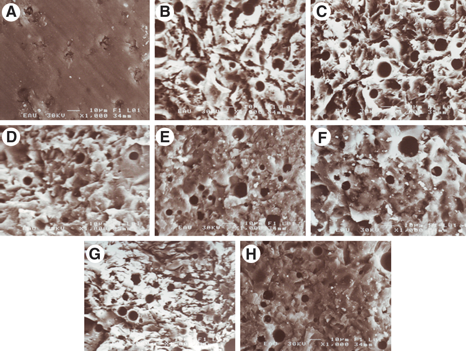
Representative scanning electron micrographs of the ceramic surface after the various surface treatments.
Discussion
The intention of this in vitro study was to investigate the influence of different surface treatment methods of a feldspathic ceramic on the microtensile bond strength of resin cement. Of all the surface treatments of feldspathic ceramic, it was shown that the highest bond strength was achieved after HF acid etching. The finding of the present study that HF acid etching results in a strong microtensile bond between resin cement and a feldspathic ceramic was in agreement with the findings of other groups. 1,14,19 –26,35 –38
In this study, the laser parameters for each laser were chosen based on a previous study in which we participated. 28 In this previous study, we evaluated the shear bond strength of a composite resin to a feldspathic ceramic (Vita VMK) irradiated with the Er:YAG laser at a power setting of 300 mJ/pulse and 10 Hz for 1 min and with the Nd:YAG laser at power setting of 100 mJ/pulse and 10 Hz for 1 min. It was found that the lowest bond strength was achieved with Er:YAG laser irradiation, and the bond strength of Nd:YAG laser irradiation was similar to that of untreated specimens. For this reason, in the present study, the laser energies for each laser were done at a higher level than those used for the previous study. The same laser equipment was used in both studies, although the energy delivered was not measured with a power meter in the previous study.
When Er:YAG laser irradiation is applied on a ceramic surface, it is expected that the laser irradiation will create a rough surface by removing the glass phase of the ceramic 27 and increase the micromechanical retention of resin. However, in this study, the Er:YAG laser irradiation actually produced the lowest microtensile bond strength. Erosion and melting were observed, but with the scanning electron microscopy (SEM) observation no fissures or cracks were seen on the surface after Er:YAG laser irradiation. Although the higher laser energy setting (400 mJ/pulse, 10 Hz) was used, this result was similar to that of the previous study. 28 Similarly, Shiu and colleagues 1 observed that Er:YAG laser irradiation of a feldspathic ceramic surface at an energy setting of 500 mJ/pulse, 4 Hz for 2 min was insufficient to cause adequate surface roughing. They also found that the surface treatment proposed with the Er:YAG laser resulted in low bond strength. The ceramics do not effectively absorb the 2940-nm wavelength energy emitted by the Er:YAG laser, and they exhibit the reflector property. 1,34,39 To increase energy absorption, a hydroxyapatite paste 1,34,39 can be applied on the ceramic surface. The lowest bond strength obtained from the Er:YAG laser–irradiated group may be explained by the lack of any application of a hydroxyapatite paste in the present study.
The results of the present study showed that the second lowest bond strength value was achieved in the group irradiated with the Nd:YAG laser. In contrast, Li and colleagues 24 observed that the shear bond strengths of composite resin to a feldspathic ceramic after Nd:YAG laser irradiation at a power setting of both 60 mJ/pulse, 15 Hz and 80 mJ/pulse, 15 Hz were similar to that of HF acid etching. However, in the previous study, 28 it was found that the shear bond strength of composite resin to a feldspathic ceramic irradiated with the Nd:YAG laser was statistically lower than that of HF acid etching. Although a higher output power was used in the present study compared to the two previously mentioned studies, it was found that the application of the Nd:YAG laser decreased the bond strength compared to that of untreated samples. This finding may be explained by the fact that the 1064-nm wavelength energy emitted by the Nd:YAG laser cannot be effectively absorbed by the ceramic. 30,31 For this reason, it is suggested that a graphite powder should be applied to cover the ceramic surface in order to increase energy absorption. 30,31,34 In the present study, the application of graphite powder might increase the bonding results obtained from the group irradiated with the Nd:YAG laser.
Although the bond strength can be used to evaluate the performance of resin cement, failure types and analysis of the causes of pretest failures of specimens should also be taken into consideration. 21 In the present study, it was found that the failure type for both Er:YAG and Nd:YAG laser–irradiated specimens was completely adhesive in nature. In addition, the highest number of pretest failures occurred in the Er:YAG and Nd:YAG laser–irradiated specimens while slicing the blocks or during thermocycling. This might be explained by the irradiation of a feldspathic ceramic surface with an Er:YAG or Nd:YAG laser having created inadequate bonding between the resin cement and the feldspathic ceramic surface due to a lack of surface roughness being produced by each laser irradiation process. Therefore, the first working hypothesis was partially accepted in that air abrasion could improve the bond strength, whereas laser irradiation could not.
The microtensile bond strength of laser-irradiated specimens after HF acid etching was higher than those found after each laser irradiation alone. In fact, it was determined that the changes to the ceramic surface after laser irradiation, observed in the scanning electron micrographs, were insufficient to create a strong adhesive bond. When the surface was HF acid–etched after laser irradiation, HF acid dissolved the glass phase of the ceramic, exposed areas of crystals, and created a more microporous surface. 1,11 The bond strength increased, probably because the acid increased the roughness of the surface. However, it was determined that the bond strengths obtained from the groups of HF acid etching after each laser irradiation were lower than that of the HF acid–etching group. This might be explained by the fact that HF acid etching was not effective on the ceramic surface irradiated with each laser because laser irradiation might cause the development of a heat-damaged layer on the ceramic surface. 25 In fact, erosion and melting were observed in SEM evaluations of laser-irradiated surfaces.
Results from previous studies showed that air abrasion was a good mechanical method for providing retention of the adhesive. 14,40 However, it was found that the microtensile bond strength following air abrasion was also high, but not as high as after HF acid etching in this study. Furthermore, other researchers reported that the Er:YAG or Nd:YAG laser irradiation combined with air abrasion could improve the bond strength between the ceramic and resin cement. 30,31,34 Ferreira and colleagues 34 suggested that the application of laser irradiation with the Er:YAG laser at a power setting of 500 mJ/pulse, 4 Hz, and with the Nd:YAG laser at a power setting of 100 mJ/pulse, 20 Hz to a feldspathic ceramic (Ceramco) after air abrasion with 50-μm Al2O3 could be an alternative bonding technique for resin cement to ceramic. The air abrasion was not combined with both types of laser irradiation in this study. However, it was found that the bond strength in the group etched with HF acid after air abrasion was lower than that of the groups etched with HF acid after Er:YAG or Nd:YAG laser irradiation, as was shown in the study by Ferreira and colleagues. 34 This may be due to excessive conditioning of the ceramic surface, which decreased the superficial resistance of the ceramic despite enhancing its surface roughness. 41,42 Therefore, the second working hypothesis was partially accepted in that HF acid etching after each laser irradiation could improve the bond strength, whereas HF acid etching after air abrasion could, in fact, decrease this bond strength.
The laser parameters are the most important factor in modifying the surface texture of a ceramic when the laser irradiation was used as a method for surface treatment. For this reason, findings obtained from future studies will need to test the different parameters of the lasers used in order to determine whether or not Er:YAG or Nd:YAG laser irradiation should be used as a tool to roughen feldspathic ceramic surfaces to achieve strong bonding of resin cement.
Conclusions
The best bonding value was achieved by HF acid etching alone. Air abrasion alone and both air abrasion and laser irradiations combined with HF acid etching promoted an improvement in adhesion between Panavia F and feldspathic ceramic. However, the Er:YAG or the Nd:YAG laser irradiation used alone within the parameters tested negatively changed the bonding of the resin cement. Erosion and melting were observed in SEM evaluations of laser-irradiated ceramic surfaces, which were undesirable surface appearances for adhesive bonding. If laser irradiation is used as a pretreatment method to improve the bonding of the resin cement to a feldspathic ceramic surface, it should be combined with HF acid etching to enhance the surface roughness.
Footnotes
Author Disclosure Statement
No competing financial interests exist.