
Abstract
Introduction
Low-level laser therapy (LLLT) has been used by physiotherapists in the treatment of ulcers, usually colonized by pathogenic microorganisms, to accelerate the healing process and reduce inflammation. However, the wound infection is a barrier to the application of laser therapy, as there is no in vivo evidence of the safety of this therapy, although some studies show the inhibitory effect of the in vitro laser applications. 3 –11
Among the few studies found in scientific literature on the effect of laser therapy in bacterial cultures, a consensus regarding the results was not found. Nussbaum et al. 4 –6 observed the effect of laser therapy using 630, 660, 810, and 904 nm wavelengths (1–50 J/cm2) on cultures of S. aureus, Pseudomonas aeruginosa, and Escherichia coli, and obtained responses of inhibition or bacterial growth, depending upon the wavelength, radiated fluence, or the species. When Dadras et al. 7 analyzed the effect of laser with 514, 532, and 633 nm wavelengths (0.015–1.13 J/cm2), they identified growth inhibition of S. aureus with 514 and 532 nm and no changes with 633 nm, unlike the P. aeruginosa, where growth occurred with all wavelengths and fluences tested. Recently, Guffey and Wilborn 8, 9 observed growth inhibition of P. aeruginosa and S. aureus with 405, 470, and 880 nm wavelengths (1–20 J/cm2). Similarly, Enwemeka el al. 10,11 identified inhibition of S. aureus, methicillin-resistant, with 405 and 470 nm wavelengths (1–60 J/cm2). These results showed that the species responded differently when irradiated by distinct wavelengths.
Atomic force microscopy (AFM) is a recent, highly versatile technique that proved to be an effective tool to explore microorganisms. It can generate ultrahigh resolution and three-dimensional topographic imaging in nanoscale. 12 This new technique has been used to evaluate the topography of bacterial cells in ambient air. 13 The samples do not require any special treatment, and the cell structure can be revealed in details, 14,15 unlike conventional scanning electron microscopy. The morphological analysis of bacteria can help in understanding the mechanisms of antimicrobial action used through the visualization of the structural changes after the treatment.
In this context, it is expected that LLLT promotes growth inhibition of S. aureus derived from changes of the bacterial structure. For this reason, the goal was to analyze the bacterial morphology by AFM after the application of LLLT in in vitro growth of S. aureus, using 660, 830, and 904 nm wavelengths at fluences of 0, 1, 2, 3, 4, 5, and 16 J/cm2.
Materials and Methods
For this study, a strain of S. aureus (ATCC 29213) was used. It was provided by the Anaerobic Microbiology Laboratory of the Microbiology Department at the Federal University of Viçosa.
Bacteria were kept in Mannitol Salt Agar (Biobras ®, Montes Claros, Brazil) and incubated at 37°C for 24 h. Following this, the cultures were grown in 9 mL brain heart infusion (BHI; Acumedia®, Indaiatuba, Brazil), and a suspension of cells was prepared in 5 mL of 1% saline solution to obtain a turbidity comparable to the 0.5 McFarland standard (1.5×10⁸ CFU/mL). This cell suspension was diluted in 104 CFU/mL and 50μL aliquot were transferred to petri dishes (80×12 mm) containing culture medium Mannitol Salt Agar which have been homogenized with the Drigalski loop. After seeding was performed on the surface of the plates, these were subjected to laser irradiation and then incubated at 37°C for 24 h.
The diode laser equipment used was Laserpulse (Ibramed®, Amparo, Brazil), with the physical parameters shown in Table 1, as proposed by Jenkins and Carroll. 16
After irradiation, the temperature of the plates was measured with a surface digital thermometer RTDF-150 (Rücken®, São Paulo, Brazil) to identify the possible thermal changes. The plates were divided into three groups: T-660 irradiated with 660 nm wavelength for 0, 2, 4, 6, 8, 10, and 32 sec; T-830 irradiated with 830 nm wavelength for 0, 4, 8, 12, 16, 20, and 64 sec; and T- 904 irradiated with 904 nm wavelength for 0, 3, 6, 9, 12, 15, and 48 sec, respectively, with all of the groups being irradiated with fluences of 0 (control), 1, 2, 3, 4, 5, and 16 J/cm2 per point. The result of multiplying the power emitted by each wavelength by the time of emission can be calculated the emitted energy, as presented in Table 2.
Irradiation occurred uniformly and perpendicularly to the surface of the culture of S. aureus at a standard distance of 1cm, with the aid of a support to position the transmitter and standardize a distance of 1 cm between points totaling 32 points, distributed over the entire surface of the plate.
The irradiation of each group was held in duplicate, with two repetitions, and incubated at 37°C for 24 h. After this period, the CFUs/mL were counted, always by the same blinded examiner. After the colonies were counted, a thin layer of cells was picked up at random and scattered on a glass slide with a total area of 1 cm2. Topographical measurements of bacterial cells were held in an atomic force microscope NT-MDT (Ntegra Prima®, Moscow, Russia).
The results of the CFU/mL count obtained were submitted to the analysis of variance (ANOVA), followed by the Tukey test with critical level of 1% (p<0.01) using the Biostatic® software.
Results
With regard to the temperature, no difference was observed between the pre (26.5±0.6°C) and post (26.3±0.5°C) LLLT application.
Table 3 shows the global average of CFUs in each wavelength, demonstrating that the laser irradiation reduced the S. aureus growth with wavelengths of 830 and 904 nm, the latter with the highest inhibition (331.1±38.19 and 137.38±21.72). Considering the reduced percentage of bacterial growth, it can be observed that there is only a pattern for 904 nm, in which the trend line represents 60% of all fluences (Fig. 1). The intragroup results of the CFU to different fluences and wavelengths, as well as the intragroup analysis,using the same amount of energy, is presented in Table 4.
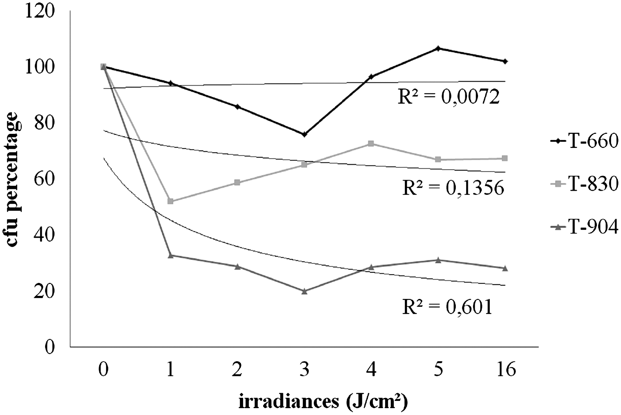
Inhibition pattern of S. aureus in wavelengths of 660, 830, and 904 nm, in control (0) and irradiated groups (1, 2, 3, 4, 5, and 16 J/cm2). n=4.
The mean followed by the same letter did not differ significantly among them.
The mean followed by the same letter did not differ significantly among them.
T-830 respective fluence (p<0.01)
T-830 and T-904 fluence 1 J/cm2 (p<0.01).
T-830 and T-904 fluence 2 J/cm2 (p<0.01).
T-830 and T-904 fluence 3 J/cm2 (p<0.01).
The AFM results for the untreated cells demonstrated a smooth surface, with grape-like clusters, typical of the species (300–700 nm tall) and without any difference in the cell texture. However, at 904 nm, some clear changes of cell conformation were observed, indicating morphological mischaracterization (Fig. 2).
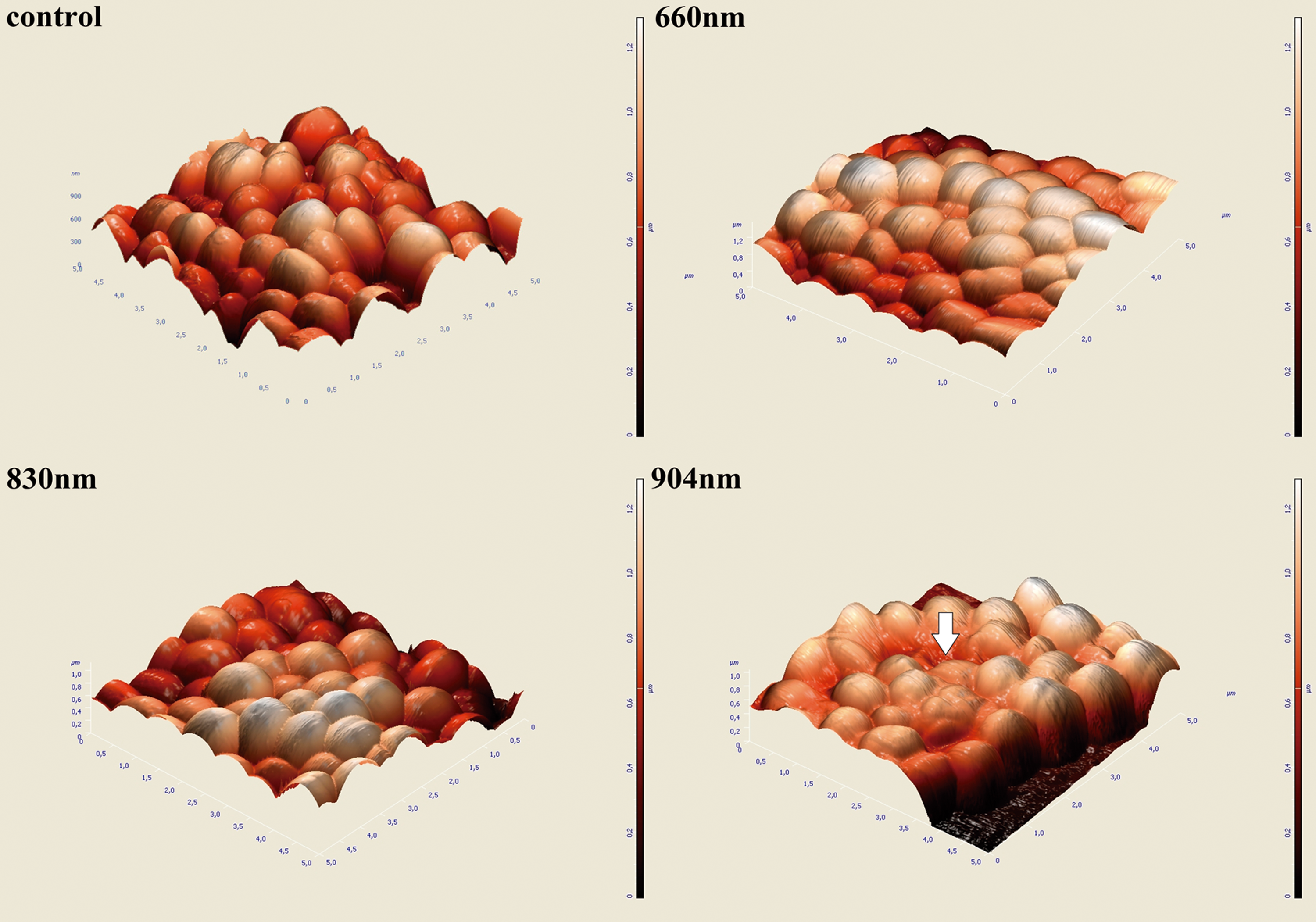
Atomic Force Microscopy (AFM) of S. aureus control and subjected to irradiation of 660, 830, and 904 nm, at 3 J/cm2. Scale in micrometers (μm). The arrow indicates changes in cell topography.
Discussion
This study showed that the S. aureus line responded differently to LLLT when the wavelengths were compared, presenting greater growth inhibition with 904 nm. This result corroborated those observed in atomic force microscopy, where changes in cell structure were observed with this wavelength.
The choice of wavelengths was based on the ones commonly used by physiotherapists, red and infrared, as well as in the range of 1–16 J/cm2, which comprised the fluence of clinical use reported in the literature. 17 In addition, several reports indicate that wavelengths between 400 and 1000 nm are bactericidal. 4,18
Nussbaum et al. 4 showed that the biomodular effects of red and infrared LLLT on E. coli, P. aeruginosa and S. aureus depended upon the wavelength, the bacterial species, the exposure time, and the fluence. A similar fact was observed by Guffey and Wilborn 8 who combined wavelengths of 405 nm (blue laser) and 880 nm (infrared), promoting a dose-dependent effect on the death of S. aureus, reducing the number of colonies 72.3% at 3 J/cm2.
This research corroborated the best result obtained in this study, which showed a bacterial inhibition of 80% at 3 J/cm2 with 904 nm wavelength. However, Guffey and Wilborn 9 developed another study with a blue laser (405 and 470 nm) with 3 J/cm2, in S. aureus obtaining a growth inhibition of 61.7% and 15.5%, respectively.
The results showed that the growth inhibition depended upon the spectrum and fluence of irradiation. Considering the wavelengths used, it became apparent that the infrared spectrum was more effective when compared to the red one, especially for 904 nm, as the 660 nm did not promote global inhibition, 830 nm inhibited 36.32%, and 904 nm inhibited 71.75%. Such results might have occurred because of higher energy absorption by bacteria or even because of a specific action in cases involving their proliferation, because the studies that used infrared light spectrum obtained, in some fluences, positive results in bacterial inhibition. 4 –6, 8
With regard to fluence, it could be observed that the bacterial inhibition did not show a positive interaction with fluence, as the results have shown that there was no difference between the fluences tested. The same was observed by Enwemeka et al. 11 when they reported a decrease in non-linear inhibition with increasing fluence.
The results demonstrated in each wavelength, an inhibition of S. aureus growth to the same fluency was of 24, 35, and 80%; with 660 nm (0.03W; 0.18 J; 3 J/cm2), 830 nm (0.03W; 0.36 J; 3J/cm2) and 904 nm (0.04W; 0.36J; 3J/cm2), respectively. It must be highlighted that the 660 nm wavelength that emitted energy was 50% less than the other two groups, which did not interfere with the results, since different fluencies with the same energy level showed different results. Both inter- and intragroup analysis reinforced the hypothesis that the wavelength is the main parameter in the results found.
In contrast, in later studies conducted by Nussbaum et al. 4 in which different wavelengths in different exposure times and fluences were tested, the authors observed inhibition or dependent-dose growth of S. aureus and P. aeruginosa in all wavelengths tested, in addition to the inhibition with 630 and 660 nm wavelengths and growth with 810 and 905 nm wavelengths (0.015 W/cm2) for E. coli.
In a parallel study, the authors verified that with 810 nm (0.015 W/cm2), there was a growth of 35% and 138% of S. aureus at fluences of 1 and 2 J/cm2, respectively. There was also an increased growth of E. coli of 121% and P. aeruginosa of 192% at 2 J/cm2. 5 In a further investigation, Nussbaum et al. 6 used the 810 nm (0.015 and 0.03 W/cm2), and there was no global effect on the growth of S. aureus. However, there was an increase of 40% for P. aeruginosa (1 and 2 J/cm2) and E. coli (5 J/cm2). 3 According to the data obtained, the conclusion was that the variables, which determined bacterial inhibition, were related to the wavelength and fluence.
In this respect, Enwemeka et al. 10,11 used the blue laser, 405 and 470 nm in two different strains of methicillin-resistant S. aureus, in which they observed a non-linear dose-dependent inhibition of bacterial growth reaching an inhibition of 94.8% with a 55 J/cm2 dose. Lower doses 1–15 J/cm2 resulted in inhibition of up to 61.2% (11 J/cm2). The results of this study were consistent with these findings with regard to obtaining good results with the use of a lower dose range.
To isolate the possible causes of bacterial growth, the temperature was quantified, before and after the irradiations; no significant change in the temperature was observed immediately after irradiation. The study conducted by Nussbaum et al. 6 indicated the same results (23°C) with the same irradiation (0.03 W/cm2), combined with high fluence (50–80 J/cm2), these results showed that the temperature should not be associated with any physiological or morphological variation of S. aureus, with a growth temperature ranging from 4° to 46°C. 19
The morphological changes of the S. aureus cells showed distinct differences between the wavelengths tested at the same fluence (Fig. 2). The literature can explain this paradox. The endogenous porphyrins of bacteria may have a greater absorption of light in a given spectrum, resulting in increased free radicals, 20 which can affect the cytoplasmic membrane proteins and DNA, 21 inactivating the cell when the resulting rate of DNA damage exceeds the rate of repair. 22
However, so far, studies were not found in the literature that would examine the absorption spectrum, inhibition, or stimulation of the synthesis of the bacterial DNA. An analysis of absorption spectra of human HeLa cells showed that the respiratory chain molecules in the membrane acted as primary photoaccelerators with 624 and 668 nm, wavelengths followed by a cascade of secondary reactions in the cytoplasm of these cells, and therefore, stimulated the DNA synthesis. 23 These photoaccelerators include the cytochrome C oxidase enzyme that absorbs red light or infrared. 23,24 However, the excitement of some chromophores changes the rate of electron flow of electrons of the transport chain, which may produce singlet oxygen that would lead to the destruction of cells. 24
Therefore, the significant levels of photo-inhibition show that irradiation with LLLT can be an alternative to control bacterial growth. However, further studies are required for the identification of optimal parameters, associating the analyses of bacteria metabolism with the rate of inhibition, as well as the morphology. Despite the significant results obtained in this study, it should be emphasized that the behavior of these cultures was evaluated only after 24 h irradiation, and therefore, the possibility of recovering from damages suffered by irradiation could not be ruled out, which may result in growth after 24 h. Another point to be considered concerns the methodology used for bacterial growth, as the use of biofilm as a means of culture would better resemble in vivo conditions, although little used in the literature.
This may be a gap presented by the experimental model, as there are no studies in the literature that can describe the bacterial behavior after this period of time, as well as the laser bactericide and/or bacteriostatic effect.
Conclusions
The study concluded that the LLLT was effective in inhibiting the growth of S. aureus ATCC 29213 with wavelengths of 830 and 904 nm, which provided morphological changes in the cellular structure of bacteria irradiated with 904 nm, which presented greater inhibition.
Footnotes
Author Disclosure Statement
No competing financial interests exist.