
Abstract
Objective:
The aim of this in vitro study was to evaluate the efficacy of photon-induced photoacoustic streaming (PIPS) activation of sodium hypochlorite (NaOCl), chlorhexidine (CHX) or ethylenediaminetetraacetic acid (EDTA) on the dentin microhardness and to assess the compositional changes of root dentin.
Background data:
It is still unclear whether PIPS activation of irrigants alters the dentin microhardness and mineral content of dentin.
Materials and methods:
Root canals of 72 extracted single-rooted teeth were prepared and teeth were fixed in microcentrifuge tubes with silicone impression material. After setting of the silicone, teeth were removed and split longitudinally in buccolingual direction. One half was used as control (pretreatment) while the other was placed into the tube (posttreatment). Then specimens were divided into six test groups (three with and three without PIPS activation). The irrigants tested were 2.5% NaOCl, 17% EDTA, and 2% CHX. Experimental tooth specimens were irrigated with 6 mL of test solution, with additional PIPS activation applied to the PIPS groups. Then specimens were subjected to Vickers microhardness testing. Percentage change of microhardness was calculated. Energy dispersive X-ray spectroscopy (SEM-EDX) was performed to measure element content.
Results:
Among the irrigant-alone groups, NaOCl and CHX did not alter the dentin microhardness, whereas statistically lower microhardness values were obtained in EDTA group. Chemical composition of dentin was affected from all irrigants used. PIPS activation led to no additional alteration in dentin microhardness. PIPS significantly increased the phosphorus level in NaOCl group.
Conclusions:
Dentin microhardness was significantly affected by the irrigation solution, not by the PIPS activation.
Introduction
Irrigation is one of the essential elements for a successful endodontic treatment due to its antibacterial activity, tissue dissolution capacity, cleaning efficacy, and chelating. Since no one solution has all these properties, a combination of irrigants is often used to enhance the irrigation efficacy. Sodium hypochlorite (NaOCl) is the most widely used irrigation solution, and it has become the gold standard in endodontic therapy. In addition, chlorhexidine gluconate (CHX) has been proposed as both an endodontic irrigant and root canal dressing based on its broad-spectrum antimicrobial activity, relatively low toxicity, and substantivity. 1 Smear layer removal, as a crucial part of irrigation, can be achieved by using the most common combination of the chelating agent–ethylenediaminetetraacetic acid (EDTA) and NaOCl. 2
Laser devices have various applications in dentistry, including caries removal, cavity and root canal preparation, root canal disinfection, tooth sensitivity treatment, and bleaching. Lasers also take part in irrigation methods with others such as sonic, negative apical pressure, and passive ultrasonic irrigation. Photon-induced photoacoustic streaming (PIPS) is a novel irrigation activation technique for cleaning and debriding the root canal system, and it uses an erbium-doped yttrium aluminum garnet (Er:YAG) laser with a radial and stripped tip at subablative energy levels. The specialized tip is localized in the pulp chamber only. 3 The mechanism of action of PIPS is based on strong agitation of the irrigant by photoacoustic shock wave. This enables irrigant powerful streaming three dimensionally throughout the root canal system. 4 This new method has been gaining popularity in endodontics since it has improved the irrigation quality. 4,5 Further, PIPS activation has been found superior to passive ultrasonic and sonic irrigation techniques with respect to elimination of biofilm, enhancing the penetration of the irrigant, and calcium hydroxide removal. 6 –8
Microhardness can be an indicator of the loss or gain of minerals in the dental hard tissue. 9 A significant decrease in dentin microhardness may point to its dissolution and degradation. 10 The effects of NaOCl, CHX, and EDTA on dentin microhardness have been previously demonstrated 11 –13 ; however, no studies have investigated the efficacy of PIPS activation of irrigants. Therefore, the aim of this in vitro study was to evaluate the efficacy of PIPS activation of NaOCl, CHX, and EDTA on dentin microhardness, and to assess the alterations in dentin mineral content.
Materials and Methods
The study protocol of this research was reviewed and approved by the Ethics Committee of the Necmettin Erbakan University, Faculty of Dentistry in Konya, Turkey (Protocol no. 2016/09).
Specimen preparation
Seventy-two single-rooted mandibular premolars extracted for periodontal reasons, without caries, cracks, previous endodontic treatment, or coronal restoration, were collected for use in this study. The teeth were stored in distilled water for <3 months until further processing. The calculus was removed using periodontal curettes. The endodontic access cavities were prepared with diamond fissure bur (SWS dental; GULSA, Izmir, Turkey) and the working length was determined by inserting a size 15 K-type file into the root canal terminus and subtracting 1 mm from this measurement. The root canals were enlarged with ProTaper Next files to size #X3 (30/0.07; Dentsply Maillefer, Ballaigues, Switzerland) under irrigation with 1 mL of distilled water between each file. The apical parts of the roots were closed using flowable composite resin (Tetric Flow; Ivoclar-Vivadent, Schaan, Liechtenstein) to prevent irrigant leaks. A small cotton pellet was placed into the access cavity, and each tooth was fixed in a 1.5 mL plastic microcentrifuge tube with silicone. After the impression material was set, the specimens were removed, and the teeth were longitudinally sectioned into two halves in the buccolingual direction. One half was again placed into the impression material, and the other half was used as a control and incubated in distilled water. The specimens were randomly divided into six experimental groups as follows (n = 12):
Group 1, 2.5% NaOCl: Six milliliters of 2.5% NaOCl (Imicryl, Konya, Turkey) was administered with a 27-gauge syringe for 60 sec. The needle was placed at a distance of 1 mm from the root apex and moved up and down.
Group 2, 2% CHX: The specimens were irrigated with 6 mL of 2% CHX (Imicryl, Konya, Turkey) for 60 sec as in Group 1.
Group 3, 17% EDTA: A total of 6 mL of 17% EDTA (Imicryl, Konya, Turkey) was applied for 60 sec as in Group 1.
Group 4, 2.5% NaOCl+PIPS activation: The irrigation activation was performed by using an Er:YAG laser with a wavelength of 2.940 nm (Fotona, Ljubljana, Slovenia). A 14-mm-long, conical, 0.4-mm-diameter PIPS tip (Fotona, Ljubljana, Slovenia) was used with the following settings: 0.9 W, 30 mJ/pulse, and 30 Hz. The pulse duration was 50 μsec (Table 1). The laser unit air and water sprays were set to off. Initially, 0.5 mm of NaOCl was placed into the space between tooth half and the silicone. Then, the PIPS tip was inserted to the coronal level of the tooth, and laser irradiation was started. The NaOCl was refreshed during the application. The laser activation was applied for 60 sec with a total of 6 mL NaOCl.
Laser and Treatment Parameters in Photon-Induced Photoacoustic Streaming-Activated Groups
Group 5, 2% CHX+PIPS activation: The same irrigation protocol as described in Group 4 was performed using 2% CHX. The total activation time was 60 sec, and the volume of 2% CHX was 6 mL.
Group 6, 17% EDTA+PIPS activation: A total of 6 mL of 17% EDTA was continuously activated for 60 sec, similar to Group 4.
After treatment, all the specimens were irrigated with 2 mL of distilled water to prevent a prolonged effect of the tested irrigants, and samples were kept moist until testing. They were immediately dried using absorbent paper points (Sure Dent Corporation, Seongnam-si Gyeonggi-do, Korea) before hardness testing.
Hardness testing
Each test and control specimen were sectioned perpendicular to the long axis at the midroot level using a low-speed diamond saw (Fig. 1). The apical parts of the specimens were embedded vertically in autopolymerized acrylic resin, and the coronal surfaces were ground flat and polished using #500, #800, #1000, and #1200 grit abrasive papers under distilled water. Then, the specimens were subjected to Vickers microhardness test device (LHV 1D; Bursam NDT, Bursa, Turkey). Three indentations were made on each specimen at depths of 100 μm from the edge of the canal lumen using a 300-gram load and a 20-sec dwell time. Representative hardness values were obtained for each control and test specimen, and the hardness reduction percentages were calculated using the following formula:
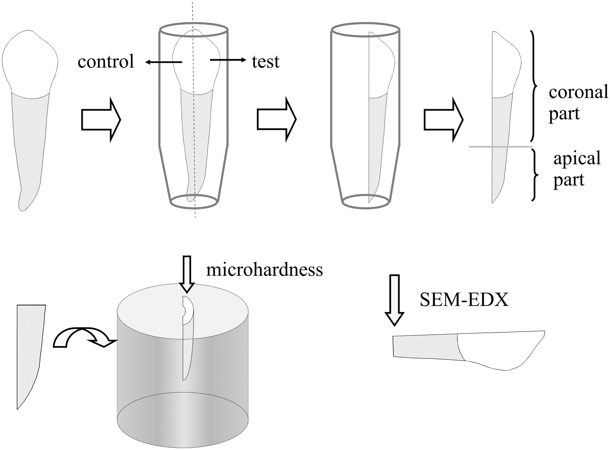
Schematic illustration of experimental design.
where initial microhardness is the microhardness value of the control specimen (pretreatment) and final microhardness is the microhardness value of the test specimen (posttreatment).
Energy dispersive X-ray spectroscopy
The coronal parts of three randomly selected test and control specimens from each group were subjected to elemental analysis under high vacuum at 5000 × magnification using an energy dispersive X-ray system (EDX) (Oxford INCA X-Act), which is attached to an environmental scanning electron microscope—Hitachi SU-1510 (Hitachi High-Technologies Corp., Tokyo, Japan). Three surface areas on each specimen were selected, the element content in weight % of calcium (Ca) and phosphorus (P) was measured; later the Ca/P ration was calculated for each group.
Scanning electron microscopy
The SEM analysis was performed at the Science and Technology Research and Application Center of Necmettin Erbakan University. One representative root half was randomly selected from each group for SEM analysis to observe any morphological changes in the root canal surface. The samples were fixed in a 2.5% glutaraldehyde solution and sputter coated for 3 min with gold/palladium. The observations were performed with a SEM under 20 kV and 700 × magnification. SEM micrographs were taken from representative areas on the root canal surface (Fig. 2).
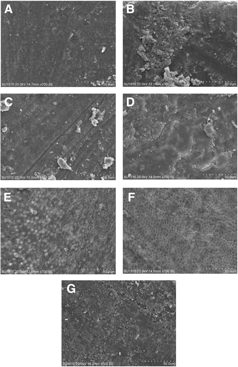
Representative SEM micrographs of root canal dentin treated with
Statistical analysis
IBM SPSS Statistics for Windows version 22.0 (IBM Corp., Armonk, NY) was used to analyze the results. A normal data distribution was confirmed using the Shapiro–Wilks test. One-way analysis of variance and Tukey's honest significant difference test for multiple comparisons were used to assess the statistical differences between the groups. A paired-samples t test was used to compare the pre- and posttreatment microhardness values in each group. In addition, the Student's t test was applied as a specific comparison of the means to determine the statistical difference between the nonactivated and PIPS-activated groups. The significance level was set at p < 0.05.
Results
Microhardness
The pre- and posttreatment microhardness values (mean ± standard deviation) among the different irrigant groups are shown in Table 2. No statistically significant differences were found between the initial microhardness values in any of the groups (with or without PIPS) (p > 0.05). Among the posttreatment samples, the CHX group showed higher microhardness when compared with the EDTA group (p < 0.05). However, there was no statistically significant difference between the NaOCl and EDTA groups (p > 0.05). The paired-samples t test revealed that the NaOCl and CHX groups exhibited no statistically significant differences when the microhardness values were compared before and after the irrigation procedures within the groups (p > 0.05). However, a significant reduction in the microhardness was observed between the values before and after irrigation in the EDTA group (p < 0.05).
Microhardness Values (Mean and Standard Deviation) of Root Dentin Treated with NaOCl, CHX, and EDTA
One-way ANOVA test.
Paired-samples t test.
p < 0.05.
CHX, chlorhexidine; EDTA, ethylenediaminetetraacetic acid; NaOCl, sodium hypochlorite.
No statistically significant differences were found between the NaOCl versus NaOCl+PIPS, CHX versus CHX+PIPS, and EDTA versus EDTA+PIPS groups in either the posttreatment microhardness values or the percentage changes (p > 0.05).
Atomic analysis
Figure 3 shows the Ca (weight %), P (weight %), and Ca/P ratio (mean ± standard deviation) of the experimental groups and the control group. The SEM-EDX analysis revealed a reduced Ca content in the root dentin specimens subjected to the EDTA and CHX solutions (p < 0.05), whereas the P content was decreased in the NaOCl and CHX groups (p < 0.05) when compared with the control group. Moreover, only the EDTA group exhibited a lower Ca/P ratio than the control group (p < 0.05).
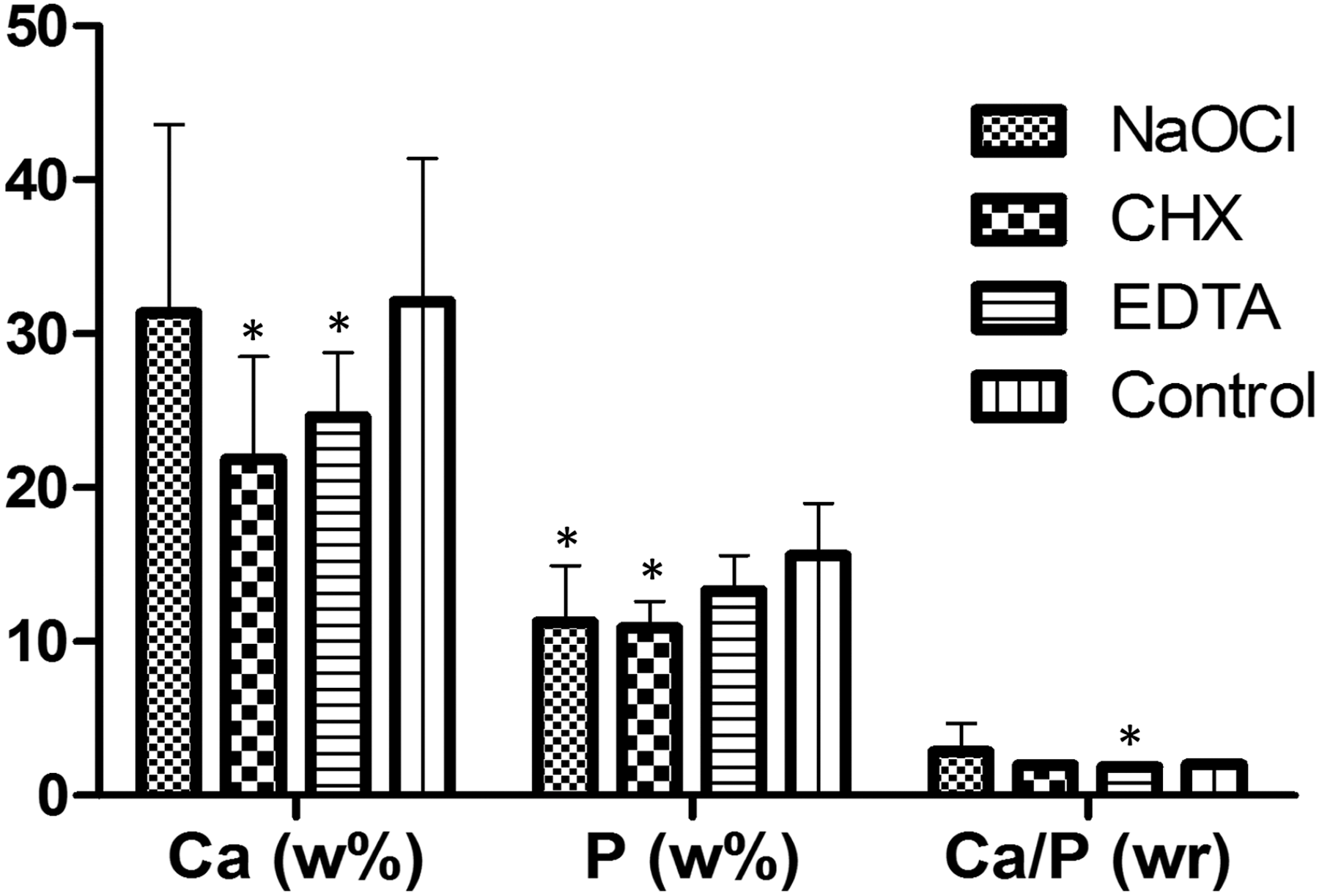
Histogram of Ca (weight %) and P (weight %) contents and Ca/P (weight ratio) of root dentin samples in experimental groups and control group. *Significantly different from control group to Student's t test (p < 0.05).
The elemental compositions of the treatment groups according to the PIPS activation are listed in Table 3. No statistically significant differences in the Ca component or the Ca/P ratio were found between the PIPS-activated and nonactivated irrigant groups (p > 0.05). When considering the P composition, the NaOCl+PIPS group revealed a higher P percentage than the NaOCl group (p < 0.05), while no significant PIPS effect was found in the CHX and EDTA groups (p > 0.05).
Means ± and Standard Deviations of Element [Ca (w %), P(w %)] Content and Ca/P Ratio of Root Dentin Subjected to Nonactivated (−) and PIPS-Activated (+) Irrigation Solutions
Student's t test.
p < 0.05.
PIPS, photon-induced photoacoustic streaming.
Scanning electron microscopy
Apparent open dentin tubules were observed on the root dentin in the EDTA and EDTA+PIPS groups. However, a smear layer and debris could be seen on the root dentin treated with the NaOCl and CHX and their PIPS-activated combinations.
Discussion
Root canal irrigants may lead to undesirable effects in the chemical and physical properties of dentin, including microhardness changes. 10 Different teeth may possess different structural variations; 14 therefore, the hardness values were calculated before and after treatment within the same root sample. With regard to the different dentin depths, there was a decrease in the microhardness from the superficial to the deep regions of the dentin. 15 A 100 μm penetration depth was chosen for this study to better show the effect of the solutions and the PIPS on dentin microhardness because the inner part of dentin is more susceptible to hardness reduction. With respect to the duration of irrigation, 1-min contact time with irrigants was selected based on previous studies. 6,8,16,17 With regard to methodology, the Vickers indenter method was preferred in this study for determining microhardness of dentin. Due to the suitability and practicality of Vickers microhardness test, this method has been used previously for evaluating surface changes of dental hard tissues treated with chemical agents. 11 –13,18,19
The results of the present study indicated that the EDTA solution reduced the microhardness of the root canal dentin, whereas the NaOCl and CHX did not alter the microhardness. Confirming our results, the softening effect of EDTA has been demonstrated previously 11 –13 and could be attributed to the chelating action and demineralizing capacity of EDTA, 12,13 Our findings are also in agreement with previous work, which revealed that CHX had no effect on root dentin microhardness. 11 Although the CHX concentration was lower than that used in the present study, those researchers applied the CHX for a prolonged period of time. In addition, it has been reported that CHX absorption could positively affect the structure of dentin. 11 Contrary to our results, NaOCl has been found to negatively alter dentin microhardness. 20,21 These conflicting results may be explained by the low NaOCl concentration and reduced application time. However, when the post-treatment values were compared among the groups, the NaOCl and EDTA groups showed similar and lower microhardnesses than the CHX group.
PIPS, as a novel technique, has been previously investigated regarding its antimicrobial activity, smear and debris removal, resin sealer adhesion to dentin, and so on. 4,5,16,22 However, to the best of the authors' knowledge, this is the first study to evaluate the effects of PIPS activation on dentin microhardness. Based on the results of the present study, PIPS activation did not cause an additional decrease in dentin microhardness, which could be attributed to the absence of thermal damage to PIPS-treated samples. 4 PIPS has been found to cause a minimal temperature rise (<1.5°C) at the external root surface. 4 Because of the harmless effects of PIPS on root dentin microhardness, it may be used as an appropriate and safe irrigation activation technique.
The SEM-EDX analysis was performed to evaluate surface changes of root canal dentin for alterations in the Ca, P, and Ca/P ratio. Further, the SEM-EDX analysis not only enabled a comprehensive understanding of the surface changes but also provided complementary information about dentin microhardness. The findings of the present study revealed that EDTA reduced both the Ca content and Ca/P ratio of dentin. These results are in agreement with previous investigations that showed significant EDTA effects on dentin mineral content. 23,24 Our results also indicated reduced Ca and P contents in the root dentin subjected to CHX. This finding is consistent with previous results showing decreases in both the Ca and P levels after treatment with 0.2% CHX for 15 min. 23 Moreover, CHX has been shown to induce crucial changes in the inorganic components of dentin. 25 However, the Ca/P ratio did not change in the CHX group due to proportional decreases in Ca and P levels. Regarding NaOCl, a significant decrease was observed in P content. Similarly, an earlier Raman spectroscopy study determined that NaOCl treatment increased the amount of carbonate, while reducing the amount of phosphate. 26
As mentioned above, no study has evaluated the PIPS activation effects on dentin mineral content, other than the present study. However, Er:YAG lasers, with which PIPS is applied, have been investigated with respect to this topic. 27 –30 An earlier study researching Er:YAG laser effects on morphological changes of human dentin indicated that Er:YAG laser did not significantly alter the surface structure. 29 Moreover, neither surface cracking nor carbonization was observed in the Er:YAG lased dentin. 29 Another study revealed that Er:YAG laser did not change the mineral content of dentin. 30 Contrarily, some reports have shown increased Ca and P levels after Er:YAG laser treatment due to organic component vaporization. 27,28 The findings of the present study indicated no alteration in dentin mineral content between the nonactivated and PIPS-activated groups, except for the NaOCl+PIPS group, in which the PIPS-activated NaOCl significantly increased P level. The reaction between the hydroxyapatite and sodium hypochlorite could result in the production of calcium hypochlorite, sodium phosphate, and water, which could lead to the production of unbound hydroxyapatite crystals and a mineral surface rich in hydroxyl, carbonate, and phosphate groups. 31,32 Therefore, PIPS could induce NaOCl to react with hydroxyapatite molecules or accelerate this chemical reaction, resulting in a higher P content. However, the extent of compositional and morphological changes depends on the absorption characteristics of dentin. 27
The SEM images of the EDTA and EDTA+PIPS groups showed open tubules, with no evidence of a smear layer. Contrary to our expectations, the other PIPS groups were not able to remove the debris and smear layer. Due to the methodological setup, half of the root specimens were excluded in this study to compare the nontreated and treated halves of the dentin. The model makes use of half the root against silicone during irrigation and agitation. Therefore, PIPS was applied to a wide surface area. Such large areas may not reveal the real action of PIPS because the larger bubble volume may be ineffective. In addition, this could change the fluid dynamics. However, PIPS could work in confined spaces, such as an intact root canal.
Conclusions
Within the limitations of this in vitro study, it can be concluded that EDTA reduced dentin microhardness significantly, whereas CHX and NaOCl did not change it. All the irrigant groups subjected to additional PIPS activation exhibited microhardnesses similar to those in which no PIPS activation was applied. However, PIPS activation only significantly increased P percentage in the NaOCl group.
Footnotes
Acknowledgments
The authors thank Ahmet Burcin Batibay for his generous help with the SEM and SEM-EDX analysis and Bozkurt Kubilay Isik for assistance with the illustration. This study was presented at the 18th Biennial Congress of ESE in Brussels, Belgium.
Author Disclosure Statement
No competing financial interests exist.