
Abstract
Objective:
To investigate the effects of Er:YAG laser on the attachment of human periodontal ligament fibroblasts (hPDLFs) to denuded root surfaces simulating delayed replantation cases.
Background data:
Dental avulsion is one of the most severe dental traumas, which is often treated with replantation. In delayed replantation scenarios, poor prognosis, including root resorption, usually occurs due to poor root surface conditioning and nonviable hPDLF attachment.
Methods:
Thirty-six root fragments (5 × 5 × 2 mm) were obtained from periodontium tissue-free premolar root surfaces. Specimens were randomly and equally assigned to the following: Group A, untreated control; Group B, 25 J/cm2 and 10 Hz of Er:YAG laser irradiation; and Group C, 50 J/cm2 and 10 Hz of Er:YAG laser irradiation. Some specimens in each group were then prepared for surface topography visualization under SEM, others were subjected to coculture with hPDLF suspension, and cell adhesion was further evaluated by SEM.
Results:
Group A presented homogenous smooth root surface, with fewer and round-shaped cells attached; Group B and C exhibited rather rough and irregular morphologies, and spindle-shaped fibroblasts were firmly attached by numerous lamellipodia and extensions. After a 3-day coculture, the number of fibroblasts attached in Group A was significantly lower compared with the other two laser-treated groups (p = 0.008 < 0.05). No significant alterations were observed between the two laser groups (p = 0.135 > 0.05).
Conclusions:
Er:YAG laser-treated root surfaces are compatible for the attachment of PDLFs, which suggests that Er:YAG laser irradiation may be used as a promising strategy for root surface conditioning in delayed replantation cases.
Introduction
Dental avulsion that is present in 0.5–3% of all dental injuries is the complete displacement of a tooth from its socket in alveolar bone owing to trauma. 1 Numerous studies have shown that avulsion is one of the most severe dental injuries, which in most cases is treated with replantation. Nevertheless, according to an examination done by Andreasen, 2 –5 the survival rate of 400 avulsed and replanted permanent teeth was 70%, with 36% of pulpal healing and only 8% of periodontal ligament healing. The overall prognosis is very much dependent on the storage media and the length of the extra-alveolar period. 6 If excessive drying occurs before replantation, the damaged periodontal ligament cells eventually elicit inflammatory resorption or replacement resorption, 7 which relatively commonly occurs since appropriate management cannot always be immediately performed.
In cases of delayed replantation, the presence of necrotic periodontal ligament might compromise the survival rate of the replanted tooth. Nonetheless, the replantation of avulsed teeth should always be considered and encouraged, since the replanted teeth might hold up the alveolar bone and remain functional for years before further prosthetic treatment is requested. 7 In addition, the root surface conditioning is of paramount importance especially in delayed replantation cases, since they attempt to slow down the replacement process. 8 So far, several conditioning substances have been widely investigated, namely sodium fluoride, 9 bisphosphonate, 10 and enamel matrix derivative (Emdogain®). 11 However, the existing data are controversial, especially those related to promotion of periodontal ligament healing. 12,13
With respect to periodontal tissue healing, nowadays the erbium-doped:yttrium, aluminum, and garnet (Er:YAG) laser are widely considered as important tools for the improvement of periodontal disease treatment, due to their ability to induce a root surface that supports a better biocompatibility for soft tissue attachment and regeneration. 14,15 Since the prognosis of avulsed and replanted teeth is closely related to the viability of periodontal ligament cells, it is possible that enhancement in the attachment and viability of periodontal ligament fibroblasts to the conditioned root surfaces could further improve the survival rate of replantation in the long run. Yet whether Er:YAG laser irradiation can be successfully applied in root surface conditioning procedures and management of delayed replantation cases still remains unclear. Thus, the aim of this in vitro study was to investigate the effects of Er:YAG laser on the attachment of human periodontal ligament fibroblasts (hPDLFs) to denuded root surfaces simulating delayed replantation scenarios.
Materials and Methods
Sample collection
Human single-rooted premolars (n = 15) extracted for orthodontic treatment reasons were collected from healthy patients (eight donors, aged 12–20 years) at the Oral and Maxillofacial Surgery Department of Peking University School and Hospital of Stomatology, following the protocol approved by the Ethics Committee of Peking University Health Science Center (PKUSSIRB-201417105). Immediately upon extraction, each tooth was soaked into sterile alpha modification of Eagle's medium (αMEM; Gibco, Grand Island, NY) supplemented with 100 U/mL penicillin-G and 100 mg/mL streptomycin (Gibco) and stored at 4°C.
Specimen preparation
To evaluate changes of the surface topography and of the adhesion and growth of hPDLFs on the root surface, 36 root fragments were prepared. Briefly, after removing the crown and the apical third area, each root was soaked into 5.25% NaOCl for 5 min, followed by sterile saline solution for 30 min, so as to remove all the periodontium tissues. Root fragments on either mesial or distal surface of the roots were cut with diamond burs and handpieces (NSK LTD, Japan) to dimensions of 5 × 5 mm and to a thickness of ∼2 mm, 2 mm apically from the cementoenamel junction (CEJ). Later, specimens were stored in sterile saline solution at 4°C and were ready for experiments.
Experimental groups
The 36 specimens were randomly divided into three experimental groups as follows (n = 12/group): Group A (control group): no laser irradiation was performed; Group B: root fragments were treated with Er:YAG laser irradiation (25 J/cm2; 10 Hz; 15 sec; from 4 mm above); and Group C: specimens were treated with Er:YAG laser irradiation (50 J/cm2; 10 Hz; 15 sec; from 4 mm above). In each group, three root fragments were prepared for surface topography visualization under scanning electron microscopy (SEM), while the other nine were for cell specimen coculture assay (Table 1).
Experimental Groups
Er:YAG laser irradiation
Er:YAG laser (DEKA Smart 2940D Plus, Electronic Engineering, Italy) was used as the laser source. The emission wavelength was 2940 nm, and laser parameters used in this study are as shown in Table 2. Specimens were irradiated with the laser tip in a perpendicular position at a focal distance of 4 mm from the surface, and manual irradiation was performed by scanning the entire surface in both vertical and horizontal directions. In addition, all of the irradiations were performed on a clean bench at room temperature. The control group (Group A) was processed under the same conditions, except without laser irradiation.
Laser System Description and Summary of the Laser Parameters
Cell culture
hPDLFs were isolated according to previously reported method. 16 Briefly, middle third of the periodontium tissues was scraped from the extracted teeth, avoiding scraping of apical or CEJ area. Tissues were minced and enzymatically digested by collagenase type I together with dispase (Sigma-Aldrich, St Louis, MO) for 60 min at 37°C. The isolated cells were then seeded and cultured in αMEM (Gibco) supplemented with 15% fetal bovine serum (FBS; Gibco), 2 mM L-glutamine, 100 U/mL penicillin G, and 100 mg/mL streptomycin and maintained in 5% CO2 at 37°C. Cell identification has been completed previously, indicating fibroblast origin. 17 Cells at passage 5 (p5) were used for further experiments.
Cell specimen coculture
Nine specimens from each group were soaked with 2 mL antibiotic–antimycotic solution for 1 h to reduce potential bacterial and fungal contamination. After thorough phosphate-buffer solution (PBS) rinse, specimens were placed into 24-well plate with three specimens/well and three wells/group, followed by hPDLFs (p5) seeding into the same plate at 5 × 10⁴ cells/well. After coculture in 5% CO2 at 37°C for 3 days, all of the samples were gently rinsed with PBS and further analyzed by scanning electron microscopy in terms of cell adhesion.
Scanning electron microscopy
All of the 36 specimens were fixed for 12 h in glutaraldehyde (2.5% in PBS, pH = 7.4) at 4°C. The specimens were then dehydrated in increasing concentrations of ethanol (from 25% to 100%) and subjected to chemical drying in hexamethyldisilazane (Sigma). After coating with gold (BioWhittaker, Cambrex Bioscience), SEM analysis was carried out using a scanning electron microscope (Quanta 200F, FEI, OR).
Cell counting
For cell counting, 27 root fragments collected after coculture were observed with a scanning electron microscope (500 × ) from a 20-mm distance. Nine fields (from four imaginary evenly spaced horizontal and vertical lines) of each root fragment were taken and selected for further counting.
Statistical analysis
All of the data were collected from three independent experiments. Data were analyzed with the statistical software SPSS version 21.0 and expressed as the mean ± standard deviation. p < 0.05 was considered as statistically significant.
Results
Topography of root surface under SEM
Root surfaces under different magnifications of SEM in three experimental groups are shown in Fig. 1. The control group (Group A) presented homogenous smooth root surfaces, without obvious pits or cracks. Specimens with 25 J/cm2 Er:YAG laser irradiation (Group B) exhibited a rather rough and irregular morphology. Cluster-like structures were visible, while no smear layer or cracks were observed. Root surfaces treated with 50 J/cm2 Er:YAG laser (Group C) indicated a similar nonhomogeneous rough structure with Group B, however, with more stereoscopic and more cluster-like topography, especially under higher magnifications.
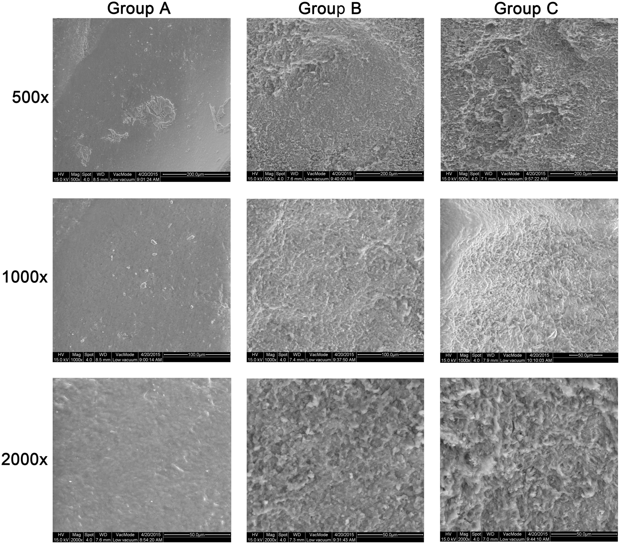
Root surface topography under different magnifications of SEM in different experimental groups. Group A: the control; Group B: 25 J/cm2 and 10 Hz of Er:YAG laser irradiation; and Group C: 50 J/cm2 and 10 Hz of Er:YAG laser irradiation.
hPDLF distribution and adhesion
After cell-specimen coculture, fibroblast distribution and morphology on root surfaces in different experimental groups were investigated under SEM (Fig. 2). The untreated root surfaces appeared to have fewer cells with attachment processes, round and oval in appearance. In Group B (25 J/cm2 Er:YAG laser irradiation) and Group C (50 J/cm2 Er:YAG laser irradiation), fibroblasts firmly attached to rough-surfaced specimens by numerous lamellipodia and extensions. Meanwhile, cells were well spread and exhibited as either stellate or spindle-shaped.
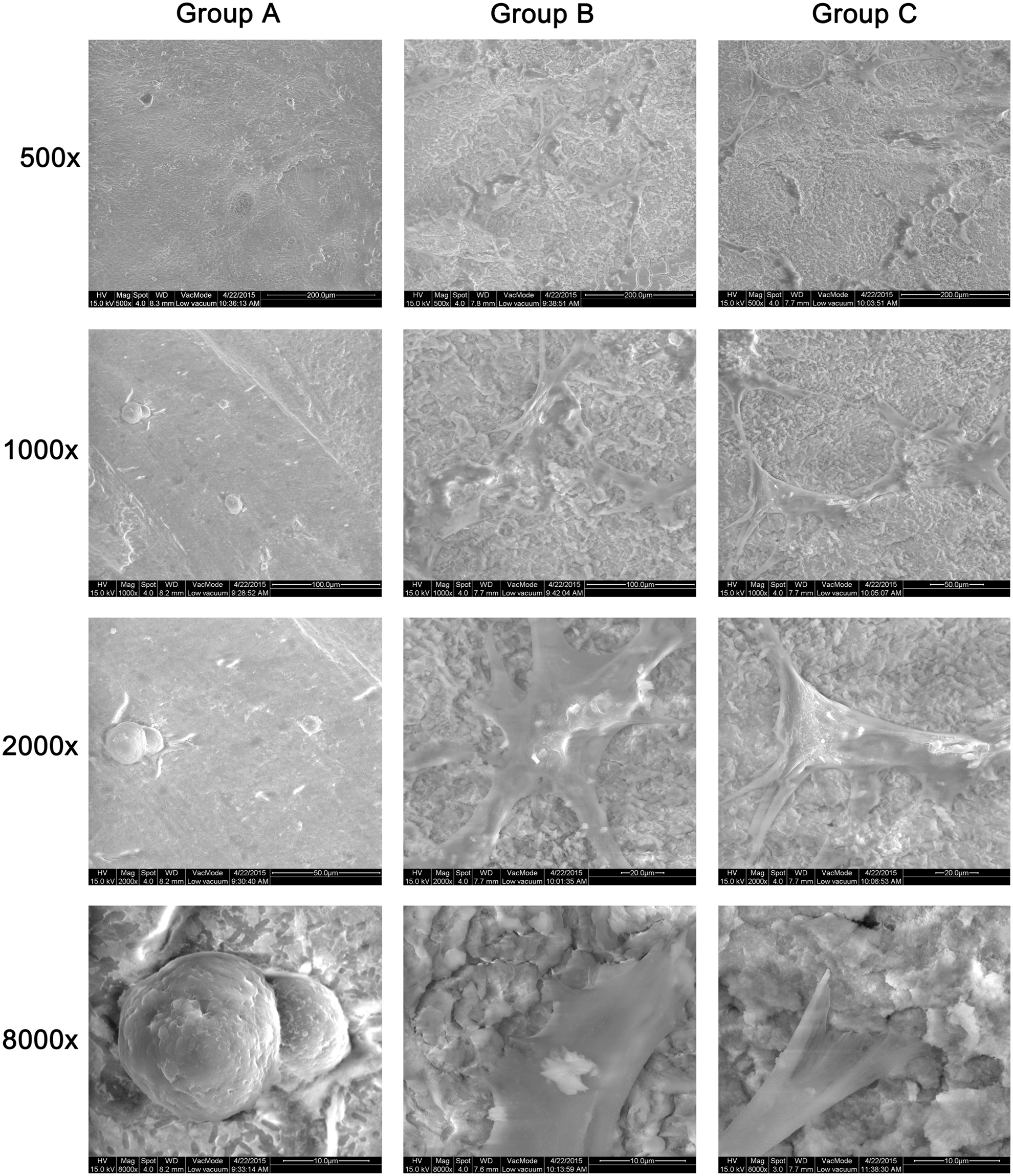
SEM graphs of the hPDLF attachment throughout root fragments in different experimental groups after coculture. Group A: the control; Group B: 25 J/cm2 and 10 Hz of Er:YAG laser irradiation; Group C: 50 J/cm2 and 10 Hz of Er:YAG laser irradiation.
Cell counting
Cell numbers in all experimental groups increased during the 3-day coculture. The number of fibroblasts attaching to the root surfaces in control group (15.55 ± 2.64) was significantly lower (p = 0.008 ≤ 0.05) compared to the other two laser-treated groups (29.66 ± 5.84 in Group B, 27.23 ± 4.31 in Group C). In addition, no significant (p = 0.135 ≥ 0.05) alterations were observed between the two laser groups (Fig. 3).
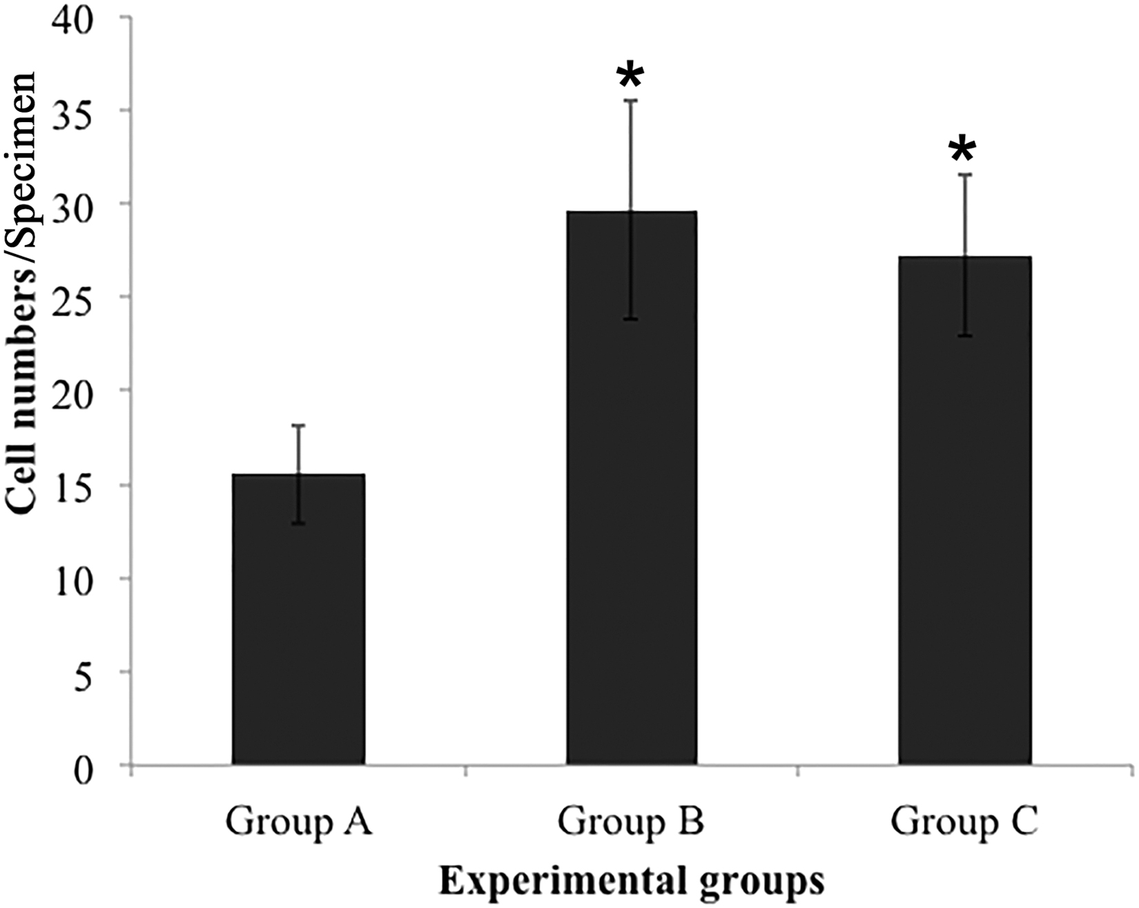
Bar graph showing the counting results (mean ± SD) of the cell adhesion in different experimental groups after a 3-day coculture (*p < 0.05). Group A: the control; Group B: 25 J/cm2 and 10 Hz of Er:YAG laser irradiation; Group C: 50 J/cm2 and 10 Hz of Er:YAG laser irradiation.
Discussion
The length of extra-alveolar period and the storage media appears to have crucial roles in the overall prognosis of teeth replantation. 6 Previous study has shown that the PDL healing percentage of replanted teeth drops to 10% after 20-min dry storage. 3 What's worse, survival of PDL cells is unlikely to happen if dry storage time is longer than 60 min. 1,18 In such cases, necrotic PDL cells can further induce root resorption, which is the main cause of replantation failure. 3,19 Therefore, the first and foremost step in delayed replantation protocol should be the proper and complete removal of attached nonviable periodontal soft tissue, so as to prevent the root resorption. 7 After that, the promotion of the reattachment of periodontal tissue should be further considered. In the present study, roots were soaked into 5.25% sodium hypochlorite for 5 min to remove the periodontal soft tissue, meanwhile leaving little or no harm to the root surface structure. So, the root surface model adopted in this research was used to simulate the avulsed teeth in delayed replantation scenario. This approach was very different from those studies in which Er:YAG lasers were used to treat periodontally involved root surfaces. 15,20
As described in the present study, the morphological roughness and irregularity of Er:YAG laser treated root surfaces could have helped in the enhancement of hPDLF adhesion, which could be concluded from the higher cell counting number observed in both laser-treated groups. Acting as a direct consequence of root conditioning, those minor surface alterations caused by Er:YAG laser irradiation may induce chemical substance exposure in the dentin, which could further promote the fibroblast migration. 21 However, this raises a question whether it's possible for hPDLFs to adhere and proliferate in every case as long as it has rough and uneven root surfaces treated by lasers. Various studies have indicated that CO2 laser irradiation could produce severe thermal damage like distinct multiple affected layers, together with melted and re-solidified structures and carbonization. 22,23 This is very detrimental and may have a negative effect on the absence of PDL cell attachment. 24 Besides, cyanamide and cyanate, both of which are cytotoxic chemicals, were detected on damaged surfaces. Previous studies have shown that these cytotoxic residues prevent cell attachment, migration, and proliferation. 25 Existing studies have also indicated that Neodymium-doped:yttrium aluminum garnet (Nd:YAG) laser can modify the biocompatibility of the cementum surface, making it incompatible for fibroblast attachment. 26 In contrast, no similar damaging phenomenon was reported with Er:YAG lasers. The underlying mechanisms may be related to the difference of laser wavelengths, leading to different interactions with the root surface. With a wavelength of 10.60 μm, energy from most of the CO2 lasers cannot be absorbed by the root surface; thus, the converted heat could induce severe charring and melting. 27 Likewise, Nd:YAG laser at a wavelength of 1.06 μm also has much lower absorption in water and hydroxyapatite compared to the Er:YAG laser. 28 Er:YAG laser has a wavelength of 2.94 μm, and most of the generated energy gets absorbed by water and by the hydroxyapatite on the root surface. 27 This better absorption allows for a more efficient photothermal effect on hard tissue. Consequently, the thermal damage or side effects induced by Er:YAG laser could be much smaller.
Other than cell number, fibroblast distribution and morphology are also very important and valuable. 29 In this context, cells observed in Er:YAG laser-treated groups were flat or spindle-shaped, attaching firmly to the root surfaces by means of numerous attachment extensions and lamellipodia. In contrast, cells in the control group were more of round or oval shape, with relatively poor attachment. These findings are in accordance with results from previous studies which have shown that spindle-shaped fibroblasts were predominantly found on all Er:YAG laser specimens, indicating that such treatment approaches seem to produce more favorable root surface conditions to cellular attachment. 28,29
In the present study, no exposed dentinal tubules were observed, which indicated that only the cementum layers were affected or slightly modified by Er:YAG lasers. Almehdi et al. have shown that in terms of the thickness of altered cementum, the thickness after Er:YAG laser irradiation (21.10 ± 2.40 μm) was markedly lower compared to that obtained after CO2 laser irradiation (143.00 ± 19.70 μm). 22 Since the retention and integrity of the cementum structure are crucial for the reattachment of periodontal tissues, 30 Er:YAG laser appears as an appropriate option when dealing with delayed replantation cases. Of course, appropriate parameters should be selected when using it clinically to avoid excessive energy use and adverse effects.
Conclusions
The present study shows that Er:YAG laser-treated root surfaces are compatible for the attachment of human PDL fibroblasts. Such findings imply that Er:YAG laser irradiation may be used as a promising strategy for root surface conditioning in delayed replantation cases.
Footnotes
Author Disclosure Statement
The authors declare no potential conflicts of interest with respect to the authorship and/or publication of the article.
Funding Information
There was no funding provided for this article.