
Abstract
Objective:
This study sought to compare enamel surface morphology and orthodontic bracket re-bonding strength after phosphoric acid- or erbium-doped yttrium aluminum garnet (Er:YAG) laser-mediated re-etching.
Methods:
A total of 81 extracted premolars were obtained from patients undergoing orthodontic procedures. Conventional etching with 35% phosphoric acid was first used to bond brackets to the enamel surface. Then brackets were de-bonded 1 week later. These samples were then separated randomly into three groups (n = 27 teeth each group) and re-bonded with new brackets after one of the following re-etching manners: Group A—35% phosphoric acid, Group B—Er:YAG laser (200 mJ, 30 Hz), and Group C—Er:YAG laser (250 mJ, 30 Hz). The enamel surface and the interface of enamel and adhesive were then analyzed through scanning electron microscopy. Shear bond strength (SBS) and adhesive remnant index (ARI) were also measured.
Results:
Samples in Group A exhibited significant residual adhesive at the enamel surface, whereas samples in Groups B and C showed a cleaner surface with more distinct and evenly distributed honeycomb-like structures. Further, samples in Group C displayed a larger average SBS value between the two laser-etching groups, although there were no significant differences in SBS values or ARI scores between the acid and laser re-etching groups (p > 0.05).
Conclusions:
Er:YAG laser-based enamel re-etching (250 mJ, 30 Hz) produces an uniform honeycomb-like structure and a trend of similar SBS compared with 35% phosphoric acid-mediated re-etching. Er:YAG laser-mediated re-etching seems to be a promising alternative approach for bracket re-bonding.
Introduction
The use of phosphoric acid for the preparation of tooth enamel surface before orthodontic bracket bonding has been a standard practice for >60 years since it was first detailed by Buonocore. 1 However, de-bonding due to eating during fixed orthodontic treatment is a common problem, and in many cases an individual tooth may need to undergo repeated re-bonding. Previous studies 2,3 reported that the initial bonding typically fails in 2–4% brackets in a period of 4–5 days, re-bonding is associated with a significantly higher rate (10–15%) of bracket bonding failure. Indeed, Bishara et al. 2 have determined that re-bonding is associated with a roughly 33% reduction in shear bond strength (SBS). The presence of residual adhesive on the enamel surface after the use of phosphoric acid etching could be one of the reasons leading to the significant reduction in re-bonding strength. In addition, phosphoric acid-mediated etching can also result in substantial decalcification of the surrounding enamel and leave the area surrounding these orthodontic brackets more susceptible to acid-mediated damage. 4 –6 Therefore, it is important to explore better strategies for enamel surface etching to increase the re-bonding strength and reduce the decalcification.
Erbium-doped yttrium aluminum garnet (Er:YAG) lasers have a 2940 nm emission wavelength, which coincides with the primary absorption band for water and OH− groups in hydroxyapatite, thus leading to the frequent use of these lasers for ablation of hard surfaces such as dentin and enamel. 7 Notably, the laser-etched enamel surface has been shown to be more resistant to the development of caries. 8 –11 Previous reports 12 have validated the use of Er:YAG lasers to ablate enamel and dentin without inducing any thermal damage to the pulp and any cracking or carbonization on the enamel surface when combining with water cooling.
Previous studies focused primarily on whether Er:YAG laser can serve as a viable alternative to phosphoric acid for enamel etching of initial bonding. Studies 13,14 reported that laser etching yields lower SBS than acid etching. Akin et al. 15 also failed to detect any advantage to the use of an Er:YAG laser (80 mJ/4 Hz) relative to phosphoric acid etching as a means of improving orthodontic bracket bonding strength. In contrast, Aglarci et al. 16 found that Er:YAG (120 mJ/10 Hz; LightWalker; Fotona, Slovenia) and acid-based etching approaches yielded comparable SBS values [7.49 ± 3.4 and 7.55 ± 2.8 megaPascals (MPa), respectively]. Sallam and Arnout 17 similarly observed no significant differences in outcomes when they used phosphoric acid (8.988 ± 1.6194 MPa) or a 1.5 W Er:YAG (LightWalker; Fotona) laser (9.600 ± 1.1072 MPa) to facilitate orthodontic bracket bonding. Notably, the use of different laser parameters might be the main cause for the discrepancies among different studies. In our previous research, 18 the SBS between the acid-mediated primary etching group and the Er:YAG-mediated primary etching with three different parameter settings (200 mJ/30 Hz, 250 mJ/30 Hz, and 300 mJ/20 Hz) was compared, and the result displayed that only 250 mJ/30 Hz Er:YAG laser-mediated etching generated a similar SBS with the acid-mediated etching (23.37 ± 4.28 and 25.09 ± 5.19 MPa, respectively). The SBS after 200 mJ/30 Hz and 300 mJ/20 Hz laser-mediated etching (20.63 ± 4.94 and 16.17 ± 3.63 MPa, respectively) were significantly lower than that of the acid-mediated etching. This proves that laser with different parameters can obtain different enamel etching effects. Thus, it is important to obtain suitable laser parameters for the optimal enamel adhesion effect.
However, the use of Er:YAG laser for enamel re-etching and related bond strength have not been evaluated. Whether Er:YAG laser-mediated re-etching suites for bracket re-bonding has not been reported either. This study, therefore, sought to compare the SBS after the re-bonding of orthodontic brackets through either conventional phosphoric acid or Er:YAG laser-mediated etching of the enamel surface. In addition, the morphological changes of enamel surface and the resin–enamel interface were further analyzed through scanning electron microscope (SEM).
Materials and Methods
Tooth selection and storage
The research material consisted of 81 anatomically normal human premolars that had been extracted from patients during orthodontic procedures. These teeth were free of caries, cracking in the enamel, fillings, or endodontic treatment. The study was approved by the human subjects ethics board of Xuanwu Hospital, Capital Medical University (Ethical Approval: LYWS[2019]008) and was conducted in accordance with the Helsinki Declaration of 1975. After collection, these teeth were incubated for 24 h in 1% Thymol, followed by storage in the 37°C water bath (HH-US-A; HerryTech, China). All samples were collected within a 6-month period, and the water bath was replaced weekly to avoid bacterial growth.
Initial tooth preparation, bonding, and de-bonding
Stored teeth were cleaned with pumice and a fluoride-free paste (Proxyt; Ivoclar, Liechtenstein) for 20 sec with a rubber cap (P-C505, TPC, China), followed by rinsing using water and air drying. Thirty-five percent phosphoric acid gel (Unitek; 3M, USA) was then used to etch all teeth for 60 sec, after which they were rinsed for 5 sec and dried again for 15 sec. Next, Transbond XT primer and Transbond XT (Unitek; 3M) was used to bond the stainless steel brackets (Unitek; 3M). Then samples were put into a 37°C water bath for 24 h, after which the brackets were removed from all teeth by de-bonding pliers. Remnant adhesive was cleaned with an adhesive removal clamp and fluoride-free paste.
Secondary tooth preparation
The 81 teeth were randomly assigned to three different treatment groups (n = 27/group): Samples in Group A underwent re-etching using 35% phosphoric acid gel. Samples in Groups B and C underwent re-etching using an Er:YAG laser (LiteTouch™; Syneron, Israel). A 1.3 × 1.9 mm tip coupled with a water spray was used for laser emission, with the laser positioned 2 mm from the tooth surface at a 60° angle. The parameters of Er:YAG laser were derived from our initial study
18
and detailed in Table 1.
SEM assessment of enamel surface morphology
Adhesive carbon paper was used to attach a total of six teeth (two per group) to a testing ring. Samples were gold-coated (FC-1100; The Island Ferry, Japan) before analysis through SEM (EVO 18; ZEISS, Germany).
Treatment Group Parameters
E, energy; Er:YAG, erbium-doped yttrium aluminum garnet; F, frequency; W, water.
SBS and adhesive remnant index testing
The re-bonding process was identical to the initial bonding process, and was conducted on all remaining teeth (n = 25/group). Samples were then again stored in a 37°C water bath for 24 h, after which a universal testing machine (3367; Instron, USA) was used to test shear bonding force using a flat metal blade. The crosshead had a 1 mm/min loading speed, with the load being applied vertically to the surface between base and wing of the bracket. At the moment of de-bonding, a computer recorded the shear force magnitude (in Newtons), after which this value was divided by the bracket surface area (9.99 mm2) to calculate SBS in MPa. In addition, the adhesive remnant index (ARI) was assessed through stereo-microscope (220670; Olympus, Japan) at 10 × magnification to understand the mechanism of failure with scoring from 0 to 3 as follows:
ARI 0: No remaining adhesive was present on the enamel.
ARI 1: <50% of adhesive remained present on the enamel.
ARI 2: >50% of adhesive remained present on the enamel.
ARI 3: All adhesive remained present on the enamel together with a clear bracket mesh impression. 19
SEM cross-sectional observation of interface between the enamel and adhesive
After the de-bonding, six teeth (n = 2/group) were cut along the long buccal axis with a dedicated slicer (IsoEet Low Speed SAW; Buehler, Germany). Samples were then immersed in 15% HCl for 15 sec, followed by ultrasonic washing with distilled water for 30 sec, after which they were placed in a dryer for 24 h. Samples were then attached to a testing ring and gold-coated before the SEM analysis.
Statistical analysis
SPSS 17.0 was used for all statistical testing. SBS data for the three groups were subjected to normality testing using the Kolmogorov–Smirnov test. Normal distributions were observed; therefore, the one-way analysis of variance was used to determine the significance of differences of SBS among groups. The Student–Newman–Keuls test (α = 0.05) was performed to determine differences among groups, whereas ARI scores were assessed through the Kruskal–Wallis test (α = 0.05). The level of significance for the analysis was p < 0.05.
Results
Enamel surface analysis by SEM
The research began by assessing the enamel surface of teeth after the three re-etching procedures through SEM (Fig. 1). The result showed that acid-mediated re-etching (Group A) was associated with significantly more residual adhesive on the enamel prisms (Fig. 1A-a). In contrast, samples subjected to laser-mediated re-etching (Groups B and C) exhibited prism center hollowing consistent with a uniform honeycomb-like structure 20 (Fig. 1B-b, C-c). These laser-etched samples also exhibited areas of roughness in the enamel, with the peripheries of prisms being clearer in Group C relative to Group B.
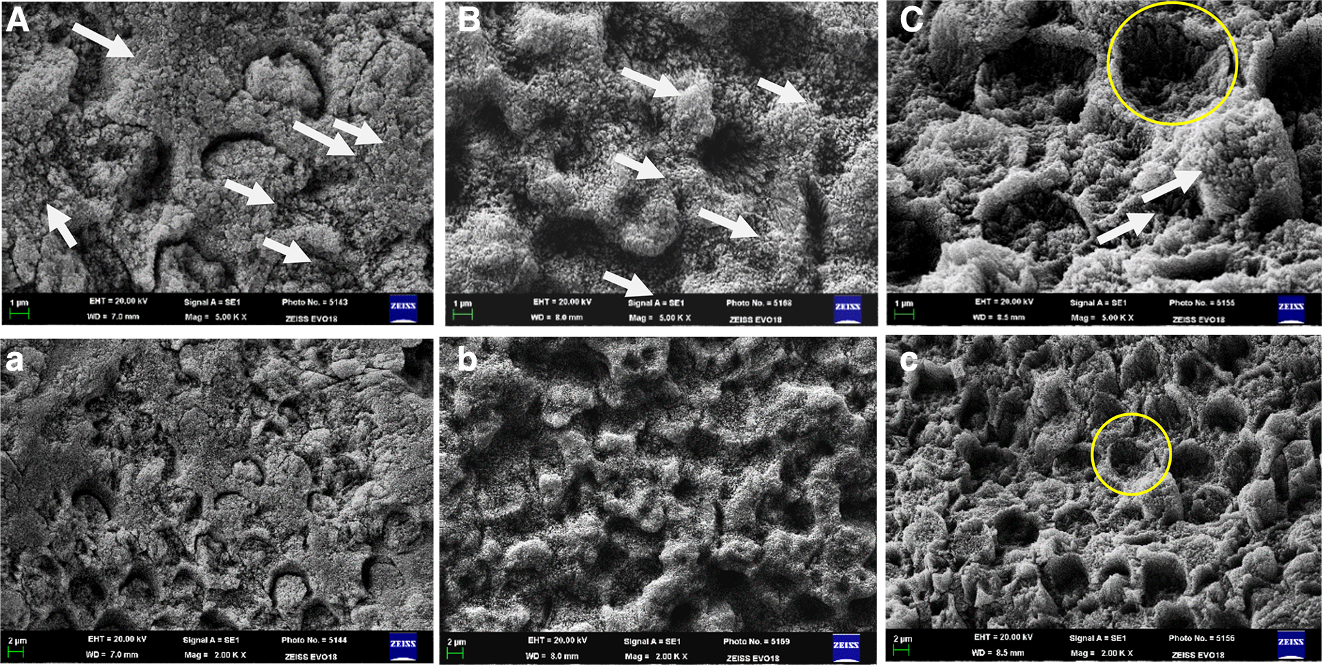
SEM images of the surface of the enamel in the three study groups ( × 5000, × 2000).
Assessment of SBS and ARI
The SBS values after re-bonding after the implementation of the three re-etching procedures are shown in Table 2. Samples in Group C (Er:YAG laser; 250 mJ) had an SBS value (19.79 ± 4.45 MPa), samples in Group B (Er:YAG laser; 200 mJ; 17.09 ± 4.92 MPa) and Group A (phosphoric acid; 17.91 ± 4.83 MPa). However, no significant difference (p > 0.05) was shown between the acid-etched and laser-etched groups, and the difference between Groups B and C was not significant (p > 0.05) either. The ARI values in the three treatment groups are shown in Table 3. In Group A, 48% of the specimens had an ARI score of 0 and 1, whereas 52% had an ARI score of 2 and 3. In Group B, 60% of the specimens had an ARI score of 0 and 1, which indicates the presence of less adhesive on the enamel surface after bracket de-bonding. In Group C, 48% of the specimens had an ARI score of 2 and 3, which was close to Group A.
Shear Bond Strength Values
p > 0.05; no statistically significant difference.
SBS, shear bond strength; SD, standard deviation.
Adhesive Remnant Index Scores
p > 0.05 no statistically significant difference.
ARI, adhesive remnant index.
SEM cross-sectional analysis
Upon SEM-mediated analysis of the interface of bonding, we found that after phosphoric acid re-etching the bonding interface presented more gaps, and uneven cross chimerism; whereas the laser groups presented tight bonding and fewer gaps, and the enamel–adhesive interface presented more uniform cross chimerism with disordered enamel rods under this interface layer with ∼15–20 μm thickness (Fig. 2).
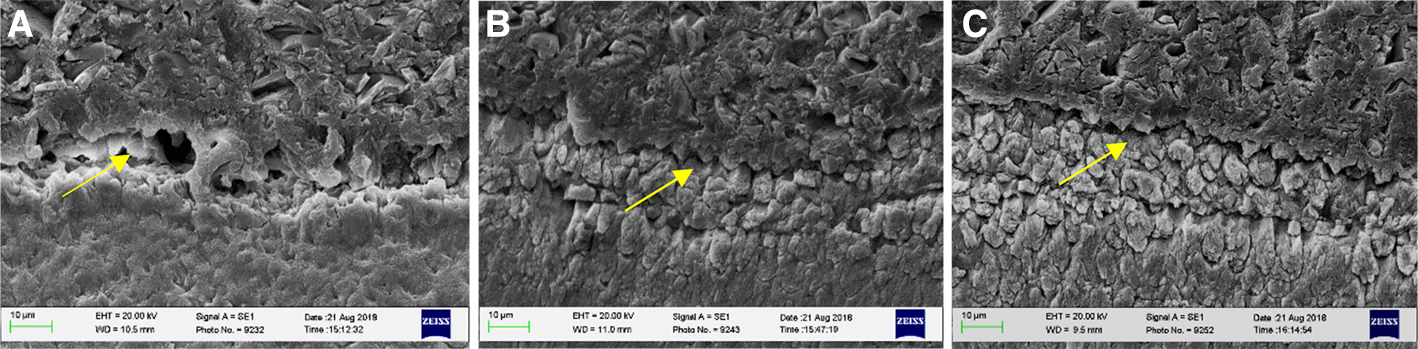
SEM images of enamel cross section in the three study groups ( × 1000).
Discussion
The repeated use of 35% phosphoric acid to etch teeth may increase susceptibility to caries. Moreover, after re-etching with phosphoric acid, the adhesive shedding rate of brackets increased obviously. 2 Given these significant disadvantages, many groups have recently explored the application of laser-mediated etching of the enamel surface. Laser-mediated etching was originally predicted to result in reduced demineralization and damage relative to acid-mediated etching. 5 Several researchers have provided different results about Er:YAG laser-mediated etching. 13 –17 This might be due to the differences of laser working parameters in different studies. Recent research has largely suggested that, Er:YAG laser-mediated etching with appropriate optimization could achieve similar results to those produced by acid etching. 16 –18 This led us to hypothesize that laser re-etching may achieve similar or superior efficacy in the context of bracket re-bonding relative to acid-mediated re-etching.
The difference of enamel surface morphology between acid- and ErYAG laser-mediated re-etching
Primary phosphoric acid etching is associated with an overall increase in enamel surface roughness, with a distinct etching pattern defined by the hollowing of prism centers with relatively intact peripheral regions being observed in most cases. 20 In this study, teeth subjected to laser-mediated re-etching exhibited a clear and evenly distributed etching pattern with prism center hollowing consistent with a honeycomb-like etching pattern. But the depth of the prism center varies in laser groups may be related to unstable hand speed and repeated irradiation.
The difference of re-bonding efficacy between acid- and Er:YAG laser-mediated re-etching
In addition, acid re-etching was associated with a similar SBS to laser re-etching (200 mJ, 30 Hz/250 mJ, 30 Hz), and the difference between these groups was not significant. Bishara et al. 2 previously found that bond strength (4.1 ± 2.3 Mpa) decreases significantly after acid-mediated re-etching owing to the presence of residual adhesive on the enamel surface and associated morphological changes. Although Er:YAG lasers are capable of removing residual adhesive after de-bonding, acid re-etching fails to remove resin, leading to the presence of many residual resin tags attached to the enamel surface. In our study, the ARI score was mostly 1 and 2 in the three groups, this means that bond failure happened at bracket–adhesive and adhesive–enamel interface. And in laser group (200 mJ/30 Hz), 60% of the specimens had an ARI score of 0 and 1, this means that bond failure was mostly at the adhesive–enamel interface, but the SBS values of Group B was the lowest. In laser group (250 mJ/30 Hz) and acid group, close to half of specimens had an ARI score of 0 and 1, and the SBS values were higher than Group B. Maybe that the number of these two bonding failure mode was closely associated with the higher SBS values.
The difference of the enamel-adhesive surface
Further, we conducted a cross-sectional examination of the enamel-adhesive surface and found that teeth subjected to acid re-etching had loose bonding and uneven cross chimerism in the interface relative to teeth subjected to laser re-etching, which was associated with shorter but more densely packed resin tags and a more compact interface layer. This means that the depth of laser re-etching enamel is shallow and the roughness of enamel surface is larger compared with acid re-etching. Laser re-etched teeth exhibited a clearer and more evenly distributed etching pattern than did acid re-etched teeth, with the higher power laser yielding clearer enamel rod boundaries. This may indicate that the higher laser parameter could effectively remove residual adhesives. Delfino et al. 21 found that a 250 mJ Er:YAG laser resulted in a more regular interface layer with good tag formation mainly in the total etching system, but when the energy was increased to 300 mJ, the Er:YAG laser yielded a more irregular interface with amorphous enamel and fused areas. They concluded that lasers with higher energy had a more profound influence on the enamel–adhesive interface, and the result was completely consistent with our experiment about the primary etching. 18 Therefore, the higher energy parameter (300 mJ) was not selected in this study. Again, it must be emphasized the importance of selecting appropriate energy parameters when using lasers for re-etching.
In this study, we also found that enamel rods beneath the interface layer were disordered with ∼15–20 μm thickness by SEM observation. This might be because laser re-etching has led to rearrangement and could potentially result in increased acid resistance, but additional research will be needed to confirm this.
Conclusions
The use of an Er:YAG laser (200 mJ, 30 Hz or 250 mJ, 30 Hz) to facilitate enamel re-etching resulted in similar SBS relative to the use of 35% phosphoric acid. And the ARI score had no significant difference between laser groups and the acid group. Also, laser-based re-etching was associated with a more uniform honeycomb-like structure on the enamel surface and a more uniform cross chimerism in enamel–adhesive interface when a 250 mJ laser irradiation was employed. Combining these results, Er:YAG laser-mediated re-etching seems to be representing a promising alternative approach for bracket re-bonding.
Footnotes
Author Disclosure Statement
No competing financial interests exist.
Funding Information
No funding was received for this article.